1

1 CASE 1


A 42-year-old man comes to his primary care physician complaining of awkwardness when running or walking.


The patient indicates that he noticed trouble with cramping and weakness in his right leg 4 months ago but attributed that to a change in his exercise habits. The weakness is progressing despite continued exercise, and now the left leg is beginning to show signs of weakness. He was last seen for a routine physical examination 3 years ago, and no abnormalities were noted at that time.


PATHOPHYSIOLOGY OF KEY SYMPTOMS


The diagnosis of ALS is achieved by eliminating the variety of other causes of muscle weakness. This diagnosis is complicated by the fact that the appearance of clinical symptoms and the time course for the progression of the disease are quite variable. One diagnostic approach involves beginning with skeletal muscle function and assessing function from the periphery back to the central nervous system.


Skeletal muscles elsewhere in the body are functioning normally. This indicates that the muscles are capable of maintaining a normal resting membrane potential and that cellular and extracellular electrolyte concentrations are within the normal range. The muscle biopsy rules out any protein-related defects in the muscle contractile and structural proteins. Electromyography confirms that the muscle is still able to contract when directly electrically stimulated. The defect in nerve traffic, however, eventually leads to atrophy of the skeletal muscle and exacerbates the weakness associated with the progression of the disease.


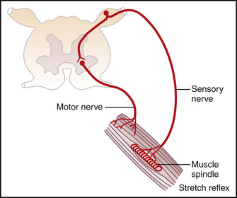
An appropriate patellar tendon reflex requires afferent input from the muscle spindles, transmission of the afferent action potential to the spinal cord, a monosynaptic reflex within the spinal cord, efferent action potential transmission by the alpha (α)-motor neuron, transmission across the neuromuscular synapse, activation of the skeletal muscle, and, finally, contraction (Fig. 1-1). This patient shows a hyperreflexive response in the right leg. Normally, the α-motor neurons receive descending inhibitory input from upper motor neurons and both inhibitory and stimulatory input from interneurons (Figs. 1-2 and 1-3). Hyperreflexia can be an indication that the normal descending input is diminished, which is characteristic of an upper motor neuron disease.


< div class='tao-gold-member'>

Stay updated, free articles. Join our Telegram channel

Jul 4, 2016 | Posted by in PHYSIOLOGY | Comments Off on 1

Full access? Get Clinical Tree

Get Clinical Tree app for offline access