CASE 1
A 35-year-old man, a moving company employee for 15 years, complained of back pain and neurological symptoms after a full day of work moving a family to their new home. He went home and took some aspirin for the pain, but the symptoms persisted the next morning. Concerned, he visited his physician who conducted a neurological examination and ordered imaging studies. The MRI revealed a herniated intervertebral disc at L4/L5 (Fig. 1-1).
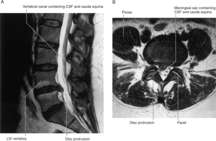
FIGURE 1-1 MRI of a herniated disc between L4 and L5 vertebrae. A, Sagittal plane. B, Axial plane.
(Drake R, Vogl W and Mitchell A: Gray’s Anatomy for Students. Churchill Livingstone, 2004. Fig. 2-33.)
WHERE ARE INTERVERTEBRAL DISCS FOUND?
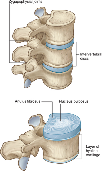
Intervertebral discs are found between the bodies of adjacent vertebrae from the axis to the sacrum (Fig. 1-2). A disc is not found between the atlas and the axis. The discs are thinnest between the cervical vertebrae and progressively thicken as they descend the vertebral column. In the secondary curvatures of the vertebral column (cervical and lumbar regions), the intervertebral discs are thicker anteriorly. This produces the anterior convexity of the cervical and lumbar segments of the vertebral column.
WHAT IS THE HISTOLOGY OF THE INTERVERTEBRAL DISC?
The intervertebral disc is composed of an outer ring, anulus fibrosus, and an inner core, nucleus pulposus (Fig. 1-2). The anulus fibrosus has an outer thin layer of dense collagenous tissue and an internal wider layer of fibrocartilage. The nucleus pulposus is derived from the notochord. It has a gelatinous consistency due to the presence of glycosaminoglycans (mucopolysaccharides) and proteins.


