Ischaemic Heart Disease
This typically presents as the tight or crushing central chest pain of angina or myocardial infarction. Less commonly, it presents as an arrhythmia or conduction defect, or heart failure.
Myocardial ischaemia is normally caused by atherosclerosis, but cardiac pain is also produced by:
- aortic dissection
- paroxysmal tachycardias
- severe anaemia, cardiomyopathy, coronary artery embolism and vasculitis – all rare causes.
Coronary Artery Disease
Examination of atherosclerotic plaques indicates an interaction between blood constituents and cellular elements of the arterial wall. Alteration of normal endothelial cell function may allow accumulation of macrophages, which form foam cells and provoke proliferation of smooth-muscle cells and connective tissue. Cholesterol crystals and other lipids accumulate at the base of plaques, which are covered by a fibrous cap. Plaque rupture leads to thrombosis.
Factors associated with coronary artery disease include:
- Sex: it is more common in men than women, particularly before the menopause.
- Age: there is a steady increase with age.
- Smoking: is a powerful risk factor for coronary heart disease (CHD). Cessation is associated with significant reduction in risk, which decreases by half after 1 year and approaches that of never-smokers after several years.
- Hypertension: the risk of coronary heart disease rises progressively with increasing blood pressure. Although most antihypertensive trials have shown a lower than expected reduction in risk, this may reflect the short duration of the trials.
- Obesity: central adiposity is a better marker for CHD risk than the overall level of obesity.
- Hyperlipidaemia: CHD is associated with raised total cholesterol and high ratio of total cholesterol:high-density lipoprotein (HDL) cholesterol. Hypertriglyceridaemia appears to be associated more with risk of myocardial infarction than coronary atherosclerosis, possibly because it affects coagulation.
- Diabetes mellitus: hyperlipidaemia in diabetics is a powerful risk factor. Men with diabetes have 2–3 times and women 4–5 times the background population risk of CHD.
- Alcohol: the association between CHD and alcohol is J-shaped – the lowest risk is associated with moderate alcohol intake.
Angina Pectoris
Diagnosis
The diagnosis of angina is clinical, based on the characteristic history:
- site: central chest
- character: usually tight, heavy, crushing
- radiation: to arms, epigastrium, jaw or back
- precipitation: by effort or emotion, particularly after meals or in the cold
- relief: within minutes by rest or sublingual or buccal glyceryl trinitrate.
There are no specific physical signs. A non-cardiac cause is favoured by continuation for several days, precipitation by changes in posture or deep breathing, the ability to continue normal activities, and lack of relief by rest. The more common alternatives in the differential diagnosis are oesophageal pain and musculoskeletal pain.
Unstable angina refers to:
- angina of effort of recent onset with no previous history
- increased frequency and/or severity of pre-existing angina
- angina at rest.
Investigation
Electrocardiogram
The electrocardiogram (ECG) is usually normal between attacks, but may show evidence of old myocardial infarction, T-wave flattening or inversion, bundle branch block or signs of left ventricular hypertrophy. ST segment depression is usually seen during attacks and may be provoked by exercise testing. A negative exercise test, in which there is no chest pain, no ST depression, no arrhythmia and no sustained fall in blood pressure, indicates a good prognosis. Radionuclide studies can be performed if the patient is physically unable to exercise. Images at rest are compared with images obtained after pharmacological stimulation of coronary flow to evaluate the presence of local ischaemia or infarction.
In unstable angina there is ST depression or T-wave change during typical anginal pain but without diagnostic elevation in cardiac enzymes. Nitrates can reverse ST segment elevation.
Coronary arteriography may provide unequivocal evidence of arterial narrowing and define its site to guide revascularisation procedures.
Management of Stable and Unstable Angina
Risk factors should be identified and advice given about stopping smoking, losing weight and taking regular exercise. Treat hypertension and hyperlipidaemia. Anaemia should be investigated and treated.
Sublingual glyceryl trinitrate remains the mainstay of symptomatic treatment. The major side effect is headache. It should be taken for pain, and prophylactically before known precipitating events. Long-acting nitrates such as isosorbide-5 mononitrate in a 60-mg sustained release formulation given once daily in the morning can give therapeutic plasma nitrate concentrations during the day, and allow a gradual fall during the night to prevent nitrate tolerance without a pre-dose rebound in angina. β-blockers reduce morbidity as well as control symptoms in stable angina. If necessary a dihydropyridine calcium-channel blocker such as amlodipine (not verapamil or diltiazem) can be added. If a β-blocker is contraindicated or not tolerated, diltiazem or verapamil can be used. Nicorandil, a potassium-channel activator, can also be beneficial. ACE inhibitors should be used in patients who also have left ventricular dysfunction or diabetes, unless there are contraindications. Low-dose aspirin (75 mg/day) reduces the risk of acute coronary events and myocardial infarction in patients with stable angina. Clopidogrel, an adenosine diphosphatase (ADP) receptor antagonist, may confer additional benefits but increases the risk of bleeding (see Trials Box 10.1). Diet and statins should be used to reduce LDL cholesterol with a target of < 2.0 mmol/l.
 Trials Box 10.1 Anti-platelet and Anti-coagulant Agents in Ischaemic Heart Disease and Myocardial Infarction
Patients with unstable angina (evidence of ongoing myocardial ischaemia without evidence of infarction) should be admitted to hospital and precipitating factors (e.g. anaemia, arrhythmia) identified and treated. Non-ST segment elevation myocardial infarction (NSTEMI) is a closely related condition in which there is sufficient myocardial damage to release a marker of cardiac injury such as troponin or the MB isoenzyme of creatine phosphokinase (creatine phosphokinase has two subunits, which are either B (brain) or M (muscle) types; CK-MB containing B and M subunits is found in cardiac muscle – page 77). Treatment involves bed rest with ECG monitoring whilst pain is ongoing, and escalation of anti-ischaemic therapy. This should include low-dose aspirin, intravenous unfractionated or subcutaneous low-molecular-weight heparin (see Trials Box 10.1) and sublingual nitroglycerin, followed by intravenous nitrates. β-blockade can be added if there is ongoing pain and no contraindication. If β-blockers are contraindicated a non-dihydropyridine calcium antagonist (e.g. verapamil or diltiazem) can be used in the absence of severe left ventricular dysfunction or other contraindications. An ACE inhibitor can be added if hypertension persists despite the above measures. Pain should be controlled with morphine if not relieved, and supplemental oxygen administered if needed to maintain SaO2 > 90%.
Coronary Angiography and Revascularisation
Indications for coronary angiography differ between units, but angiography with a view to percutaneous coronary intervention or cardiopulmonary bypass surgery should be considered in all patients with evidence of recurrent ischaemia (angina or ST- segment changes at rest or with minimal activity) or a strongly positive stress test despite medical therapy.
Coronary revascularisation can be achieved by coronary artery bypass grafting (CABG) or percutaneous coronary intervention (PCI) using catheter-borne devices (usually a balloon or laser) to open stenotic areas within coronary arteries (see Trials Box 10.2). CABG tends to be recommended in patients with three vessel disease, significant left main coronary artery disease or two vessel disease with significant proximal left anterior descending coronary artery disease and abnormal left ventricular function. PCI was initially only used for more proximal one vessel disease. Improved techniques, the advent of drug eluting stents and improved pharmacological therapies following PCI have reduced the risk of restenosis or occlusion, and PCI is now used in more complex situations. Patients should receive dual antiplatelet treatment with aspirin and clopidogrel following PCI with stent placement. The recommended duration of clopidogrel therapy depends on the type of stent.
 Trials Box 10.2 Revascularisation for Coronary Artery Disease
Myocardial Infarction
The European Society of Cardiology, the American College of Cardiology Foundation, the American Heart Association and the World Heart Federation published a universal definition of myocardial infarction in 2007 (Thygesen et al. 2007).
The criteria for diagnosis of acute myocardial infarction are met if there is a rise in biomarkers of cardiac injury (preferably troponin) together with one of the following:
- symptoms of myocardial ischaemia
- ECG changes indicative of new ischaemia – new ST segment or T-wave changes, or new LBBB (p. 79)
- development of pathological Q waves (p. 77)
- evidence of new loss of viable myocardium or new regional wall motion abnormality on imaging.
Aetiology
The most common cause is thrombosis in association with an atheromatous plaque that has cracked or ruptured. Necrosis of muscle supplied by the vessel is followed by scarring.
Rare causes (consider in young patients without risk factors) are:
- coronary artery embolism: from thrombus in left atrium or ventricle, or mitral or aortic valve lesions
- congenital abnormalities, such as anomalous origin of coronary artery from pulmonary artery
- coronary artery vasculitis: consider Kawasaki disease in children (p. 287)
- dissecting aneurysm with coronary artery occlusion.
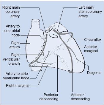
The size and location of the infarct depend on which artery is involved (Fig. 10.1) and the presence of any collateral supply. Occlusion of:
- left anterior descending affects the anterior wall of the left ventricle, and sometimes the septum
- right coronary artery involves the inferior part of the left ventricle, as well as part of the septum and right ventricle
- left circumflex involves the lateral or posterior walls of the left ventricle.
The infarct may extend from endocardium to epicardium (transmural) or involve only the subendocardial region.
Figure 10.1 Anatomy of the coronary arteries. Note that the right coronary artery supplies both the SA and AV nodes.
There may be a
- past history of hypertension, stroke, intermittent claudication, diabetes mellitus, hyperlipidaemia, smoking
- family history of cardiovascular disease, hyperlipidaemia.
Symptoms
- Pain: onset, duration (usually over 20 min), character (often tight or compressing), site and radiation (usually chest going to arms or neck). Associated sweating, breathlessness, nausea and vomiting are common. There may be a previous history of angina or myocardial infarction.
NB: Intensity is no guide to the extent of the infarct, especially in the elderly and in diabetics where pain may be absent. If there is interscapular pain associated with a ‘myocardial infarction’ syndrome, consider dissection of the thoracic aorta.
Examination
Once any distress has been alleviated by pain control there may be no signs. Examine:
- for associated diseases:
 xanthelasmata and xanthomata of hyperlipidaemia
 evidence of diabetes, thyroid disease, diabetes mellitus, gout, cigarette smoking (smell and finger staining)
- pulse for small volume (low cardiac output), arrhythmia
- blood pressure (hypotension usually indicates low cardiac output; hypertension may not be long-standing)
- jugular venous pressure (JVP) is usually normal – elevation suggests heart failure
- listen to heart for:
 fourth heart sound (common); third heart sound if there is heart failure
 pericardial friction rub
 mitral regurgitation (papillary muscle dysfunction or ventricular dilatation)
 ventricular septal defect (VSD) caused by a ruptured septum (rare)
- listen to lungs for basal crackles of heart failure.
Investigations
ECG (see p. 78 and Box 10.1)
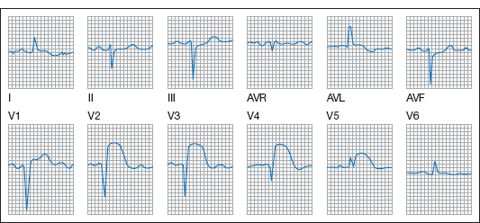
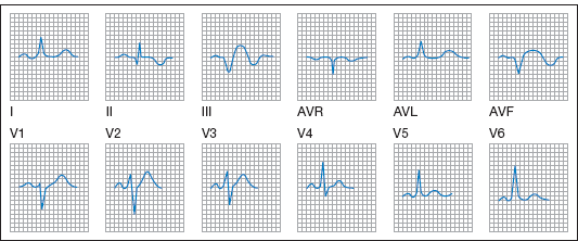
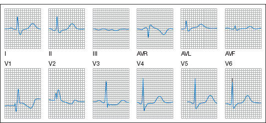
Serial ECGs typically show ST segment elevation and T-wave inversion, with the development of Q waves indicating full-thickness myocardial necrosis. Changes occur in the anterior chest (V) leads in anterior myocardial infarction (Fig. 10.2) and in leads II, III and augmented voltage foot (AVF) in inferior myocardial infarction (Fig. 10.3).
 Box 10.1 ECG Patterns in Common Clinical Conditions
- First few minutes – peaked T waves.
- First few hours – ST segment elevation, inversion of T waves, development of Q waves.
- After a few days the ST segment returns to normal (persistent elevation raises the possibility of left ventricular aneurysm).
- The T waves may eventually become upright, but in full thickness untreated myocardial infarction Q waves persist indefinitely.
- Rhythm abnormalities are common.
- Left bundle branch block (Fig. 10.8, p. 79) may occur at any stage, making further interpretation of the site, timing or extent of an infarct impossible.
Figure 10.4 Acute pulmonary embolism with S1, Q3, T3 pattern, a mean frontal QRS axis towards the right (+90°) and right ventricular (RV) strain pattern in leads V1–3.
- Tachycardia and transient arrhythmias (particularly AF).
- Right axis deviation.
- Right ventricular strain pattern – dominant R wave and inverted T waves in V1–4.
- RBBB.
- Occasionally S1, Q3, T3 pattern (S wave in lead I, Q and inverted T in III).
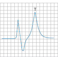
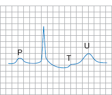
Figure 10.7 Hypocalcaemia. Normal complexes apart from a prolonged QTc (QT interval corrected for heart rate). Rate 95/min, QT 0.4, QTc 0.5.
- The QRS complex is greater than 0.12 s.
- In left bundle branch block (LBBB) the complex is negative (V- or W-shaped) in V1 (Fig. 10.8). Causes include ischaemic heart disease, myocardial infarction, cardiomyopathy, hypertension and aortic stenosis.
- In right bundle branch block (RBBB) the complex is positive (M-shaped) in V1 (Fig. 10.9). In addition to occurring in conditions associated with LBBB, RBBB occurs in conditions that put a strain on the right ventricle (e.g. COPD, pulmonary embolism). RBBB can occur in an otherwise normal heart.
Figure 10.8 Left bundle branch block. RSR (M-shaped QRS complex) is visible in some of the left ventricular leads, I, AVL and V4–6, and notched QS complexes in the right ventricular leads, V1–2.
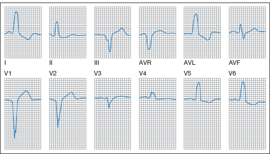
Figure 10.9 Right bundle branch block. RSR in the right ventricular leads V1–2 and slurred S waves in the left ventricular leads I, AVL and V4–6.
- In left anterior hemiblock there is left axis deviation.
- In left posterior hemiblock there is right axis deviation.
Sub-endocardial myocardial infarction leads to ST segment and T-wave changes, but not Q waves. A normal ECG does not exclude myocardial infarction. Posterior infarction is rare and does not produce Q waves, but gives a tall R wave in V1.
Right ventricular infarction is usually associated with inferior infarction, and produces ST elevation which can be detected if right ventricular leads (V3R and V4R) are recorded by placing chest electrodes on the right side of the chest in positions equivalent to V3 and V4.
Cardiac Biomarkers
Troponin T, from cardiac muscle breakdown, is cardiac specific and the marker of choice.
Creatine kinase (CK) is formed by dimerisation of two polypeptide chains, B and M, giving rise to three different isoenzymes. The predominant isoenzyme in skeletal muscle is MM, whereas in brain it is BB. Cardiac muscle contains both MM and MB, and the MB isoenzyme is used in diagnosis of myocardial infarction, although it is also elevated in skeletal muscle disease and by muscle trauma.
Management
Early mortality (within 4 weeks) is chiefly within the first 2 h and usually from ventricular fibrillation. Any patient suspected of having a myocardial infarction requires:
- pain relief – usually in the form of opiates plus antiemetics
- oxygen
- aspirin 300 mg to chew immediately
- nitrates should be administered for relief of ongoing ischaemic pain and can be used to control blood pressure and treat heart failure
- transfer to specialist facilities for reperfusion therapy.
Reperfusion Therapies
Shortening the time between recognition of symptoms and seeking medical help, and initiation of reperfusion treatment with thrombolysis or primary percutaneous coronary intervention is a key element of management (see Trials Box 10.3).
 Trials Box 10.3 Myocardial Infarction
Several studies in the late 1980s showed that intravenous streptokinase reduced mortality in patients reaching hospital with myocardial infarction from just over 10% to around 8%. ISIS-2 (Second International Study of Infarct Survival) showed that aspirin gives additional benefit, and subsequent trials showed that alteplase had similar effects.
Streptokinase 1,500,000 units is given by intravenous infusion over 1 h. It is cheaper than alternatives but can cause allergic reactions. Its potential for repeated use is limited by antibody formation. Alteplase (recombinant tissue plasminogen activator, rtPA) is not antigenic but is expensive. It is critically important to start thrombolysis within 12 h of myocardial infarction; additional benefit is obtained if started within 6 h.
Contraindications to thrombolysis are recent bleeding, severe hypertension (blood pressure > 200/120 mmHg), active peptic ulceration, recent stroke (within the last 2 months), proliferative diabetic retinopathy, severe liver or renal disease, pregnancy/lactation, bacterial endocarditis and acute pancreatitis.
Primary percutaneous coronary intervention (PCI) is the preferred treatment if it is rapidly available (Box 10.3). The European Society of Cardiology and the American College of Cardiology/American Heart Association Task Forces all recommend a target for the time from arrival in the emergency department to angioplasty (door-to-balloon time) of 90 min.
β-Blockade
ISIS-1 (First International Study of Infarct Survival) showed that intravenous β-blockade in the early stages of myocardial infarction may confer benefit, but is contraindicated if there is bradycardia, hypotension, heart failure, asthma, sick-sinus syndrome or heart block (second- or third-degree or bifascicular). Long-term β-blockade post-infarction is routine in the absence of contraindications.
Angiotensin-Converting Enzyme Inhibitors
Angiotensin-converting enzyme (ACE) inhibitors are beneficial, particularly in patients with anterior myocardial infarction, but their use may be limited by hypotension.
Anticoagulant and Antiplatelet Therapy
NICE Technology Appraisal 230 recommends the thrombin inhibitor bivalirudin in combination with aspirin and clopidogrel for the treatment of adults with ST-segment-elevation myocardial infarction (STEMI) undergoing primary percutaneous coronary intervention (PCI).
Complications
- Heart failure (p. 88).
- Shock: the patient is hypotensive, pale, cold, sweaty and cyanosed. Arrhythmias are common. Treatment is with:
 oxygen
 diuretics if pulmonary oedema
 vasodilators – ACE inhibitors (arterial and venous) or nitrates (venous) if blood pressure allows
 inotropes – dopamine and dobutamine increase cardiac contractility by stimulating β1-receptors in cardiac muscle. Low doses of dopamine (< 5 mcg/kg/min) induce vasodilatation and increase renal perfusion, whereas higher doses cause vasoconstriction and may exacerbate heart failure.
- Arrhythmias (see p. 84 and Table 10.1).
- Sinus tachycardia: usually requires no treatment.
- Supraventricular extrasystoles: common, but rarely requires treatment.
- Supraventricular tachycardia: arise from the atria or atrioventricular junction. Management depends on the nature of the tachycardia.
- Sinus or nodal bradycardia: may be caused by sedation, particularly with opiates. If the rate is < 50 beats/min and the patient is hypotensive, give atropine 0.6 mg intravenously and repeat twice if necessary. If unsuccessful, consider cardiac pacing.
- Heart block: all degrees of heart block are more serious if they complicate anterior rather than inferior infarcts.
 first-degree: requires no therapy
 second-degree: monitor and consider atropine. Many physicians would consider cardiac pacing for Mobitz type II with anterior infarcts (p. 88)
 third-degree (complete heart block): atropine and isoprenaline may be helpful while awaiting cardiac pacing.
Table 10.1 Cardiac arrhythmias
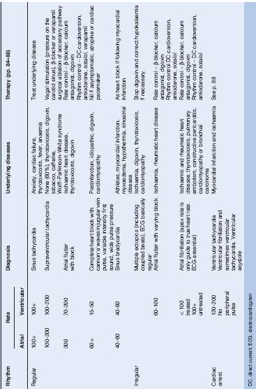
NB: Complete heart block is more common in inferior myocardial infarctions because the atrioventricular nodal artery is a branch of the right coronary artery; complete heart block complicating anterior infarction is ominous because it implies a large muscle infarction.
- Ventricular tachycardia: this can respond to intravenous amiodarone, but proceed to direct current (DC) cardioversion without delay if no success or there is haemodynamic collapse.
- Ventricular fibrillation: this is frequently within 6 h of myocardial infarction (see Cardiorespiratory arrest, p. 88).
- Electromechanical dissociation (complexes on ECG with no pulse – see Cardiorespiratory arrest, p. 88).
Late Complications
Ventricular aneurysm can cause heart failure, angina, arrhythmias and emboli from thrombi within the aneurysm. There may be cardiac enlargement and abnormal cardiac pulsation (e.g. an impulse at the left sternal border). Its presence is suggested by ST-segment elevation persisting in convalescence. The aneurysm may be demonstrated by echocardiography, radionuclide studies or left ventriculography. Surgical removal is indicated for heart failure or arrhythmias. Anticoagulants reduce the risk of emboli.
Papillary muscle dysfunction or rupture may cause heart failure. There is a pansystolic or late sysytolic mitral regurgitant murmur. Echocardiography confirms the diagnosis. Surgery may be indicated.
Ruptured ventricular septum is rare. Urgent surgical repair may be required for severe heart failure.
Myocardial rupture leads to death from tamponade (unless immediate surgery is available). Dressler (post-myocardial infarction) syndrome occurs weeks or months after myocardial infarction or cardiac surgery. It is characterised by fever, pleurisy and pericarditis, and the presence of antibodies to heart muscle.
Invasive and Non-Invasive Assessment Post Myocardial Infarction
Patients with ongoing angina (or other evidence of ischaemia) at rest or on minimal exertion or left ventricular dysfunction should undergo coronary angiography to evaluate the need for coronary revascularisation. Patients in whom angiography is not planned should undergo exercise testing towards the end of the hospital admission or early after discharge to assess functional capacity and look for evidence of inducible ischaemia. Echocardiography should be performed to assess left ventricular function. Dipyridamole or adenosine stress myocardial perfusion imaging can be used in patients unable to exercise or if baseline abnormalities compromise ECG interpretation during exercise testing.
Rehabilitation
Cardiac rehabilitation programmes reinforce the importance of secondary prevention and promote adoption of a healthy lifestyle. Dietary education is given to achieve normal BMI and reduce dietary cholesterol and saturated fat. Active involvement in self-rehabilitation should be encouraged. The importance of stopping smoking must be stressed and strategies to help smokers used.
Patients should be encouraged to increase activity gradually over 1–2 months, when return to work can be considered. Patients should be advised about driving restrictions (p. 88).
Long-Term Pharmacological Treatments
Unless contraindicated, aspirin, β-blockade, angiotensin blockade and statins (Trials Box 10.4) should be started in all patients and continued indefinitely. Target LDL cholesterol should be less than 2.0 mmol/l.
 Trials Box 10.4 Statins
Arrhythmias
(See Table 10.1.)
Supraventricular Tachycardias
Supraventricular tachycardias arise from the atria or atrioventricular junction. QRS complex is normal unless there is also bundle branch block.
Sinus Tachycardia
In sinus tachycardia the rate is >100 beats/min. An underlying cause (anxiety, exercise, fever, anaemia, heart failure, thyrotoxicosis) can usually be identified.
Atrial Fibrillation
Atrial fibrillation (AF) is predominantly a disease of the elderly, occurring in 0.2% of men aged 47–56 and 3% of those aged 77–86 (Framingham study, 1949). Rheumatic AF occurs in association with rheumatic valve disease, whereas in non-rheumatic AF there is an underlying metabolic disorder or other form of cardiac disease. Lone AF occurs in the absence of valvular, myocardial or other identifiable disease. The common causes are:
- ischaemic heart disease
- thyrotoxicosis
- mitral valve disease
- cardiomyopathy.
Management
Check serum potassium, echocardiogram and thyroid function. The aims are to restore sinus rhythm or control the ventricular rate and minimise the risk of embolisation.
Direct current (DC) cardioversion can establish sinus rhythm in 90% of patients, but relapse is common. Long-term amiodarone reduces the frequency of relapse, although side effects can limit its use. Anticoagulant treatment is recommended for 3 weeks before and at least 4 weeks after successful cardioversion (see Box 10.2).
 Box 10.2 Anti-thrombotic Therapy in Patients with Atrial Fibrillation, Including Paroxysmal Atrial Fibrillation, and Atrial Flutter
NB Side effects of amiodarone are reversible corneal microdeposits, photosensitivity, skin discolouration, hypothyroidism, hyperthyroidism, diffuse pulmonary alveolitis and fibrosis, peripheral neuropathy and myopathy.
Medical Therapy
Quinine, flecainide and amiodarone have all been used to restore and maintain sinus rhythm. Digoxin does not restore sinus rhythm, but is effective in controlling the ventricular rate, either alone or in combination with β-blockers.
The incidence of ischaemic stroke (embolic or thrombotic) is increased in patients with AF, and anti-thrombotic treatment is recommended (Box 10.2 and Table 10.2).
Table 10.2 Summary of recommendations for antithrombotic therapy in atrial fibrillation
| Associated conditions | Recommended antithrombotic treatment |
| Prior ischaemic stroke, TIA or systemic embolisation | Warfarin2 – INR target 2.5 (range 2.0–3.0) |
| Two or more risk factors1 | Warfarin2 – INR target 2.5 (range 2.0–3.0) |
| One or more risk factor1 | Warfarin2 – INR target 2.5 (range 2.0–3.0) or aspirin 75–325 mg/day |
| Mitral stenosis | Warfarin2 – INR target 2.5 (range 2.0–3.0) |
| Prosthetic valve | Warfarin2 – INR appropriate for the specific type of prosthesis |
| 1Risk factors: age > 75 years, history of hypertension, diabetes mellitus, moderately or severely impaired left ventricular systolic function and/or heart failure. | |
| 2Or other oral vitamin K antagonist. | |
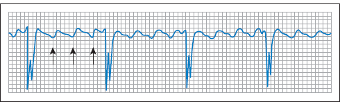
Atrial Flutter
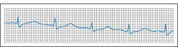
The atria discharge at around 300/min, giving the characteristic ‘saw-tooth’ baseline on ECG (see Fig. 10.10). There is usually atrioventricular block, leading to a ventricular rate of 150/min (2 : 1) or 100/min (3 : 1). The rate is basically regular but is affected by 2 : 1, 3 : 1 and variable block. The causes are similar to AF, although atrial flutter is less common.
Stay updated, free articles. Join our Telegram channel

Full access? Get Clinical Tree


