Veins, Capillaries, and Lymphatics
Lily Marsden, MD
Key Facts
Embryology and Macroscopic Anatomy
Paired vessels form basis of early venous system that returns blood to sinus venosus of primitive heart
Umbilical veins carry oxygenated blood from chorion (primitive placenta)
Vitelline veins drain poorly oxygenated blood from yolk sac
Cardinal veins drain poorly oxygenated blood from body of embryo
Umbilical veins (UVs)
Right and proximal left UVs regress at ˜ 7 weeks gestation
Distal left persists as single UV that drains into liver
Vitelline veins (VVs)
Hepatic veins form from right and left proximal VVs
Portal vein forms from anastomosis of remaining distal right and left VVs
Cardinal veins (CVs)
Common CVs form paired anterior, posterior, subcardinal, and supracardinal veins
Anterior CVs form superior vena cava and internal jugular veins
Posterior, subcardinal, and supracardinal veins all contribute to the inferior vena cava
Posterior CVs form the common iliac veins
Subcardinal veins form renal and gonadal veins
Supracardinal veins form intercostal, hemiazygos, and azygos veins
Lymphatic system develops between 5 and 6 weeks gestation, ˜ 2 weeks after arteriovenous system
6 primary lymph sacs are initially formed
2 jugular lymph sacs at site of future internal jugular veins
2 iliac lymph sacs at site of future common iliac veins
1 retroperitoneal lymph sac at mesenteric root along abdominal inferior vena cava
1 cisterna chyli dorsal to retroperitoneal lymph sac at the level of the adrenal glands
Lymphatic vessels extend from the lymph sacs, following pathways of major veins
Cisterna chyli connects to jugular lymph sacs through right and left thoracic ducts
By 9 weeks gestation, a single thoracic duct is formed from caudal portion of right thoracic duct and cranial portion of left thoracic duct
Cranial portion of right thoracic duct becomes right lymphatic duct
Microscopic Anatomy
Veins are composed of 3 tissue layers: Tunica intima, tunica media, and tunica adventitia
Large veins include vena cavae and portal vein
Intima consists of flat endothelial cells overlying scant subendothelial connective tissue and smooth muscle
Media is composed of a thin layer of smooth muscle and collagen
Adventitia is thick and consists of collagenous connective tissue, elastic fibers, and fibroblasts
Medium-sized veins include majority of named veins
Intima has endothelial cells, thin subendothelium, and smooth muscle
Media consists of concentric smooth muscle and collagen
Adventitia is composed of collagen that blends into surrounding connective tissue
Small veins and venules vary significantly in their histologic appearance
Intima consists of only an endothelial cell lining
Media varies from 1-3 smooth muscle layers in thickness but can be absent in post capillary venules
Adventitia blends into surrounding connective tissue
Capillaries are small in diameter, only allowing single red blood cells to pass at a time
A single layer of endothelial cells and basement membrane attach to each other through tight junctions
Lymphatics vessels are lined by a single layer of flattened endothelial cells
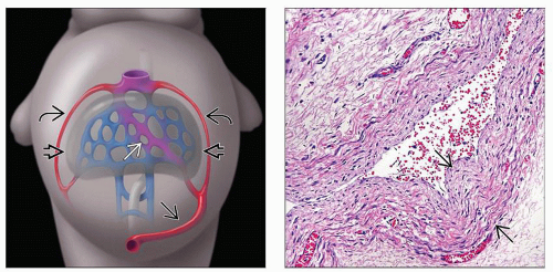 (Left) Right and cranial left UVs
 regress. Single UV regress. Single UV  drains into primitive liver drains into primitive liver  and anastomoses with vitelline system (blue) to form the ductus arteriosus and anastomoses with vitelline system (blue) to form the ductus arteriosus  , which connects to inferior vena cava. (Right) Medium-sized vein contains a thick muscular media , which connects to inferior vena cava. (Right) Medium-sized vein contains a thick muscular media  . Vein can be distinguished from arteries by the lack of elastic laminae. . Vein can be distinguished from arteries by the lack of elastic laminae.Stay updated, free articles. Join our Telegram channel
Full access? Get Clinical Tree
 Get Clinical Tree app for offline access

|