Ureter: Margins
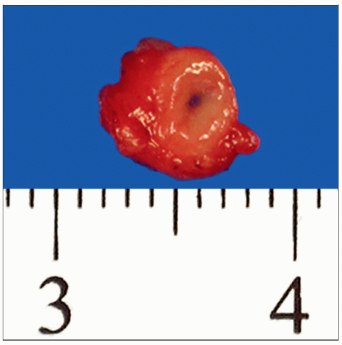 A small length of ureter received for frozen section should be oriented and embedded in such a way that a complete cross section of the lumen is obtained. (Courtesy A. Joiner, MD.) |
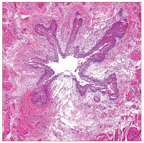 A complete cross section of the ureter is necessary to adequately evaluate the entire urothelium for the presence of carcinoma in situ (CIS). The lumen often has a star-shaped morphology on cross section. |
SURGICAL/CLINICAL CONSIDERATIONS
Goal of Consultation
Determine if ureteral margins on cystectomy specimen are free of carcinoma and atypia
Change in Patient Management
Additional ureter may be resected to achieve tumor-free margins
Clinical Setting
Cystectomies are usually performed for muscle-invasive urothelial carcinomas (pT2) and therapy-refractory urothelial carcinoma in situ
Patients with positive margins are at higher risk for recurrence within remaining upper urinary tract
SPECIMEN EVALUATION
Gross
Small length of ureter is usually provided
True margin should be identified if specimen is too long to be entirely frozen
Often identified by presence of stitch or clip at 1 end of ureter segment
Ureter may be received inverted with mucosal lining on outer surface
Frozen Section
Cross section of margin is embedded with true margin face up so that 1st frozen section will be true margin
If complete cross section of urothelium is not seen on initial sections, deeper levels should be obtained
MOST COMMON DIAGNOSIS
Urothelial Carcinoma In Situ (CIS)
Urothelium appears crowded, with loss of polarity and nuclear overlapping
Cells are enlarged, hyperchromatic, and pleomorphic
Full-thickness atypia is not required
Due to loss of cell cohesion, prominent denudation may be present
Underlying connective tissue may have associated vascular congestion and inflammation
REPORTING
Frozen Section
Positive for CIS &/or invasive carcinoma
Atypical cells present
PITFALLS
Umbrella Cells
Can be mistaken for tumor cells
Reactive Epithelial Changes
Nuclei are enlarged but appear more uniform than CIS and have vesicular nuclei with single small nucleolus
Occasional mitotic figures may be present but should not be atypical
Acute or chronic inflammation is often present within urothelium
Frozen Section Artifact
Urothelial cells artifactually appear larger and more hyperchromatic on frozen section than on permanent sections
Subepithelial Involvement
In rare instances, soft tissue and muscle layers of ureter may contain invasive urothelial carcinoma whereas urothelial mucosa is uninvolved
RELATED REFERENCES
1. Gakis G et al: Sequential resection of malignant ureteral margins at radical cystectomy: a critical assessment of the value of frozen section analysis. World J Urol. 29(4):451-6, 2011
Image Gallery
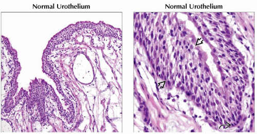
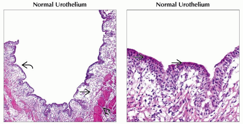 (Left) The normal urothelial surface  has an undulating contour. The urothelium is uniform in thickness. The underlying loose connective tissue has an undulating contour. The urothelium is uniform in thickness. The underlying loose connective tissue  may appear edematous or fibrotic, especially if previous treatments or procedures have been performed. Bundles of smooth muscle make up the muscularis layer may appear edematous or fibrotic, especially if previous treatments or procedures have been performed. Bundles of smooth muscle make up the muscularis layer  . (Right) Normal urothelium has small uniform cells. The cells have round to ovoid nuclei and lightly eosinophilic cytoplasm. A single superficial layer of umbrella cells . (Right) Normal urothelium has small uniform cells. The cells have round to ovoid nuclei and lightly eosinophilic cytoplasm. A single superficial layer of umbrella cells  is present. is present. |
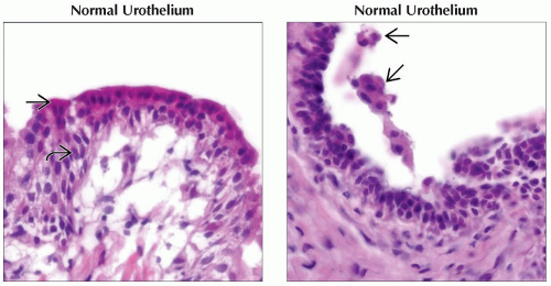 (Left) The nuclei within the intermediate layer  of the urothelium are arranged longitudinally, perpendicular to the basement membrane. A single overlying layer of larger umbrella cells of the urothelium are arranged longitudinally, perpendicular to the basement membrane. A single overlying layer of larger umbrella cells  lines the luminal surface. (Right) Detached clusters of umbrella cells lines the luminal surface. (Right) Detached clusters of umbrella cells  are present within the lumen of a normal ureter. This finding should not be confused with dyscohesive tumor cells. Umbrella cells are characterized by their large size due to abundant eosinophilic cytoplasm and may be binucleated. are present within the lumen of a normal ureter. This finding should not be confused with dyscohesive tumor cells. Umbrella cells are characterized by their large size due to abundant eosinophilic cytoplasm and may be binucleated. |
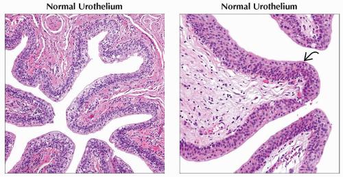
 (Left) This normal ureter appears to have a thickened urothelium. However, the cells have small, uniform nuclei and normal polarity. The lamina propria is edematous and contains inflammatory cells. (Right) Although urothelium is normally 3-7 cell layers in thickness, tangential sectioning can artificially give an appearance of a thickened hyperplastic lining  . The cells are uniform in size and shape with normal polarity. Small basal cells . The cells are uniform in size and shape with normal polarity. Small basal cells  can be seen adjacent to the basement membrane. can be seen adjacent to the basement membrane. |
 (Left) A permanent section of a ureter margin shows the typical star-shaped appearance of the lumen on cross section. The urothelium has an overall orderly appearance on low power, with no nuclear enlargement or loss of normal polarity. (Right) The cells of normal urothelium have lightly eosinophilic cytoplasm and uniform round to ovoid nuclei. A single umbrella cell layer
 is present at the luminal surface. The subepithelial connective tissue contains scattered lymphocytes. is present at the luminal surface. The subepithelial connective tissue contains scattered lymphocytes.Stay updated, free articles. Join our Telegram channel
Full access? Get Clinical Tree
 Get Clinical Tree app for offline access

|