Tumors of Specialized Lower Genital Tract Mesenchyme


Tumors of Specialized Lower Genital Tract Mesenchyme


INTRODUCTION

A number of cellular and myxoid soft tissue lesions of the skin can be encountered in the vulva or scrotum, including dermatofibrosarcoma protuberans (see Chapter 4), vascular lesions (see Chapters 17 and 18), and superficial myxoid tumors (see detailed discussion in Chapter 20). A host of sarcoma types has been described in the female genital tract, especially alveolar soft part sarcoma1 (see Chapter 12). However, a group of myxoid lesions is often considered somewhat site specific, the prototype of which is aggressive angiomyxoma (AAM). All of these myxoid lesions are also found in men but were first described in women.

In some respects, classification of the various angiomyxoid lesions of the genital region is much ado about very little. They are benign but, when infiltrative (AAM), they can be locally aggressive. They all tend to express hormone receptors with variable desmin, actin, and cluster of differentiation (CD)34 and may form a spectrum despite many publications delineating their distinguishing features. In reality, some cases are not clearly recognizable as AAM, angiomyofibroblastoma (AMF), cellular angiofibroma, or pseudosarcomatous fibroepithelial stromal polyps of the lower genital tract. In such a situation, it is probably most important to simply assure that the lesion is not a rhabdomyosarcoma. In some instances, it is most practical to simply diagnose the lesion as a “benign genital stromal tumor.” Many of the lesions in this category express desmin, so use of a specific skeletal muscle marker (MyoD1 or myogenin) is recommended in evaluating such tumors. Of these genital region lesions, AAM is the most likely to be overtly myxoid, but any such case always has a differential diagnosis that includes the other lesions in this group of neoplasms (Table 8.1).


DEEP (AGGRESSIVE) ANGIOMYXOMA

AAM is a well-vascularized, myxedematous spindle cell tumor that presents in the deep soft tissues of the pelvic and perineal regions. It is prone to recurrences owing to its infiltrative growth pattern but does not metastasize.

This distinctive tumor was first delineated by Steeper and Rosai in 1983.2 Subsequent series of AAM have reported nearly identical histologic findings and have confirmed the tumor’s proclivity for local recurrence in both female and male patients.3,4,5,6,7,8,9


TABLE 8.1 Differential Diagnosis of Selected Lesions Presenting in the Genital Region


Lesion


Gross Features


Morphologic and Immunolabeling Features


Aggressive angiomyxoma


Usually large (>5 cm), infiltrative


Infiltrative, myxoid. Medium to large-sized vessels with clusters of smooth muscle cells “spinning off” from the main vessel wall. Variable ER, PR, desmin, actin, CD34.


Angiomyofibroblastoma


Well marginated and small


Plump, epithelioid cells concentrated around vessels. Variable ER, PR, desmin, actin, CD34.


Cellular angiofibroma


Well marginated and small


Uniform short spindleshaped cells proliferating in edematous to fibrous stroma, wiry collagen akin to that in spindle cell lipomas. Variable ER, PR, desmin, actin, CD34.


Fibroepithelial polyp


Under mucosal surfaces, small


Spindled and stellate with pale tapering cytoplasmic processes. Multinucleate cells common. Variable ER, PR, desmin, actin, CD34.


Superficial myofibroblastoma of the lower female genital tract


Small subepithelial lesions


Bland ovoid, stellate, or spindled cells in a delicately collagenized stroma just under the epithelium.


Mammary-type myofibroblastoma


Inguinal region, buttock, abdominal wall


Painless, slowgrowing, rarely >6 cm diameter


Circumscribed. Irregular fascicles of spindle cells, collagenous bands, adipose tissue, mast cells. Resembles spindle cell lipoma. Atypical and epithelioid variants occur.


CD34+, desmin+, SMA+/−, h-caldesmon negative.


ER, estrogen receptor; PR, progesterone receptor; CD, cluster of differentiation; SMA, smooth muscle actin.


Clinical Features

AAM shows a striking female predominance with a female-to-male ratio of about 6:1.2,3,4,5,6,7,9,10,11 The age of patients at presentation ranges from the teens to 70 years with a peak incidence in the fourth decade of life. In females, the vast majority of tumors present in the deep soft tissues of the vulvovaginal and pelviperineal regions. Less common sites include the buttock, retroperitoneum, and inguinal region. The scrotum and spermatic cord, inguinal area (including presentation within hernia sacs), perianal area, and pelviperineal region are primary sites for AAM in men.6,12 Imaging shows a characteristic laminated appearance and minimal metabolic activity.13

The most common presenting signs and symptoms are pain or pressure in the vulvovaginal or pelviperineal areas, dyspareunia, and increased frequency of urination. In males, AAM presents in a similar fashion to a hernia. AAM is benign but may locally infiltrate adjacent soft tissue, including fascia and muscle. The reported recurrence rate of AAM ranges from 36% to 72%.7 Late and multiple recurrences are not uncommon and presumably reflect inadequate initial resection. The ability of computed tomography, ultrasound, and magnetic resonance imaging to better assess invasion of AAM into adjacent organs and soft tissue make these radiologic techniques important in the planning of a surgical approach. The recommended treatment of AAM is to surgically excise as much of the tumor as technically possible. There has been some success with hormone-directed medical therapy as well.14,15


Pathologic Features

AAM range in size from 3 cm to as large as 60 cm with the majority of lesions measuring at least 5 cm in greatest dimension.2,3 On gross examination, AAM is a large, lobulated mass that generally appears deceptively well-circumscribed. The tumor is soft to rubbery in consistency. The cut surface has a glistening, gelatinous appearance and a gray-white to pinktan color. Small cystic areas and foci of hemorrhage are occasionally observed.

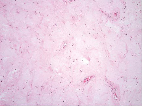
Microscopically, AAM exhibits low to moderate cellularity and is composed of haphazardly arranged, short-spindled, and stellate-shaped cells set in a myxoid-appearing, edematous stroma containing fine strands of collagen and a prominent component of variably sized vessels (Fig. 8.1, e-Figs. 8.1 to 8.3). Multinucleated cells are infrequently observed. An increase in the cellularity is occasionally observed around vessels or at the periphery of the lesion where microscopic evidence of infiltration into surrounding tissue with entrapment of fat, peripheral nerve, or muscle is common. Mitotic activity is negligible.


FIGURE 8.1 Aggressive angiomyxoma. At low magnification, the lesion is hypocellular, myxoid, and features large vessels.

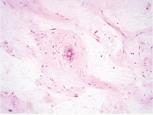
Medium to large-sized vessels with clusters of smooth muscle cells “spinning off” from the main vessel wall are found (Fig. 8.2). Capillarysized thin-walled vessels are found as scattered units or organized into tight collections. Extravasation of red blood cells is commonly noted around these fragile capillary structures.

The stroma consists of a loose meshwork of delicate, wavy collagen fibers set in a myxedematous matrix. Thin bundles or fascicles of mature
smooth muscle can be identified within the stroma, generally running parallel to vessels or nerves. Microcysts occasionally form within the stroma.


FIGURE 8.2 Aggressive angiomyxoma. Note the appearance of strands of cells emanating from the vessel in the center of the field.


Ancillary Investigations

Histochemical stains demonstrate that the noncollagenous stroma consists of a small quantity of acidic mucopolysaccharide, chiefly hyaluronic acid.7

Only gold members can continue reading. Log In or Register to continue

Stay updated, free articles. Join our Telegram channel

Jun 18, 2016 | Posted by in PATHOLOGY & LABORATORY MEDICINE | Comments Off on Tumors of Specialized Lower Genital Tract Mesenchyme

Full access? Get Clinical Tree

Get Clinical Tree app for offline access