Tricholemmoma
David Cassarino, MD, PhD
Key Facts
Terminology
Tricholemmoma (TL)
Benign clear cell adnexal proliferation with external root sheath differentiation
Subtype: Desmoplastic tricholemmoma (dTL)
Etiology/Pathogenesis
Some cases are associated with Cowden syndrome (PTEN hamartoma syndrome)
Characterized by multiple tricholemmomas, hamartomas, and visceral tumors including breast and thyroid carcinomas
Mutation of PTEN gene on 10q23.31
Microscopic Pathology
Lobular proliferation of mature squamoid cells with pale- to clear-staining cytoplasm
Peripheral palisading of basaloid cells
Cells are surrounded by thickened, glassy-appearing basement membrane
Multiple broad connections to epidermis and follicles
Desmoplastic tricholemmoma
Variant with prominent desmoplastic stroma
Typically in center of tumor, surrounded by conventional-appearing TL
Top Differential Diagnoses
Tumor of follicular infundibulum (TFI)
Clear cell acanthoma
Tricholemmal carcinoma
Clear cell squamous cell carcinoma in situ (clear cell Bowen disease)
Basal cell carcinoma (BCC)
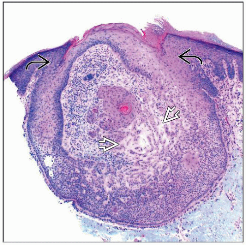 Scanning magnification of a small tricholemmoma shows a symmetric, well-circumscribed, lobular neoplasm with epidermal attachments  . Centrally, the lesion shows desmoplastic TL features . Centrally, the lesion shows desmoplastic TL features  . . |
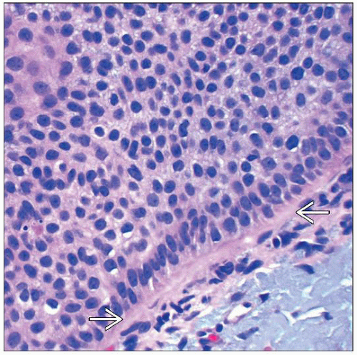 High magnification of a TL shows bland-appearing small cells with pale to clear-staining cytoplasm and small nucleoli. There is a thickened peripheral basement membrane  . . |
TERMINOLOGY
Abbreviations
Tricholemmoma (TL)
Desmoplastic tricholemmoma (dTL)
Synonyms
Trichilemmoma
Definitions
Benign clear cell adnexal proliferation with external root sheath differentiation
ETIOLOGY/PATHOGENESIS
Unknown in Most Cases
Some have considered TL to be related to HPV infection (“tricholemmal verrucae”)
Not generally accepted, and most PCR studies for HPV have been negative
Genetic
Some cases are associated with Cowden syndrome (PTEN hamartoma syndrome)
Characterized by multiple tricholemmomas, hamartomas, and visceral tumors including breast and thyroid carcinomas
Mutation of PTEN, a tumor suppressor gene, on 10q23.31
CLINICAL ISSUES
Epidemiology
Incidence
Relatively common tumors
Age
Usually adults, although Cowden syndrome patients present earlier
Site
Most occur on face, especially nose and upper lip
Presentation
Small papular lesion
Usually flesh-colored
Often clinically mimics basal cell carcinoma or verruca
Treatment
Surgical approaches
Usually not necessary, as these are benign tumors
Complete conservative excision is curative
Prognosis
Excellent, no malignant potential
Cowden syndrome patients have high risk of internal malignancies
MICROSCOPIC PATHOLOGY
Histologic Features
Lobular proliferation of mature squamoid cells with pale- to clear-staining cytoplasm
Peripheral palisading of basaloid cells
Cells are surrounded by thickened, glassy-appearing basement membrane
Squamous eddies and small foci of tricholemmal (pilar)-type keratinization often present
Stay updated, free articles. Join our Telegram channel

Full access? Get Clinical Tree


