Transduodenal Sphincteroplasty
Sphincteroplasty is a useful adjunct to bile duct exploration for calculous biliary tract disease. It produces a wide opening of the distal bile duct, allowing impacted stones to be removed from the ampulla of Vater. The ampullary sphincter is enlarged, and any stones that are left behind in the upper ductal system should be able to pass naturally into the duodenum. It is only performed when there is reason to believe that stones may have been left behind, or when there are impacted stones in the distal ampulla. It has been termed an internal choledochoduodenostomy. It has largely been superseded by endoscopic sphincterotomy.
A similar approach is used for excision of benign tumors of the ampulla (see references).
Occasionally, sphincteroplasty is performed for treatment of recurrent pancreatitis. Sphincteroplasty of the terminal pancreatic duct is included as part of that procedure (see references).
Steps in Procedure
Fully mobilize the duodenum
Make choledochotomy and pass probe through ampulla
Palpate location of ampulla and place two stay sutures in duodenum
Longitudinal incision over ampulla
Deliver probe and ampulla into incision and place stay sutures
Cut will be made at 10 or 11 o’clock to avoid pancreatic duct
Administer secretin intravenously
Incise ampulla with Potts scissors for about 2 mm
Place interrupted sutures on each side of incision
Incise for Another 2 mm and Suture
Continue process until ampulla widens out into bile duct
Copious clear pancreatic juice should flow from orifice of pancreatic duct, which may be visible
Place apex suture
Close duodenotomy in two layers, transversely if possible
Close choledochotomy without T-tube
Place omentum in subhepatic space and over duodenotomy
Close abdomen in usual fashion without drainage
Hallmark Anatomic Complications
Injury to terminal pancreatic duct
List of Structures
Gallbladder
Bile Duct
Intramural portion
Ampulla of Vater
Major duodenal papilla
Pancreatic duct (of Wirsung)
Duodenum
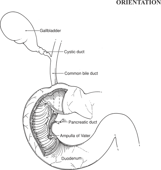
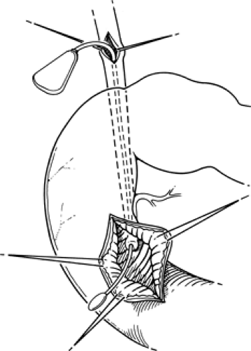
Visualization of the Ampulla (Fig. 67.1)
Technical Points
Generally, cholecystectomy and bile duct exploration will have been performed immediately before sphincteroplasty. Open the bile duct and place a probe through it to aid in subsequent dissection. This should be done even if the bile duct is not explored before sphincteroplasty.
Place a No. 3 Bakes dilator into the choledochotomy and pass it through the ampulla. Confirm that the dilator is in the duodenum by visualizing the “single steel” sign. This refers to the manner in which the shiny stainless steel tip of the Bakes
dilator is easily seen through a single layer of tissue (the duodenal wall). Fully mobilize the duodenum by performing a wide Kocher maneuver. Place stay sutures of 3-0 silk on the lateral aspect of the second portion of the duodenum in the approximate area where the ampulla is palpable over the Bakes dilator. Make a longitudinal duodenotomy approximately 4 cm in length and deliver the Bakes dilator into the duodenotomy. The ampulla should be visible in the incision. Extend the incision along the duodenum proximally, or distally if necessary, to achieve good visualization of the ampulla.
dilator is easily seen through a single layer of tissue (the duodenal wall). Fully mobilize the duodenum by performing a wide Kocher maneuver. Place stay sutures of 3-0 silk on the lateral aspect of the second portion of the duodenum in the approximate area where the ampulla is palpable over the Bakes dilator. Make a longitudinal duodenotomy approximately 4 cm in length and deliver the Bakes dilator into the duodenotomy. The ampulla should be visible in the incision. Extend the incision along the duodenum proximally, or distally if necessary, to achieve good visualization of the ampulla.
 |
 Figure 67-1 Visualization of the Ampulla
Stay updated, free articles. Join our Telegram channel
Full access? Get Clinical Tree
 Get Clinical Tree app for offline access

|