Thyroid
Staci Bryson, MD
Larissa V. Furtado, MD
Key Facts
Embryology
Endodermal thyroid diverticulum forms on floor of pharynx/base of tongue opposite the 1st and 2nd pharyngeal pouches during weeks 3 and 4
Bilobed, solid thyroid diverticulum forms in close proximity to aortic sac and mirrors its caudal descent during weeks 4 and 5
Stalk of thyroid diverticulum forms thyroglossal duct, which breaks down by end of week 5
By week 5, thyroid has distinct left and right lobes connected by isthmus
Thyroid continues to descend in neck to its final position just inferior to cricoid cartilage by week 7
Ultimobranchial body (4th pharyngeal pouch) fuses with thyroid and gives rise to parafollicular “C” cells (neural crest derived)
Vascular mesoderm invades endoderm of thyroid diverticulum to form plates
Plates form cells grouped around a lumen (follicle unit) by week 10
Thyroid gland grows rapidly during first 16-18 weeks of gestation
Colloid production begins between weeks 10 and 12; achieves mature appearance by week 20
Thyroxine secreted by week 18
Microscopic Anatomy
Thin fibrous capsule delimits thyroid
Thyroid follicles are made up of a single layer of cuboidal to low columnar epithelium with round, basally oriented nuclei around a central lumen containing colloid
Parafollicular or “C” cells have pale cytoplasm and oval nuclei; not readily identified in hematoxylin and eosin-stained sections
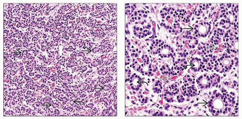 (Left) By 15 weeks gestation, the thyroid follicles  have formed. While the central lumen have formed. While the central lumen  is apparent in many of the follicles, colloid formation is minimal. (Right) Follicular cells is apparent in many of the follicles, colloid formation is minimal. (Right) Follicular cells  are cuboidal to low columnar with round, basally oriented nuclei and are arranged around a central lumen are cuboidal to low columnar with round, basally oriented nuclei and are arranged around a central lumen  . The thyroid is richly vascular . The thyroid is richly vascular  . . |
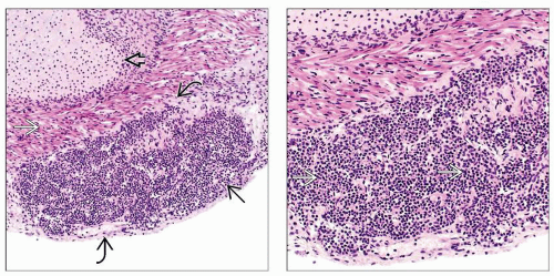 (Left) The thyroid gland
 can often be found in intimate association with the strap muscles can often be found in intimate association with the strap muscles  of the neck as shown in this example from a 16-week fetus. The thyroid gland is delimited by a thin but complete fibrous capsule of the neck as shown in this example from a 16-week fetus. The thyroid gland is delimited by a thin but complete fibrous capsule  . Laryngeal cartilage . Laryngeal cartilage  is apparent in the upper left corner. (Right) The thyroid gland often has a solid appearance is apparent in the upper left corner. (Right) The thyroid gland often has a solid appearance  early in gestation with the follicular units being inconspicuous. early in gestation with the follicular units being inconspicuous.Stay updated, free articles. Join our Telegram channel
Full access? Get Clinical Tree
 Get Clinical Tree app for offline access

|