Thymoma
Ming Guo, MD
Key Facts
Cytopathology
Biphasic pattern of epithelial cells and small lymphocytes
Epithelioid &/or spindle cells
T cells with thymic cortical immunophenotype
Ancillary Tests
Immunohistochemistry
Expression of cytokeratins and p63 in epithelial cells
CD5 usually negative in epithelial cells except for atypical thymoma or thymic carcinoma
TdT, CD1a, and CD99 expression in T lymphocytes
Top Differential Diagnoses
Lymphoblastic lymphoma
Thymic carcinoid tumor
Seminoma
Spindle cell tumors
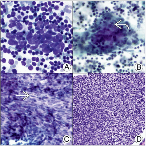 (A) Diff-Quik stained smear shows biphasic pattern of thymoma. (B) Pap-stained smear shows epithelioid tumor cells with moderate cytoplasm  in a background of small lymphocytes. (C) Pap-stained smear shows spindle thymoma tumor cells in a background of small lymphocytes. (C) Pap-stained smear shows spindle thymoma tumor cells  with slender nuclei and fine chromatin. (D) H&E-stained cell block section shows epithelioid and spindle tumor cells mixed with small lymphocytes. with slender nuclei and fine chromatin. (D) H&E-stained cell block section shows epithelioid and spindle tumor cells mixed with small lymphocytes. |
TERMINOLOGY
Definitions
Primary thymic epithelial neoplasm associated with a variety of nonneoplastic T lymphocytes
CLINICAL ISSUES
Epidemiology
Incidence
Accounts for ˜ 25% of all primary mediastinal neoplasms
Age
Stay updated, free articles. Join our Telegram channel

Full access? Get Clinical Tree


