Thoracoscopic Surgery of the Esophagus
This chapter describes two procedures: thoracoscopic esophagomyotomy and esophageal mobilization for resection. These are used to illustrate the thoracoscopic appearance of the mediastinum. As with open esophageal surgery (see Chapter 30), only the distal one third of the esophagus is accessible through the left chest. Access to the proximal two thirds requires a right thoracoscopic approach and is preferred for resection. Both are described here, and references at the end of the chapter give information about other procedures.
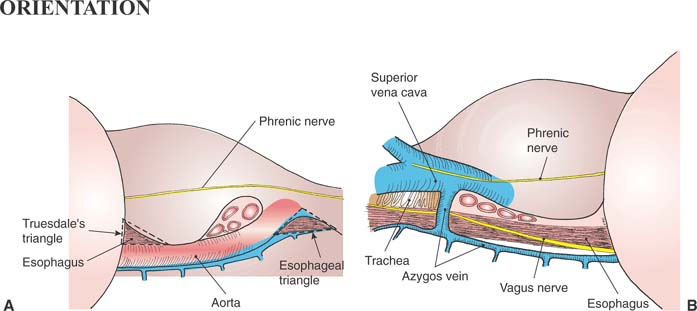
A thoracoscopic schematic view of the left posterior mediastinum is shown in part A of the orientation figure. Generally, only the lower part is accessed for esophageal surgery. Part B of the orientation figure shows the corresponding view of the right posterior mediastinum.
Steps in Procedure
Thoracoscopic Myotomy
Single lung ventilation, position as for left thoracotomy
Ports placed in four to seven interspaces, diamond-shaped configuration
Divide inferior pulmonary ligament and retract lung cephalad
Incise pleura overlying esophagus
Have an assistant pass esophagoscope into esophagus, deflect tip if necessary to aid dissection
Gently dissect around esophagus
Encircle with Penrose drain and pull cephalad
Begin myotomy at convenient point in thickened portion
Expose epithelial tube completely in region of myotomy
Extend cephalad and caudad through entire thickened portion
Confirm adequacy of myotomy by direct visualization with esophagoscope
Check for perforation (bubbles under saline)
Place chest tube, if desired, and close port sites
Esophageal Mobilization for Resection
Single lung ventilation, position patient as for right thoracotomy
Ports in interspaces four through seven in diamond-shaped configuration
Incise inferior pulmonary ligament and retract right lung cephalad and medial
Incise mediastinal pleura overlying azygos vein
Gently dissect vein and divide it with a vascular stapler
Incise pleura cephalad and caudad to expose esophagus
Elevate esophagus and encircle it with a Penrose drain
Dissect entire length of esophagus, with associated lymph nodes
Hallmark Anatomic Complications
Thoracic duct injury
Full-thickness injury to esophagus (esophagomyotomy)
Inadequate myotomy
Injury to thoracic duct
Injury to vagus nerve
Injury to membranous portion of trachea
List of Structures
Esophagus
Vagus nerves
Inferior pulmonary ligament
Mediastinal pleura
Lower lobe pulmonary vein
Diaphragm
Muscular portion
Central tendinous portion
Hiatus
Pericardium
Aorta
Arch of aorta
Left subclavian artery
Bronchial arteries
Inferior phrenic nerve
Phrenoesophageal Membrane
Endothoracic fascia
Phrenoesophageal fascia
Transversalis fascia
Peritoneum
Azygos vein
Hemiazygos vein
Thoracic duct
 |
Thoracoscopic Esophagomyotomy: Initial Exposure and Mobilization of Esophagus (Fig. 31.1)
Technical Points
After adequate single-lung ventilation has been achieved, place the patient in the thoracotomy position with the left side up. Place Thoracoports as shown (Fig. 31.1A).
Divide the inferior pulmonary ligament with ultrasonic shears and retract the collapsed lung cephalad with a lung retractor. Take care not to extend this incision up into the lower lobe pulmonary vein.
Incise the mediastinal pleura between pericardium and aorta to expose the esophagus (Fig. 31.1B). Have an assistant pass an esophagogastroscope into the esophagus. Gentle deflection of the tip will elevate the esophagus from the groove behind the pericardium and facilitate dissection (Fig. 31.1C).
Stay updated, free articles. Join our Telegram channel

Full access? Get Clinical Tree


