Therapeutic Neuromodulation


Therapeutic Neuromodulation


Neuromodulation involves the induction of electrical current in nervous tissue through various means. Therapeutic effects occur through regulation of functional disturbances in distributed neural circuitry. Further, focal stimulation can use frequencies that excite or inhibit different areas or the same area in varying patterns. Unlike medications, neuromodulation has an episodic impact on the brain, generally lacks systemic effects, and may avoid problems with adaptation over time (1,2). The prototypic example is electroconvulsive therapy (ECT) which remains the most effective treatment for severely ill, often psychotic, highly suicidal, medication-refractory or -intolerant patients (3,4). It is, however, plagued by problems in terms of misuse, inadequate access, a complicated and expensive administration process, cognitive adverse effects, high relapse rates, and a negative public image. Given these issues and a substantial proportion of depressed patients who do not adequately benefit from existing treatments, the development of more effective, safer, and relatively costefficient alternatives is essential. In this light, other approaches are engendering increased interest. Neuromodulation approaches can be categorized as follows (5,6,7,8,9 and 10a):


  • Noninvasive procedures


    • Seizurogenic


      • Electroconvulsive therapy (ECT)


      • Magnetic seizure therapy (MST)


      • Focal electrically administered seizure therapy (FEAST)


    • Nonseizurogenic


      • Transcranial magnetic stimulation (TMS)


      • Transcranial direct current stimulation (tDCS)


  • Invasive procedures


    • Nonseizurogenic


      • Vagus nerve stimulation (VNS)


      • Deep brain stimulation (DBS)

While not an exhaustive list, these areas provide a perspective on the variety of approaches that fall into this category

Figure 8-1 outlines the potential role for therapeutic neuromodulation in an overall treatment strategy for depression.


Electroconvulsive Therapy

ECT remains the most effective treatment for older patients with severe depressive disorders, often characterized by melancholic and/or psychotic features (4). Further, those with or without atypical features appear to benefit equally (11). Although primarily used for an acute episode, ECT may also be a useful maintenance strategy for patients with frequent relapses despite adequate pharmacotherapy (12,13).

The use of electrical stimulation to induce therapeutic seizures is the safest and most efficient form of convulsive therapy (e.g., as compared with pharmacoconvulsive therapy). In 1938, Cerletti and Bini (14) first attempted this approach, and until the introduction of effective pharmacotherapy, ECT remained the primary treatment for more severe mood and psychotic episodes. Since then, however, this therapy has been relegated to a secondary role, usually attempted after trials with standard psychotropics (e.g., antidepressants, antipsychotics, lithium, and other mood stabilizers, often in multiple combinations) and psychotherapy have proved inadequate or poorly tolerated.


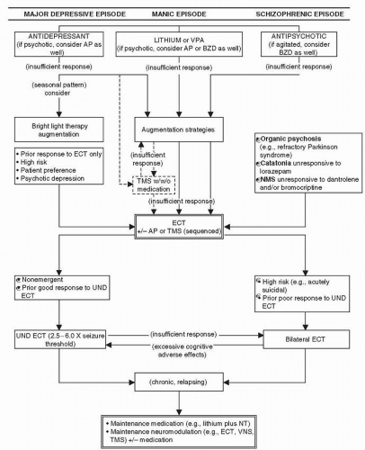
Figure 8-1 The role of therapeutic neuromodulation in the treatment of psychiatric disorders. (Adapted from Dowd SM, Janicak PG. How effective and safe is rTMS? Current Psychiatry. 2003;2:59-66.)

Uneasiness concerning the passage of electricity through the human brain to induce seizures (a phenomenon otherwise considered pathological) has contributed to the controversy surrounding this treatment. Thus, despite the lack of supporting documentation, some groups have continued to raise concerns about irreversible memory loss and possible brain damage after ECT (15,16 and 17). Further, during its zenith ECT was used in a wide range of cases now deemed inappropriate. Reports from the United States and United Kingdom also found that training was often inadequate and that a large proportion of facilities administered ECT improperly
(18,19,20 and 21). In this context, Kramer (21) and the American Psychiatric Association (APA) Task Force on ECT (22) have presented outlines for teaching ECT to address deficits in training programs.

Partly as a result of these factors, strident antipsychiatry forces have attempted to eliminate or severely curtail its use. For example, legislative restrictions were enacted in Berkeley, California, in the early 1980s and in Texas in the early 1990s. There is also data indicating racial disparity in the use of ECT, possibly due in part to physician bias in treatment decisions (23). In contrast, the attitudes of professionals regarding the use of ECT generally improve as their levels of knowledge and experience increase (24,25 and 26).

We believe ECT should be considered as a first-line treatment in patients with a previous good response to this therapy and in those who have been nonresponsive to or intolerant of standard treatments in the past. Further, for patients who present as a high risk (e.g., acute suicidality or rapid physical deterioration), this treatment may be lifesaving (27). Finally, in patients who express a preference for this efficient, rapidly acting, relatively short-term treatment, ECT may be an appropriate first-line therapy. For example, two reports found that compared with pharmacotherapy, ECT significantly shortened hospital length of stay, and decreased the overall cost for patients with a major depressive disorder (MDD) (28,29).


MECHANISM OF ACTION

Generally, theories on the mechanism of action of ECT in humans have paralleled those posited for effective mood-stabilizing pharmacotherapy, including


  • Neurotransmitters effects


  • Neuroendocrine effects


  • Neurophysiological effects


  • Neurotrophic effects

In humans, measurement of pretreatment and posttreatment changes in peripheral tissues such as platelets, lymphocytes, and amine metabolites of depressed patients has been one approach to determine ECT’s mechanism of action (MOA) (30). A different approach involves magnetic resonance spectroscopy that allows for the noninvasive study of central nervous system (CNS) neurochemical changes associated with depression and recovery, including investigations into the mechanism of action of ECT (31).


Neurotransmitter Effects

The amine hypothesis of depressive mood disorders postulates a critical disruption in one or more neurotransmitters (e.g., norepinephrine [NE], serotonin [5-HT], dopamine [DA]), culminating in a dysregulation of their activity and leading to characteristic behavioral and vegetative symptoms. Electroshock (ECS) in animals increases NE, 5-HT, and DA synthesis in the CNS. It also induces downregulation of postsynaptic β1– and β2-receptors in peripheral tissues (e.g., platelets); but, interestingly and differently from selective serotonin reuptake inhibitors (SSRIs), tricyclic antidepressants (TCAs), and monoamine oxidase inhibitors (MAOIs), ECS appears to upregulate 5-HT2 postsynaptic receptors (32,33). Results from animal and human studies, however, are inconsistent with potential reasons including


  • The use of brain tissue in animals versus peripheral tissues in humans, as well as the differential effects of ECS in animals versus ECT in humans, respectively


  • A predominance of NE β1-receptors in the CNS and of NE β2-receptors in peripheral tissues


  • Findings of receptor activity differences in normal young animals, with their own speciesspecific biochemistry and physiology, which cannot be easily generalized to baseline and posttreatment differences in normal humans or depressed patients

A further elaboration on these theories considers the modulating interactions among several neurotransmitter systems (e.g., the permissive hypothesis, the cholinergic-adrenergic balance hypothesis). Other neurotransmitters implicated include DA, γ-aminobutyric acid (GABA), and glutamate all of which subserve many of the vegetative functions disrupted in depressive states. One example is the connection postulated between DA, GABA, and major depression, in part based on the efficacy of ECT in Parkinson disease (see Chapter 7) (34,35).

Another approach considers neuronal changes subsequent to receptor-related activity. Guanosine triphosphate-binding proteins (Gproteins) transmit extracellular signals initiated
at the receptor level to a variety of intracellular effectors. Abnormalities in G-protein expression or function are implicated in various medical and neuropsychiatric disorders, and several psychotropics are known to affect G-protein activity (36). Given their ubiquitous role in neuronal function, it is plausible that G-proteins are involved in psychiatric disorders (e.g., major depression). Existing treatments and potential novel, site-specific drugs that target these proteins may help clarify pathophysiology, as well as the mechanism of action of various drug and neuromodulation therapies. In this context, studies have shown that G-proteins (inhibitory and stimulatory) in mononuclear leukocytes and platelets are decreased in depression and increased in bipolar disorder (37,38 and 39). Further, ECT has been shown to normalize G-protein function in both conditions (40). This normalization precedes and predicts improvement in depressed patients receiving ECT. One possibility is that ECT may stabilize dysregulated intracellular signaling, which explains why it is effective for both mania and depression. Of note, lithium and antidepressants can also modulate Gprotein functions (41,42) (see Chapters 7 and 10).


Neuroendocrine Effects

The neuroendocrine theory considers the effects of various ligands on their receptors located in the diencephalic and mesiotemporal areas. Cell clusters in the hypothalamus coordinate the normal regulation of the vegetative functions of sleep, appetite, and sexual drive that are typically disrupted in depression. In addition, the limbic area modulates many aspects of behavior and mood that are characteristically disturbed in affective disorders.


Neurophysiological Effects

Alterations in neurophysiological activity are subserved by many of the neurotransmitters discussed earlier in this section, as well as in “Mechanism of Action” in Chapter 7. After repeated seizures spaced over a given period of time (usually 2 to 3 times per week over a 3- to 5-week period), there is an increase in cerebral blood flow (CBF) (primarily the result of increased systemic circulation) and an acute and sustained increase in cerebral metabolism. One of the most characteristic changes is a slowing in the electroencephalographic (EEG) pattern over a series of ECT treatments associated with increased acetylcholine activity. Increases in amplitude and decreases in frequency appear to affect thalamocortical and diencephalic structures that may modulate recently acquired behavior, such as psychosis or melancholic features.

EEG interictal changes are characterized by a desynchronized resting configuration, leading to high-amplitude synchronized patterns and symmetric bursts of activity characteristic of centrencephalic seizures. With successive treatments there is progressive slowing in the mean frequency and increases in the mean amplitude of activity, both of which seem to be necessary but not sufficient for an antidepressant effect. Within 2 to 8 weeks after a course of ECT, the EEG returns to regular rhythmic baseline α-wave activity.

ECT-induced changes in sleep architecture include decreased rapid eye movement (REM) sleep, increased stage 4 sleep, and increased total sleep time. For example, we found that five depressed patients who improved clinically with ECT had a parallel normalization of all sleep parameters (e.g., total sleep time, sleep efficiency). Interestingly, REM latency initially became even shorter but then normalized by the end of the course of ECT (43). Although preliminary, we speculate that the unexpected initial decrease in REM latency might serve as a predictor of final outcome. In this context, Grunhaus et al. (44) found that sleep-onset REM periods after ECT predicted a poorer response.

The “anticonvulsant” hypothesis has been developed to explain the efficacy of ECT, as well as that of certain antiepileptics. Thus, both ECT and agents such as carbamazepine (CBZ) and valproate (VPA) have several effects on seizure activity, including


  • Increasing the seizure threshold (ST)


  • Decreasing the overall duration of an episode


  • Decreasing neurometabolic response to an episode


  • Decreasing the phenomenon of amygdaloid kindling

ECS is also known to diminish the phenomenon of amygdaloid kindling in animal models (45). Similarities between such neuroelectrical, stress-induced phenomena and the
longitudinal course of bipolar disorder have been noted and serve as a heuristic, nonhomologous model for understanding the development of certain mood disorders (46). In summary, although a given ECT treatment elicits seizure activity, the net outcome is an antiseizure effect over a course of therapy. This may be related to:


  • Enhanced GABA-ergic transmission


  • Decreased cerebral blood flow and cerebral metabolic rate (CMR)


  • Increase in neuropeptide Y (NPY) and somatostatinlike activity (47)

Finally, some have promulgated the concept of hemispheric equilibration, which attributes efficacy to the apparent ability of ECT to restore the relative balance between right and left brain functions (48,49). This observation is consistent with the modulation of aberrant neurocircuits seen in depressed states.


Neurotrophic Effects

ECS, ECT, and antidepressants have all demonstrated impact on intracellular signaling mechanisms that contribute to:


  • Alteration in neuroplasticity


  • Neurotrophic effects


  • Neurogenesis

For example, ECS in rodents can increase the growth of neurons in the hippocampus, a phenomenon that may subserve its therapeutic effects in humans and possibly reverse changes secondary to stress and steroids (50). Further, ECS can produce synaptic reorganization possibly mediated by brain-derived neurotrophic factor (BDNF) and its receptor (tyrosine kinase receptor B), which are increased in the hippocampus and dentate gyrus (51,52).


INDICATIONS

Although it is often used as a treatment of “last resort,” there are situations in which ECT may be an appropriate earlier intervention. The APA Task Force Report on ECT discusses four such specific groups of patients:


  • Those who are at high risk for suicidal behavior (27)


  • Those who are rapidly deteriorating, either physically or psychologically, or both


  • Those who have a prior history of good response to ECT or a poor history of response to pharmacotherapy


  • Those who prefer to receive this treatment rather than undergo lengthy, and possibly unsuccessful, trials of medications with their associated adverse effects (22,53)

Nonresponse to adequate pharmacotherapy is a predictor of nonresponse to ECT (54). ECT is still, however, superior to additional antidepressant trials (alone or in combination with other psychotropics) for prior drug-nonresponsive nonpsychotic or psychotic depressions (55,56).


Psychotic Depression

Schatzberg and Rothschild (57) support separating psychotic depression from other depressive disorders, in part because of its differential responsivity to various treatments. One consistent finding is that patients with psychotic depression on antidepressant monotherapy or even a combination of an antidepressant plus an antipsychotic have a lower response rate than those depressed patients without psychotic symptoms (58). By contrast, Petrides et al. (59) found that bilateral (BILAT) ECT produced earlier and higher remission rates in psychotic depressed patients than in those with nonpsychotic depression. Although evidence supports an improved response when these patients undergo treatment with ECT, this apparent superiority may be related to a selection bias (60). Thus, it may be that patients with psychotic depression are more likely to receive ECT earlier in their course of illness and therefore the extent of their true drug resistance is unknown.

The relapse rate for psychotic versus nonpsychotic depression after a successful course of ECT has also been considered. Birkenhäger et al. (61) found that relapse rates at 6 and 12 months post ECT were significantly lower in those who initially presented with psychotic depression. In another study, geriatric patients who had responded to either ECT or medication treatment were then followed longitudinally (62). Those who had initially presented with psychotic features and responded to drug treatment subsequently demonstrated a substantially higher rate of relapse than their nonpsychotic counterparts, with maintenance TCA monotherapy having limited efficacy. The overall response
rate to ECT in this group of psychotic patients, however, was 88.2% (i.e., 15 of 17), which compared favorably with the 50% response rate reported for pharmacological treatment (63).

In summary, ECT may have differential efficacy in the psychotic versus nonpsychotic depressive subgroups, both in terms of acute response and propensity for relapse. In addition, although evidence supports superior outcomes in geriatric patients, rate and extent of recovery are less clear when comparing ECT with pharmacological treatment in this age group (64).


Other Indications

In addition to depression, other indications for ECT include


  • Mania (especially manic delirium)


  • Schizophrenia


  • Catatonia associated with major mood disorders, schizophrenia, or organic mood disorders (e.g., systemic lupus erythematosus) (65)


  • Certain medical disorders

Paradoxically, ECT is equally useful in both the acute manic and depressive phases of bipolar disorder, constituting one of the most effective bimodal therapies available (see later discussion in the section “Bipolar Patient”). In schizophrenia, ECT is most effective in the acutely-ill patient with a more recent onset of illness; those who need a rapid onset of effect; or those who do not respond to antipsychotics (66,67 and 68). Catatonia, withdrawn type, characterized by immobility and an inability to interact or maintain one’s basic needs (at times posing a lifethreatening situation), can dramatically resolve with ECT. Medical disorders that may benefit include


  • Intractable seizures


  • Parkinson disease


  • Hypopituitarism


  • Neuroleptic malignant syndrome (NMS)


In particular, the use of ECT during or shortly after an episode of NMS has come under scrutiny in the case report literature. A major concern is the reported fatalities associated with its use for this problem. Davis et al. (69) reviewed the world’s literature, identifying approximately 1,000 NMS episodes in 755 patients. Using mortality rate as the major outcome variable, they compared patients who received nonspecific supportive treatment, specific drug treatment (i.e., DA agonists or dantrolene) plus supportive measures, or ECT. There was a 21% mortality rate in the nonspecific-treated group as compared with a 9.7% mortality rate in the specific drug-treated group and a 10.7% mortality rate in the ECT-treated group. Further, the ECT patients who died or experienced severe adverse effects were usually on concomitant high-potency antipsychotics. An analysis of the case-control
data indicated that ECT was clinically effective while resulting in a similar mortality rate to that with specific drug therapies and approximately half that seen with supportive treatments. Although the specific drug-supportive therapy difference was statistically significant, the sample size in the ECT group was too small (i.e., 28) to ascertain statistical significance.


CONTRAINDICATIONS

The APA Task Force Report on ECT supports the concept of differing levels of relative contraindications and no longer lists “absolute” restrictions for the use of ECT (22). In particular, the APA cautions that the following conditions may be associated with increased risk of morbidity and warrant special attention:


  • Space-occupying supratentorial cerebral lesions


  • A recent history of myocardial infarction and associated instability (<3 months)


  • Recent intracerebral bleeds


  • Bleeding or unstable aneurysm or arteriovenous malformations


  • Retinal detachment


  • Pheochromocytoma


  • American Society of Anesthesiologists (ASA) classification risk level of 4 or 5

Thus, when considering ECT in patients with any of these conditions, a careful review of the risk-to-benefit ratio must be conducted in conjunction with consultation from the appropriate specialists. There is also evidence that unipolar depressed patients with comorbid borderline personality disorder (BPD) have a poorer response to ECT than those without a personality disorder or other Axis I disorder (70).


TABLE 8-1 EFFICACY OF REAL ECT IN COMPARISON WITH SIMULATED ECT


Real ECT


Simulated ECT


Study


R


NR


R


NR


Chi Square


p Value


Ulett et al. (1956)


28


14


15


27


6.86


0.01


Brill et al. (1959)


12


6


2


7


3.13


0.08


Harris and Robin (1960)


2


2


0


4


0.67


0.41


Fahy et al. (1963)


12


5


8


9


1.09


0.30


Lambourn and Gill (1978)


8


8


8


8


0.13


0.72


West (1981)


11


0


1


10


14.85


0.0001


Chi square for statistical method of combination = 22


Composite p value = 3.5 × 10⁻⁶


Estimated difference between real ECT and simulated ECT = 33%


ECT, electroconvulsive therapy; R, responder; NR, nonresponder.


Adapted from Janicak PG, Davis JM, Gibbons RD, et al. Efficacy of ECT: a meta-analysis. Am J Psychiatry. 1985;142(3):297-302,


with permission.


EFFICACY


Acute Treatment

ECT is superior in efficacy when compared with placebo, sham ECT, and active drug therapy. Upon the introduction of effective pharmacotherapy for severe depression, the relative efficacy of drug versus ECT was often studied. Our review of the relevant literature led to an extrapolation of the data from selected studies (primarily class I or II designs) for a quantitative analysis of the efficacy of ECT versus other treatments for an acute depressive episode (71). The comparisons with ECT included simulated (or sham) ECT (72,73,74,75,76 and 77), placebo (78,79,80 and 81), TCAs (79,80,81,82,83 and 84), and MAOIs (78,79,80 and 81). We also compared the relative efficacy of the BILAT versus the UND forms of administration (55,85,86,87,88,89,90,91,92,93,94,95 and 96). A meta-analysis was computed on the data across all studies that met a priori inclusion criteria (Tables 8-1,8-2,8-3,8-4,8-5 and 8-6).

The overall efficacy of ECT was 78% that is significantly superior to that of TCAs whose overall efficacy was 64%. Further, ECT is also superior to placebo, simulated ECT, and MAOIs, whose overall response rates ranged from 28% to 38%. This difference was particularly striking because many patients in the ECT-treated groups had failed previous drug trials. The adequacy of some of these, however, was questionable. Further, in contrast to other reports, our analysis did not find a significant
difference favoring BILAT ECT over UND ECT (i.e., only a 7% overall difference). Of note, subsequent data indicate that high-energy (250% to 600% of seizure threshold) UND ECT approached efficacy rates achieved with BILAT ECT (97,98).


TABLE 8-2 EFFICACY OF ECT IN COMPARISON WITH PLACEBO


ECT


Placebo


Study


R


NR


R


NR


Chi Square


p Value


Kiloh et al. (1960)


24


3


3


25


30.56


<0.001


Greenblatt et al. (1962, 1964)


65


5


43


24


15.19


<0.001


Shepherd (1965)


49


14


23


37


18.10


<0.001


Chi square for statistical method of combination = 62


Composite p value = 4 × 10⁻⁵


Estimated difference between ECT and placebo = 42%


ECT, electroconvulsive therapy; R, responder; NR, nonresponder.


Adapted from Janicak PG, Davis JM, Gibbons RD, et al. Efficacy of ECT: a meta-analysis. Am J Psychiatry. 1985;142(3):297-302,


with permission.


TABLE 8-3 EFFICACY OF ECT IN COMPARISON WITH TCAs


ECT


TCAs


Study


R


NR


R


NR


Chi Square


p Value


Bruce et al. (1960)


21


1


16


10


5.96


0.01


Robin and Harris (1962)


12


3


3


12


8.53


0.004


Greenblatt et al. (1962, 1964)


65


5


71


28


10.36


0.001


Fahy et al. (1963)


12


5


10


6


0.015


0.90


Wilson et al. (1963)


10


0


10


0


0.0002


0.99


Shepherd (1965)


49


14


42


19


0.84


0.36


Chi square for statistical method of combination = 22


Composite p value = 3 × 10⁻⁶


Estimated difference between ECT and TCAs = 19%


ECT, electroconvulsive therapy; TCA, tricyclic antidepressant; R, responder; NR, nonresponder.


Adapted from Janicak PG, Davis JM, Gibbons RD, et al. Efficacy of ECT: a meta-analysis. Am J Psychiatry. 1985;142(3):297-302, with permission.


TABLE 8-4 EFFICACY OF ECT IN COMPARISON WITH MAOIs


ECT


MAOIs


Study


R


NR


R


NR


Chi Square


p Value


Harris and Robin (1960)


2


2


0


4


0.67


0.41


Kiloh et al. (1960)


24


3


14


12


6.38


0.01


Greenblatt et al. (1962, 1964)


65


5


30


42


39.7


<0.001


Shepherd (1965)


49


14


19


41


24.6


<0.001


Chi square for statistical method of combination = 75


Composite p value = 5 × 10-18


Estimated difference between ECT and MAOIs = 46


ECT, electroconvulsive therapy; MAOI, monoamine oxidase inhibitor; R, responder; NR, nonresponder.


Adapted from Janicak PG, Davis JM, Gibbons RD, et al. Efficacy of ECT: a meta-analysis. Am J Psychiatry. 1985;142(3):297-302, with permission.


TABLE 8-5 EFFICACY OF BILATERAL ECT IN COMPARISON WITH UNILATERAL NONDOMINANT ECT


Bilateral


Unilateral Nondominant


Individual Chi Square with Yates’ Correction


Individual p Value


Study


R


NR


R


NR


Cannicott(1962)


18


2


27


3


0.231


0.75


Strain et al. (1968)


29


17


33


17


0.008


0.90


Halliday et al. (1968)


17


1


13


5


1.800


0.25


Zinkin and Birtchnell (1968)


13


7


18


6


0.154


0.75


Abrams and DeVito (1969)


9


2


8


2


0.203


0.75


d’Elia (1970)


21


8


23


7


0.006


0.90


Flemingeretal. (1970)


10


2


7


5


1.810


0.35


Sand-Stromgren(1973)


37


11


39


13


0.000


0.95


Fraser and Glass (1980)


14


1


10


2


0.042


0.90


Janicak et al. (1989)


11


2


15


5


0.050


0.82


Heshe et al. (1978)


24


0


20


7


5.189


0.025


Sackeim et al. (1987)


19


8


7


18


7.704


0.006


Total N


222


61


220


90


Chi square for statistical method of combination


Mantel-Haenszel = 4.87


Woolf-Haldens = 2.88


Composite p value


Mantel-Haenszel = 0.03


Woolf-Haldens = NS


Estimated difference between BILAT ECT and UND ECT = 7%


ECT, electroconvulsive therapy; R, responder; NR, nonresponder; N, number; NS, not significant; BILAT, bilateral; UND, unilateral nondominant.


Adapted from Janicak PG, Davis JM, Gibbons RD, et al. Efficacy of ECT: a meta-analysis. Am J Psychiatry. 1985;142(3):297-302, with permission.


TABLE 8-6 STATISTICAL OVERVIEW OF STUDIES COMPARING UNILATERAL NONDOMINANT ECT WITH BILATERAL ECT


Mantel-Haenszel


Woolf-Haldens


Estimated Difference: BILAT > UND


Number of Studies


Number of Patients


Chi Square


p Value


Chi Square


p Value


Studies finding no difference


10


490


0.31


NS


0.25


NS


2%


Studies finding BILAT > UND ECT


2


103


14.2


0.0002


11.7


0.0006


32%


Total results


12


593


4.87


0.03


2.88


NS


7%


ECT, electroconvulsive therapy; BILAT, bilateral; UND, unilateral nondominant; NS, not significant.


Adapted from Janicak PG, Davis JM, Gibbons RD, et al. Efficacy of ECT: a meta-analysis. Am J Psychiatry. 1985;142(3):297-302, with permission.


Maintenance Treatment

Just as drug-treated patients require maintenance management after an adequate acute response, so do acute ECT responders. Unlike drug-responsive patients, however, maintenance management with ECT is a more complex matter, given that most patients receive ECT because of a poor response to or an intolerance of pharmacotherapy. In this context, Prudic et al. (99) noted that relapse rates after a successful acute trial of ECT in community settings are substantially higher than that in clinical trials. They hypothesize that residual symptoms are often unrecognized and underscore the need
to treat until optimal improvement is obtained with ECT. Maintenance strategies to consider in this group include


  • Antidepressants that have demonstrated at least partial benefit in the past


  • Lithium or lithium-antidepressant combinations, both in bipolar and unipolar disorders


  • Combined antidepressant plus antipsychotic (e.g., olanzapine plus fluoxetine for bipolar depression)


  • Second-generation antipsychotic monotherapy (e.g., quetiapine)


  • Anticonvulsants, such as lamotrigine, VPA, or CBZ, with or without other mood stabilizers


  • Maintenance ECT, possibly combined with a mood stabilizer, antidepressant, or antipsychotic


  • Possibly VNS or TMS


The Consortium on Research with ECT (CORE)

This National Institute of Mental Health (NIMH)-sponsored, multicenter trial followed acute ECT responders to determine the relative efficacy of continuation ECT versus maintenance drug therapy (i.e., lithium plus nortriptiline). The initial phase of this study demonstrated a 75% remission rate in the 217 unipolar depressed patients who completed an acute course of ECT with bitemporal electrode placement (100). Further, response rates were highest in those with psychotic symptoms and were also higher in older patients (59,101). The major focus of this trial was relapse after a successful acute ECT trial, comparing maintenance ECT (10 treatment sessions) with the maintenance medication combination over a 6-month period. In the modified ITT population (n = 184), there was no difference between groups in terms of continued remission (i.e., 46.1% for ECT; 46.3% for the medication combination) at the end of 6 months (102). The authors concluded, however, that either strategy had limited efficacy given that over 50% of patients in both arms experienced a relapse or dropped out of the trial. In particular, nonpsychotic depressed patients who had at least one failed adequate antidepressant trial prior to receiving ECT appeared most susceptible to early relapse after acute remission (103). Adding a sense of urgency to this issue are earlier reports indicating that patients who responded poorly to adequate pre-ECT pharmacotherapy may also respond poorly to ECT itself or post-ECT drug maintenance (54,104). This phenomenon, however, is not supported by all reports (105).

An important related issue is the ideal frequency of maintenance ECT treatments (e.g., weekly, biweekly, monthly). From a practical perspective, a monthly or bimonthly schedule would be desirable in terms of cost, convenience, risk, and side effects. Unfortunately, few data presently exist to guide clinicians as to the best interval between maintenance treatments, and this determination remains an empirical process with each patient. A typical maintenance ECT approach usually consists of a tapering schedule of single outpatient treatments, starting with a session every 1 to 2 weeks, and then reducing the frequency based on the patient’s response. Although compliance may also be an issue with this strategy (i.e., patients are often reluctant to return as an outpatient for treatments), reports document its efficacy and safety in an otherwise chronic, relapsing depressed group (106,107).

What is clear is that even with an effective maintenance treatment, the relapse rate is substantial. Thus, prospective trials are needed to address questions such as which factors predict the likelihood of a good response to maintenance medication, maintenance ECT, or combination strategies to prevent relapse after a successful acute ECT trial.


SPECIAL POPULATIONS


The Pregnant Patient

Certain factors slightly increase the risk of complications when ECT is administered to pregnant women. Miller (108) has summarized the modifications in technique that can usually address these issues as follows:


  • Raising gastric pH before the procedure with a nonparticulate antacid such as sodium citrate because gastric emptying is prolonged in pregnancy


  • Intubating after the first trimester with a small cuffed endotracheal tube and using a small laryngoscope and a laryngoscope blade to reduce the risk of bleeding from attempted intubation


  • Elevating of the patient’s right hip and displacing the uterus to the left to reduce the likelihood of
    aortocaval compression during the later stages of pregnancy to prevent reduction in fetal circulation


  • Pretreating with intravenous hydration without glucose to avoid osmotic diuresis


  • Avoiding hyperventilation because this may hinder oxygen unloading from maternal to fetal hemoglobin

The author also notes that ECT may be helpful in female bipolar patients who want to plan a pregnancy and discontinue mood stabilizers to minimize risks to the fetus. In addition, ECT can be remarkably effective in cases of postpartum depression and psychosis. Indeed, given the importance of that phase of child care, it can be argued that the most rapidly effective strategy is the preferred strategy. In a related issue, Walker and Swartz (109) considered the use of ECT in high-risk pregnancies, concluding that with additional precautions (e.g., monitoring of maternal and fetal well-being) such patients can safely benefit from this therapy and significant medical and obstetric complications need not prevent its use. One example is the report of a 27-year-old pregnant patient in whom NMS developed while she was on haloperidol. Although the syndrome did not respond to dantrolene, ECT was instituted at 29 weeks and the patient’s condition improved (110).


The Child or Adolescent Patient

In a review, Rey and Walter (111) found no controlled trials on the use of ECT in patients aged 18 years or younger. There were 396 patients identified, 63% of whom constituted single-case reports. The data indicated the following improvement rates in this age group:


  • 63% for depression


  • 80% for mania


  • 42% for schizophrenia


  • 80% for catatonia

These response rates parallel the reports for older age groups. Further, one small (n = 10) trial found no measurable cognitive impairment at long-term follow-up in adolescents who received ECT for a severe mood disorder (112). Unfortunately, however, many child psychiatrists lack both knowledge and experience in this area. ECT is not part of the curriculum for most fellowships in child and adolescent psychiatry, and this topic is not even discussed in some comprehensive textbooks of child psychiatry (111,113). As a result, ECT is a treatment of last resort for most patients in this age group.


The Elderly Patient

ECT continues to be used at a higher frequency in geriatric versus younger patients. This difference in usage probably reflects the decreased efficacy of medications seen in older patients, their lower tolerance to drug-related adverse effects, and the higher incidence of medical disorders in this age group (3). The CORE data also indicate that the highest remission rates are seen in unipolar depressed patients aged 65 years or older (101). Some also advocate UND ECT as comparably effective to BILAT ECT in this age group while producing lower rates of adverse effects, including cognitive impairment (114).

Although ECT is a relatively expensive form of treatment, one study in elderly depressed patients who responded to an acute course demonstrated that maintenance ECT reduced the overall cost of medical care and the relapse rate as compared with patients on maintenance medication after ECT (115). The reduced costs were evident at the 12-month follow-up, primarily through decreased hospital use. In addition, this strategy was also associated with improvements in functional status and cognition.


The Bipolar Patient

Daly et al. (116) reported that bipolar I and II depressed patients responded, had a more rapid onset of improvement than, and required fewer treatments than unipolar depressed patients. A systematic review of the literature found that bipolar mixed episode patients also responded to ECT (117). The manic phase of bipolar disorder is also responsive to ECT, with a comprehensive review of this topic reporting an overall efficacy rate of 80% (118). For patients who were selected because of drug treatment resistance, the rate of response was still approximately 58%, and for patients who did not respond specifically to lithium, the response rate was 69% (119,120). A second issue is the relative benefit of BILAT ECT versus UND ECT for the treatment of acute mania. Milstein et al. (121) reported on the initial outcome of an ongoing
controlled trial comparing ECT with lithium for acute mania, noting that the UND ECT patients did not respond. At that point, the design was changed and all subsequent patients assigned to the ECT group were given the BILAT administration. Final results of this study indicated that the BILAT ECT group tended to demonstrate greater improvement in comparison with the lithium group over an 8-week period (see Chapter 10, “Alternative Treatment Strategies: Electroconvulsive Therapy”) (122). Although these data support the preferential use of BILAT ECT over UND ECT in the treatment of acute mania, this recommendation is not accepted by all. For example, Mukherjee and Sackeim (123) reported no difference in efficacy with either method. Because the ST may be lower in manic patients, the difference in efficacy between BILAT and UND electrode placement may not be as great as for patients with major depression (123,124 and 125). Since these patients often constitute an acute emergency, starting with BILAT ECT may maximize the chances for a rapid resolution of the manic episode. In this context, Karmachary et al. (126) argue that ECT is the treatment of choice for manic delirium.

Finally, ECT may be particularly useful in older bipolar patients, who have decreased physiological reserves and/or medical comorbidity, in whom a prolonged episode of mania can be life threatening and the rapidity of response to mood-stabilizing drugs is insufficient.


ADMINISTRATION OF ELECTROCONVULSIVE THERAPY

A number of improvements in the administration of ECT have enhanced its efficacy and minimized some of its more troublesome adverse effects. These improvements include


  • Advances in anesthetic techniques


  • Selected stimulus electrode placement (e.g., high-energy UND vs. BILAT)


  • Change from sinusoidal to brief-pulse or ultrabrief-pulse inducing currents


  • A better understanding of the role played by the electrical stimulus itself and the degree to which the ST is surpassed


  • Improved assessment for the adequacy of seizure activity


Pre-ECT Workup

The standard pre-ECT workup should include


  • A complete physical and neurological examination


  • Routine hematological indices (e.g., complete blood count) and serum electrolytes


  • Review of cardiac status and electrocardiogram


  • Simple tests of cognitive function (e.g., MMSE)

If deemed necessary, radiographs (anteroposterior and lateral) to rule out spinal or other skeletal problems may be appropriate. If such xray studies are done before ECT, they should be repeated after ECT to document any changes that may have occurred.

Informed consent must be obtained before the administration of ECT. Often, the same severity of illness that necessitates the use of ECT also impairs a patient’s capacity to consent. When the patient is unable to give adequate informed consent, the clinician and the patient’s family can attempt to obtain partial conservatorship from the court allowing a family member to give substituted permission (see also Chapter 3, “Informed Consent”).


Anesthesia

Anesthetic techniques that have minimized adverse effects include the use of muscle relaxants and, more recently, nerve stimulators to assess adequacy of relaxation; the introduction of very rapid acting, short-duration anesthetic agents; and the use of atropinic agents to minimize the cardiovascular response to a combination of a seizure and anesthesia (127). In addition, 100% oxygenation (adequacy monitored by a pulse oximeter) with positive pressure ventilation can minimize related cardiac events and memory disruption.

The standard induction process used during ECT includes


  • Anticholinergic agents (e.g., atropine or glycopyrrolate)


  • Anesthetic agents (e.g., propofol, etomidate)


  • Muscle relaxants (e.g., succinylcholine)


  • β-Blockers (when indicated) to control blood pressure and heart rate changes (e.g., esmolol or labetalol) (128)

Anesthetic Agents. While methohexital was the most frequently used barbiturate anesthetic for
many years, alternative agents that may improve the effectiveness of ECT and enhance safety are increasingly employed.

Etomidate may be especially useful for geriatric patients with high STs. For example, one study reported a significant increase in seizure duration when patients were switched from methohexital to this agent (i.e., in 4 patients the mean seizure duration increased from 25 to 61.8 seconds) (129). Other studies, however, did not find a difference between these two agents (130,131). Thus, etomidate can be considered when adequate seizure duration is not obtained. Disadvantages of etomidate include


  • Delayed cognitive recovery


  • Suppression of adrenocortical function for 8 hours


  • Pain on infusion (132)

Although the use of ketamine for anesthesia induction when seizure duration is insufficient has also been recommended, other studies did not report this agent as helpful (133). Disadvantages of ketamine include

Aug 27, 2016 | Posted by in PHARMACY | Comments Off on Therapeutic Neuromodulation

Full access? Get Clinical Tree

Get Clinical Tree app for offline access