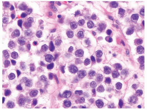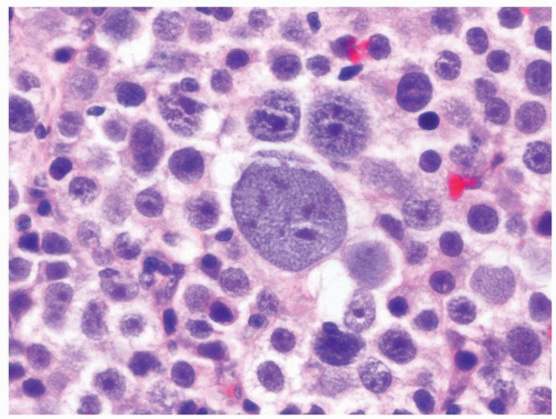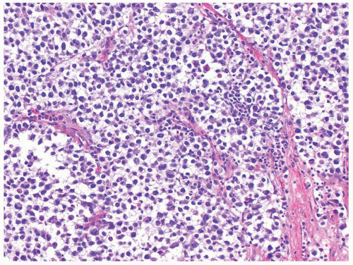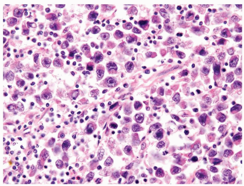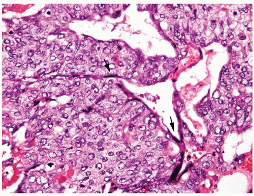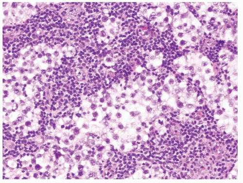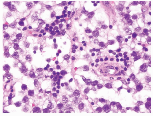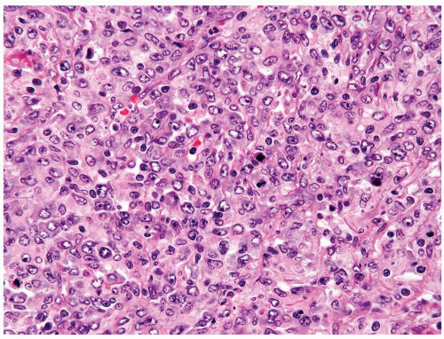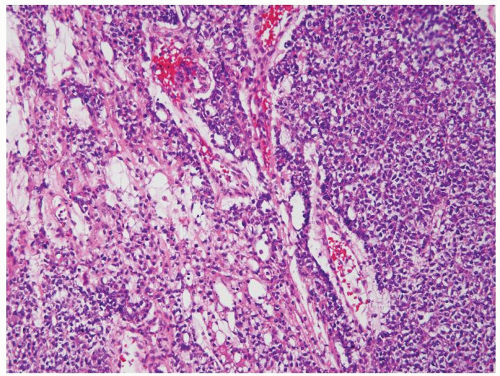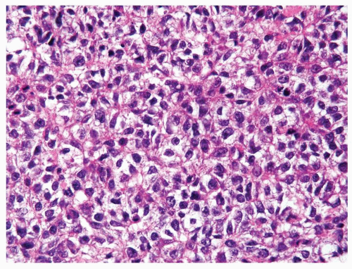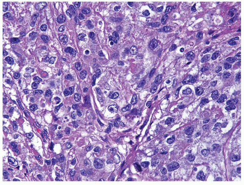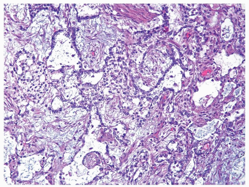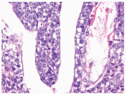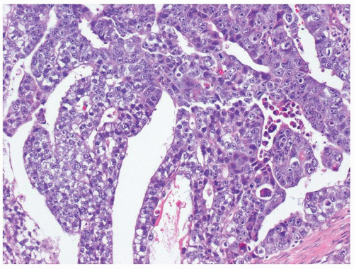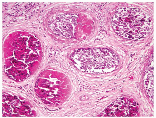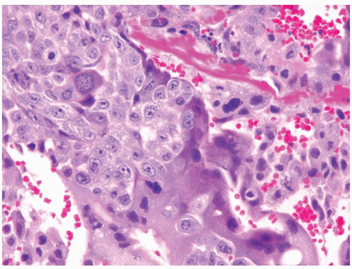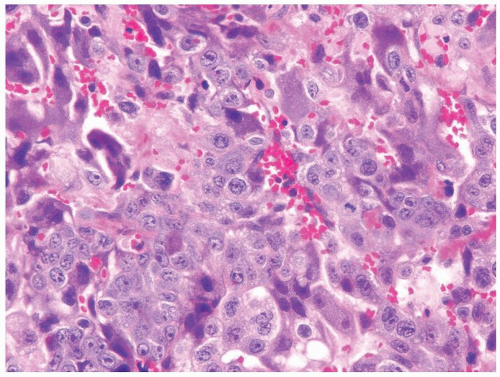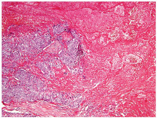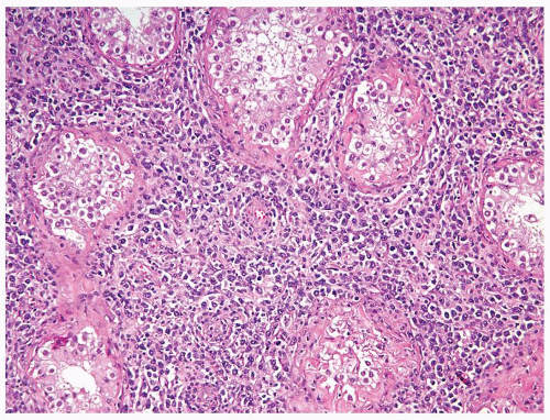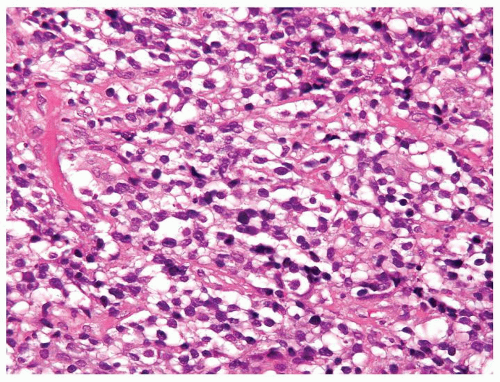
Testis


Testis


4.1 NORMAL SPERMATOGENESIS VS. HYPOSPERMATOGENESIS


Normal Spermatogenesis


Hypospermatogenesis


Age


Not applicable


Postpuberty


Location


Not applicable


Usually bilateral


Symptoms


None


Infertility


Signs


Normal serum FSH, LH, testosterone, and prolactin


Normal serum FSH, LH, testosterone, and prolactin


Etiology


None


Iatrogenic (chemotherapy or radiation), alcoholism, postinfectious, diabetes, cirrhosis, other endocrinopathies (hyperprolactinemia), congenital abnormalities (cryptorchidism), pesticide toxicity, and varicocele


Histology


  1. Complete differentiation taking place


  2. Tubules may not show complete spermatogenesis in every cross-section due to tangential sectioning at the edge of the tubule


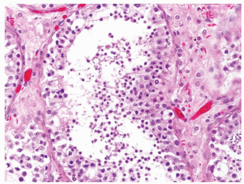
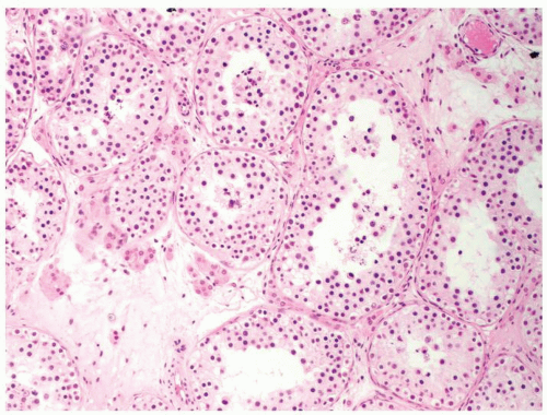
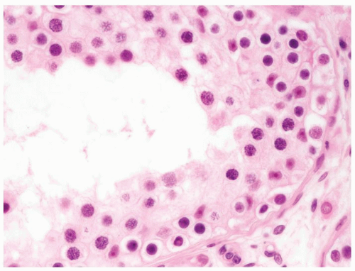
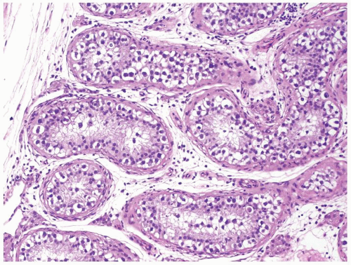
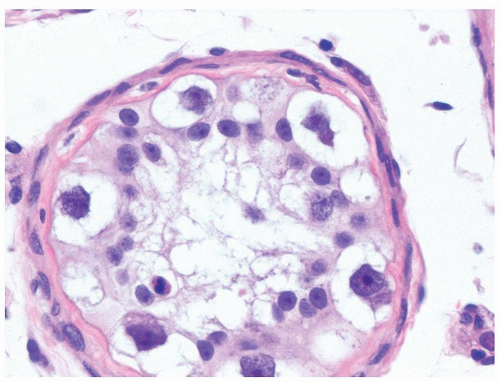
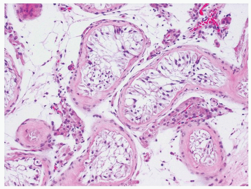
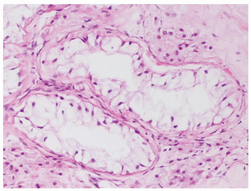
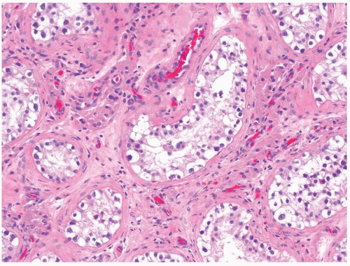
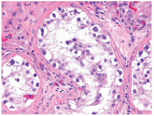
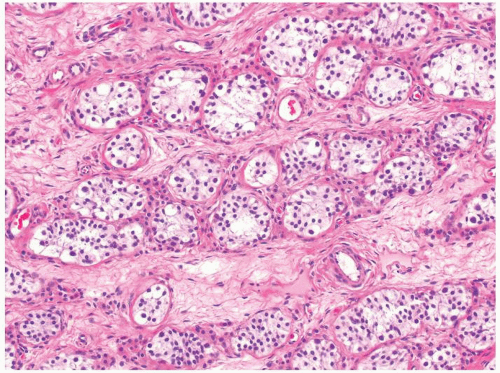
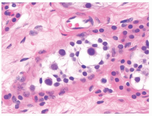
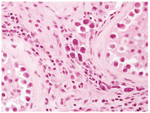
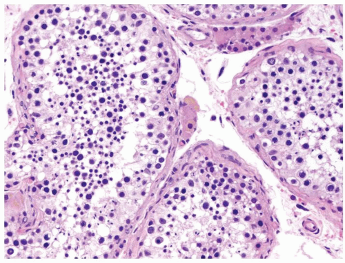
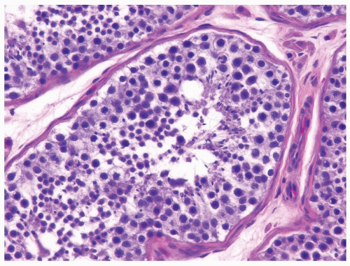
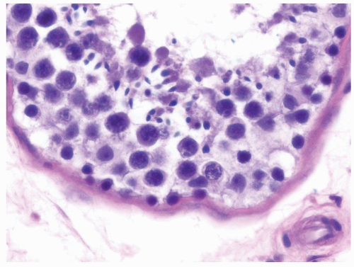
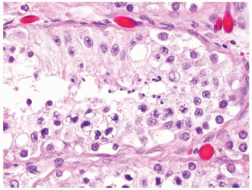
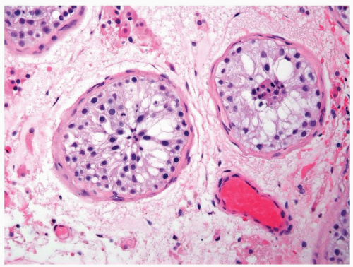
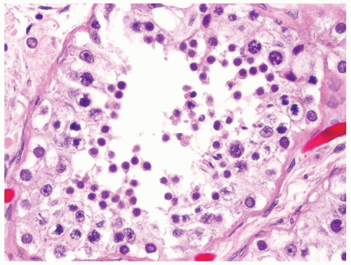
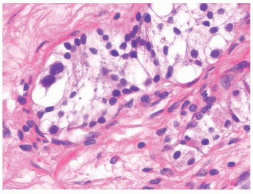
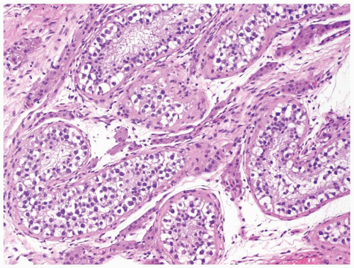
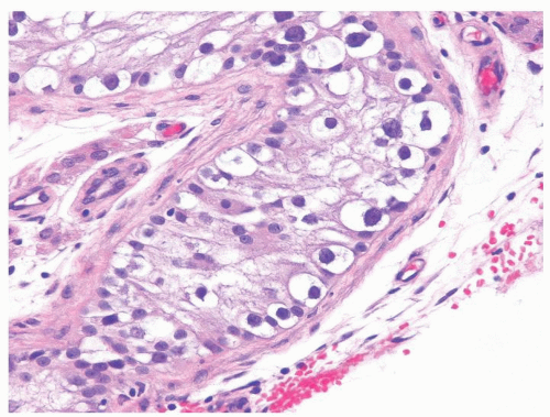
  3. Starting from the periphery of the tubule: Spermatogonium—small cell with pale nuclear chromatin and scant cytoplasm. Spermatocytes—largest and most numerous germ cells with larger nuclei and more cytoplasm. Speckled chromatin. Spermatids—smaller with darker dense chromatin and scant cytoplasm. Spermatozoon—smaller with ovoid nuclei with clearing (acrospermia) at the head. Tails not apparent on routine sections (Figs.4.1.1, 4.1.2, 4.1.3)


  4. Sloughing of germ cells not specific, possibly due to handling and fragile nature of the tissue


  5. Normal spermatogenesis is relative to age. Men 50s and under should have approximately the same thickness with decreases as age increases


  6. In order to call normal spermatogenesis, the entire biopsy should be normal


  1. Complete differentiation taking place


  2. Increased number of tubules may not show complete spermatogenesis in every cross-section due to tangential sectioning at the edge of the tubule


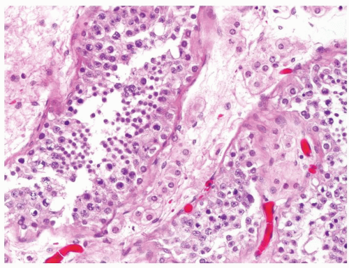
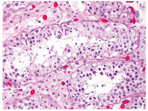
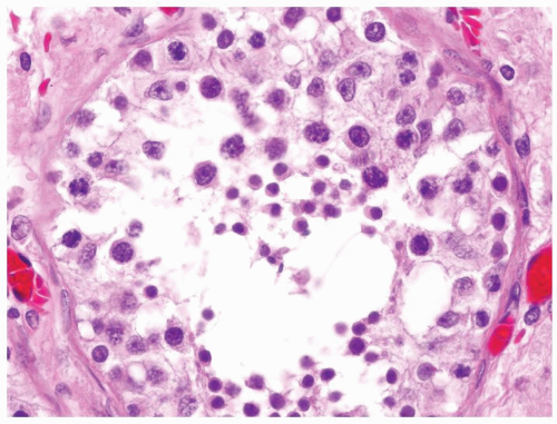
  3. Full maturation through all spermatogenic stages occurs but the total number of germ cells is decreased (Figs.4.1.4, 4.1.5, 4.1.6)


  4. Less likely to see sloughing of germ cells


  5. Should state the severity of process whether mild, moderate, or severe (i.e., only rare spermatozoa identified)


  6. The percent of each pattern of spermatogenesis should be recorded (i.e., 50% normal spermatogenesis; 30% severe hypospermatogenesis; 20% Sertoli cell-only pattern)


Special studies


Not used in this differential


Not useful in this differential


Treatment


Not applicable


Directed toward inciting etiology


Prognosis


Not applicable


Fertility can be frequently achieved


Figure 4.1.1 Normal adult seminiferous tubules showing normal spermatogenesis from spermatogonia to spermatozoa. Note the presence of primary and secondary spermatocytes and spermatids in variable proportions.


Figure 4.1.2 Normal adult seminiferous tubules showing normal spermatogenesis.


Figure 4.1.3 Normal adult seminiferous tubules showing normal spermatogenesis.


Figure 4.1.4 Adult seminiferous tubules with hypospermatogenesis. The overall total number of germ cells is decreased.


Figure 4.1.5 Adult seminiferous tubules with hypospermatogenesis.


Figure 4.1.6 Adult seminiferous tubules with marked hypospermatogenesis.


4.2 HYPOSPERMATOGENESIS VS. MATURATION ARREST


Hypospermatogenesis


Maturation Arrest


Age


Postpuberty


Postpuberty


Location


Usually bilateral


Usually bilateral


Symptoms


Infertility


Infertility


Signs


Normal serum FSH, LH, testosterone, and prolactin


Normal serum FSH, LH, testosterone, and prolactin


Etiology


Iatrogenic (chemotherapy or radiation), alcoholism, postinfectious, diabetes, cirrhosis, other endocrinopathies (hyperprolactinemia), congenital abnormalities (cryptorchidism), pesticide toxicity, and varicocele


Iatrogenic (chemotherapy, radiation, drugs, and testosterone supplementation), alcoholism, postinfectious, endocrinopathy, congenital abnormalities (cryptorchidism), and varicocele


Histology


  1. Almost all tubules with complete spermatogenesis but incomplete spermatogenesis may be present in some tubules due to tangential sectioning at the edge of the tubule. Only complete cross-sections of relatively large, round tubules (i.e., sectioned through the center) should be assessed. If tangentially sectioned the tubule, can appear identical to maturation arrest


  2. Full maturation through all spermatogenic stages occurs but the total number of germ cells is decreased. Critical to recognize mature spermatids to rule out maturation arrest (Figs.4.2.1, 4.2.2, 4.2.3)


  1. Typically, all the tubules in a biopsy will show incomplete spermatogenesis. However, this pattern may coexist with other patterns where the percent of each pattern of spermatogenesis should be recorded (i.e., 50% hypospermatogenesis; 50% maturation arrest). Should not diagnose maturation arrest on small percent of biopsy as may be an artifact


  2. Spermatogenic arrest can occur at any stage of development but most commonly at the primary spermatocyte stage. Second most common pattern is spermatogenic arrest at the spermatogonia stage with very uncommon primary spermatocytes relative to spermatogonia. Spermatids are absent (Figs.4.2.4, 4.2.5, 4.2.6)


Special studies


Not useful in this differential


Not useful in this differential


Treatment


Directed toward inciting etiology. Micro-TESE (microdissection testicular sperm extraction)


Directed toward inciting etiology. Micro-TESE (microdissection testicular sperm extraction) is attempted to achieve fertility


Prognosis


Fertility can be frequently achieved


Fertility can be frequently achieved. Patients with biopsies showing late (spermatocyte stage) maturation arrest have more favorable outcome


Figure 4.2.1 Adult seminiferous tubules with hypospermatogenesis. Full maturation through all spermatogenic stages occur, but the overall total number of germ cells is decreased.


Figure 4.2.2 Adult seminiferous tubules with hypospermatogenesis.


Figure 4.2.3 Adult seminiferous tubules with hypospermatogenesis.


Figure 4.2.4 Adult seminiferous tubules showing maturation arrest at the secondary spermatocyte phase. Note absence of spermatid and spermatozoa.


Figure 4.2.5 Adult seminiferous tubules showing maturation arrest at the secondary spermatocyte phase.


Figure 4.2.6 Adult seminiferous tubules showing maturation arrest at the spermatid phase.


4.3 IDIOPATHIC GRANULOMATOUS ORCHITIS VS. INFECTIOUS AND OTHER NONNEOPLASTIC GRANULOMATOUS ORCHITIS


Idiopathic Granulomatous Orchitis


Infectious and Other Nonneoplastic Granulomatous Orchitis


Age


19-84 y but most commonly in fifth to seventh decades


Variable


Location


Testis and occasionally secondarily involve the epididymis and spermatic cord. Typically bilateral


More frequently initiated in the epididymis and spread to the testis in later stages. More likely to be bilateral


Symptoms


Sudden testicular pain in acute cases. Rarely fever, hematuria, dysuria, and scrotal swelling


Mild testicular enlargement may be associated with systemic symptoms such as fever and weight loss


Signs


Enlarging scrotal mass with variable pain in chronic presentation. Sudden testicular swelling and tenderness in acute cases. Ultrasound shows diffuse hypoechogenicity with associated testicular enlargement, which may raise differential of germ cell neoplasm


Testicular tenderness. Fistula and abscess formation in infectious tuberculous orchitis. Most cases associated with systemic disease and signs and symptoms in other organs


Etiology


Unknown


Mycobacterial organisms, syphilis, brucellosis, leprosy, and fungal organisms. Rarely parasitic and rickettsial agents. Etiology is unknown in sarcoidosis. More common in developing countries and immunosuppressed patients


Histology


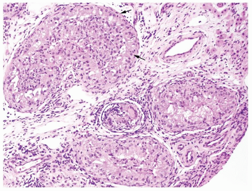
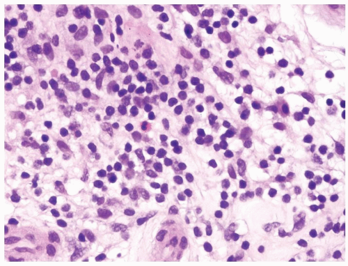
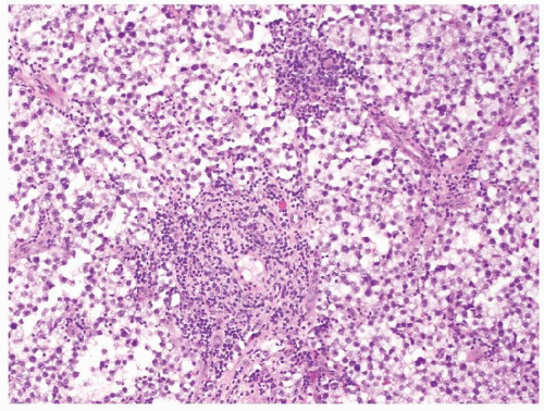
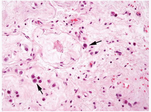
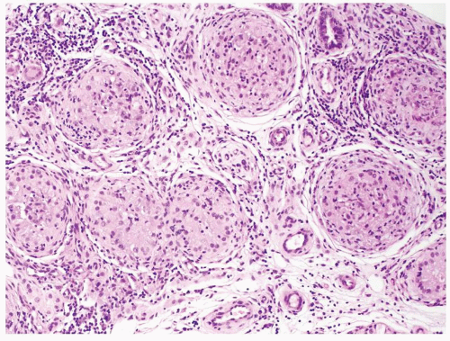
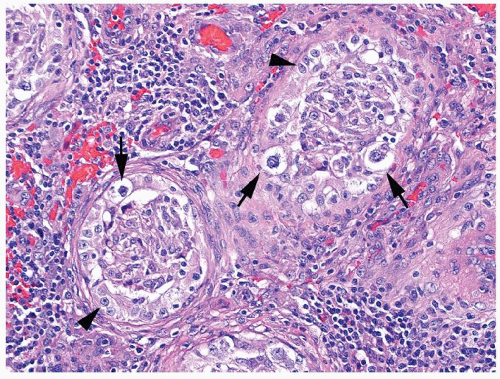
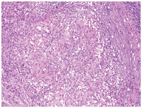
  1. Nonnecrotizing granulomas filling seminiferous tubules. Granulomas are composed of epithelioid histiocytes, giant cells admixed with lymphocytes, and plasma cells (Figs.4.3.1, 4.3.2, 4.3.3)


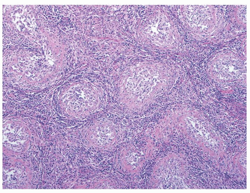
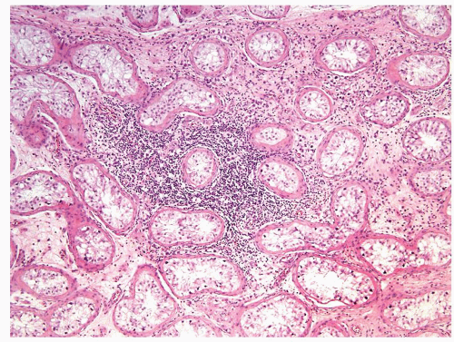
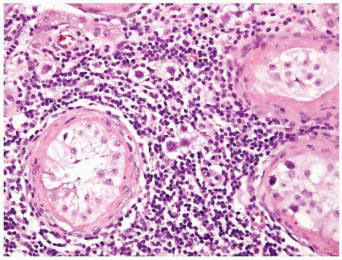
  2. Chronic inflammatory cells, including eosinophils infiltrate the interstitium (Fig. 4.3.4)


  3. In advanced stages, seminiferous tubules become atrophic and are surrounded by fibrosis (Fig. 4.3.5)


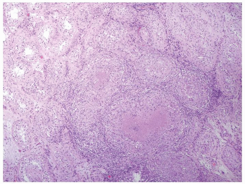
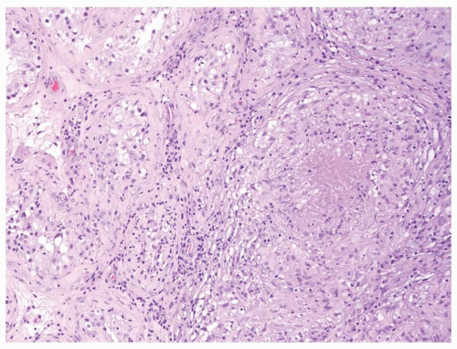
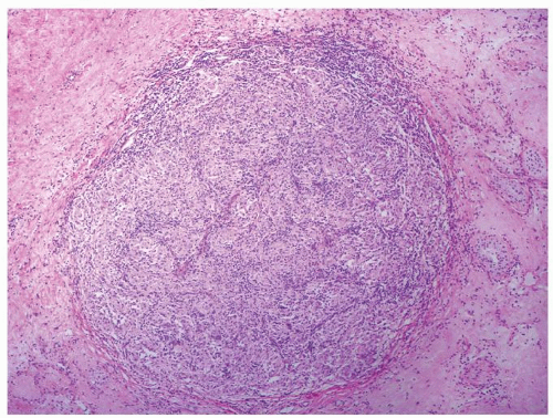
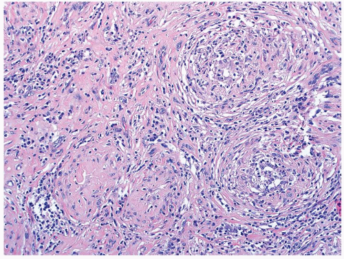
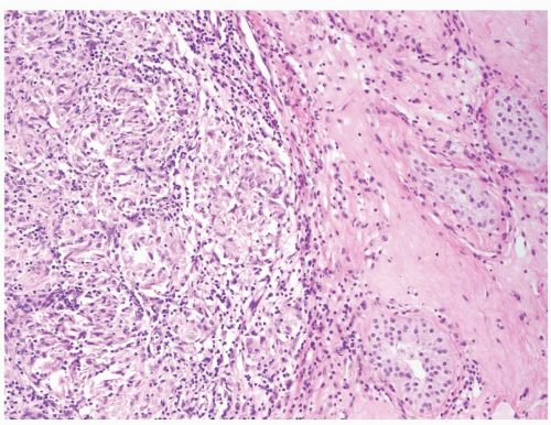
  1. In infectious processes, necrotizing and/or nonnecrotizing granulomatous inflammation composed of epithelioid histiocytes and multinucleated giant cells mainly involving the testicular interstitium (Figs. 4.3.6 and 4.3.7). The same interstitial distribution is also seen in sarcoidosis, yet nonnecrotizing granulomas are seen (Fig. 4.3.8)


  2. Interstitial chronic inflammatory infiltrate composed mainly of lymphocytes


  3. Fibrosis and scaring involving testicular parenchyma and paratesticular tissue in chronic lesions


Special studies


  • Negative histochemical stains for mycobacterial, fungal, and bacterial organisms help exclude specific infectious granulomatous orchitis


  • Reticulin stains highlight intratubular architecture


  • Histochemical stains for mycobacterial (acid-fast stains), fungal (periodic acid-Schiff and methenamine silver), and bacterial organisms will aid in establishing the infectious etiology. Negative stains for the organism can aid in pointing to the diagnosis of sarcoidosis


  • Reticulin stains demonstrate interstitial process


Treatment


Orchiectomy often performed for presumptive neoplasm. Frozen section could aid in the diagnosis and guide conservative treatment with steroids


Antibiotics to combat specific infectious agent


Prognosis


Benign inflammatory condition. Could lead to loss of spermatogenesis in the involved testis even if orchiectomy avoided


Excellent


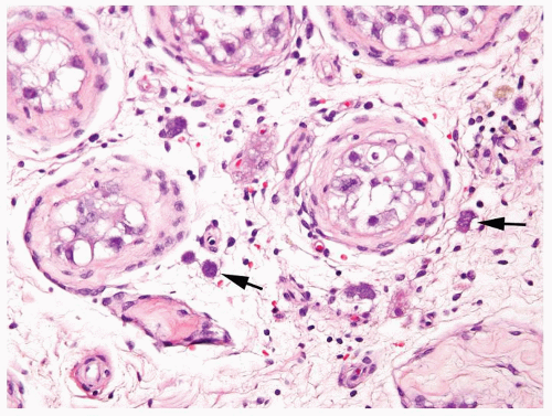
Figure 4.3.1 Idiopathic granulomatous orchitis showing nonnecrotizing granulomas filling seminiferous tubules. Granulomas are composed of epithelioid histiocytes, giant cells admixed with lymphocytes and plasma cells.


Figure 4.3.2 Idiopathic granulomatous orchitis. Granulomas are composed of epithelioid histiocytes and giant cells.


Figure 4.3.3 Idiopathic granulomatous orchitis showing nonnecrotizing granulomas filling seminiferous tubules. Residual germ cells are noted (arrows).


Figure 4.3.4 Idiopathic granulomatous orchitis. Associated chronic inflammation is illustrated with occasional eosinophils admixed with lymphocytes and plasma cells.


Figure 4.3.5 Idiopathic granulomatous orchitis with scarring.


Figure 4.3.6 Mycobacterial granulomatous orchitis. Necrotizing granulomas with palisaded central necrosis is illustrated.


Figure 4.3.7 Mycobacterial granulomatous orchitis. Necrotizing granulomas involve the interstitium with minimal involvement of atrophic seminiferous tubules seen on the left side.


Figure 4.3.8 Nonnecrotizing granulomatous orchitis in sarcoidosis. The process primarily involves the interstitium.


4.4 IDIOPATHIC GRANULOMATOUS ORCHITIS VS. INTRATUBULAR GERM CELL NEOPLASIA UNCLASSIFIED (IGCNU) WITH GRANULOMAS


Idiopathic Granulomatous Orchitis


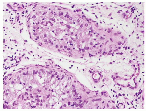
Intratubular Germ Cell Neoplasia Unclassified (IGCNU) with Granulomas


Age


Most commonly in fifth to seventh decades (19-84 y)


Typically 15-35 y


Location


Typically bilateral


Usually unilateral


Symptoms


Sudden testicular pain in acute cases. Rarely fever, hematuria, dysuria, and scrotal swelling


Asymptomatic


Signs


Enlarging scrotal mass with variable pain in chronic presentation. Sudden testicular swelling and tenderness in acute cases


None except for those of associated conditions such as cryptorchidism and infertility


Etiology


Unknown


Associated risk factors include cryptorchidism (4% risk) and infertility (IGCNU in 1%-2% of biopsies). Frequently adjacent to germ cell tumors


Histology


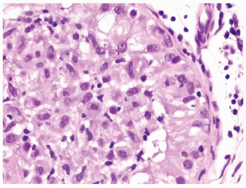
  1. Seminiferous tubules filled with epithelioid histiocytes, giant cells admixed with lymphocytes, and plasma cells (Figs. 4.4.1 and 4.4.2)


  2. Lacks atypical cells


  3. Involved tubules may show focal spermatogenesis


  4. Lymphocytes, plasma cells, and eosinophils in the interstitium


  1. Intratubular epithelioid histiocytes, fewer giant cells associated with IGCNU


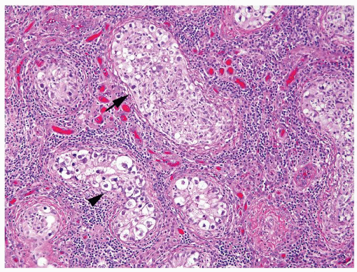
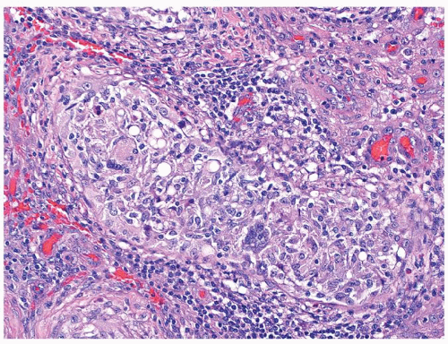
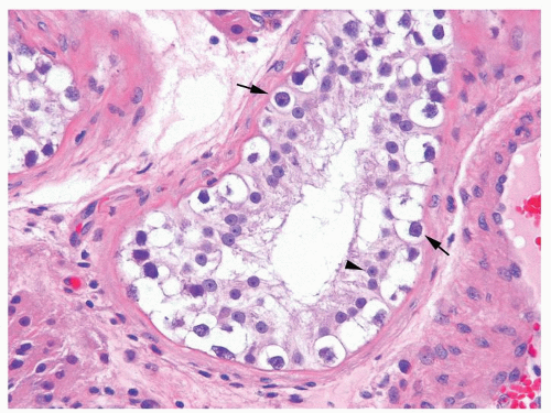
  2. IGCNU cells are enlarged, atypical germ cells residing within seminiferous tubules as isolated cells or a single row along a usually thickened basement membrane. IGCNU cells have clear cytoplasm, irregular nuclear contours, coarse chromatin, and enlarged single or multiple nucleoli. Some IGCNU cells have mummified pyknotic enlarged hyperchromatic nuclei (Figs.4.4.3, 4.4.4, 4.4.5)


  3. Seminiferous tubules containing IGCNU usually lack active spermatogenesis and contain mostly Sertoli cells


  4. Mainly lymphocytes in the interstitium


Special studies


  • Negative for PLAP and OCT4


  • SALL 4 and CD117 are expressed in normal adult spermatogonia


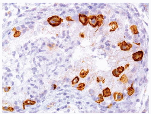
  • Positive for PLAP and OCT4 (Fig. 4.4.6)


  • SALL4 and CD117 positive but not useful as they are not specific for IGCNU


Treatment


Orchiectomy. Frozen section could aid in the diagnosis and guide conservative treatment


If IGCNU identified on biopsy, typically just close follow-up. In some countries, contralateral biopsy performed, and if positive, then sperm banking and bilateral low-dose radiation are offered


Prognosis


Benign inflammatory condition but may lead to loss of spermatogenesis in the involved testis even if orchiectomy was avoided


Up to 50% risk of developing invasive germ cell tumor within 5 y. Risk of increased testicular germ cell tumor also in the contralateral testis


Figure 4.4.1 Idiopathic granulomatous orchitis showing nonnecrotizing granulomas filling seminiferous tubules. Note lack of cytologic atypia in Sertoli cells at periphery of the tubules.


Figure 4.4.2 Idiopathic granulomatous orchitis with a seminiferous tubule filled with epithelioid histiocytes. There is a lack of atypical cells.


Figure 4.4.3 Some seminiferous tubules with IGCNU not involved with granulomatous response (arrowhead). Other tubules have granulomatous inflammation within the tubules involved by IGCNU (arrow).


Figure 4.4.4 Nonnecrotizing granulomas involving some seminiferous tubules in association with IGCNU. Note the presence of highly atypical precursor neoplastic cells at the periphery of the tubules with typical cleared cytoplasm and coarse chromatin (arrows). Contrast with Sertoli cells with smaller, uniform, round-oval shape, uniform chromatin, and a single central nucleolus (arrowheads).


Figure 4.4.5 Nonnecrotizing granulomas in association with IGCNU (same case as in Figure 4.4.4) with extensive intratubular granulomatous reaction destroying IGCNU cells, mimicking idiopathic granulomatous orchitis.


Figure 4.4.6 Nonnecrotizing granulomas in association with IGCNU highlighted by CD117.


4.5 GRANULOMATOUS SEMINOMA VS. INFECTIOUS AND OTHER NONNEOPLASTIC GRANULOMATOUS ORCHITIS


Granulomatous Seminoma


Infectious and Other Nonneoplastic Granulomatous Orchitis


Age


Second to fourth decades. Can also be seen in older men


Variable


Location


Usually unilateral


More frequently starts in the epididymis and spread to the testis in later stages. More likely to be bilateral


Symptoms


Painless swelling of one testicle; one-third of cases complain of scrotal heaviness or dull pain. One-tenth of cases can present with symptoms due to metastatic disease such as abdominal/back pain, cough


Mild testicular enlargement may be associated with systemic symptoms such as fever and weight loss


Signs


Usually palpable testicular nodule


Testicular tenderness, fistula, and abscess formation in infectious tuberculous orchitis


Etiology


Associated risk factors include cryptorchidism (10%), infertility, low socioeconomic status, and male genital malformations. No known reason why granulomatous response in some men. Seen in up to 50% of seminomas


Mycobacterial organisms, syphilis, brucellosis, leprosy, and fungal organisms. Rarely parasitic and rickettsial agents. Etiology remains unknown in sarcoidosis


Histology


  1. Nonnecrotizing interstitial granulomatous inflammation composed of histiocytes and occasional giant cells, which when exuberant can obscure neoplastic germ cells


  2. Ill-defined granulomatous inflammation, as opposed to well-formed granulomas (Fig. 4.5.1)


  3. Lymphoplasmacytic infiltrate at times with lymphoid follicles and eosinophils are typically also present and reside in fibrovascular septae dividing the sheets of neoplastic seminoma cells


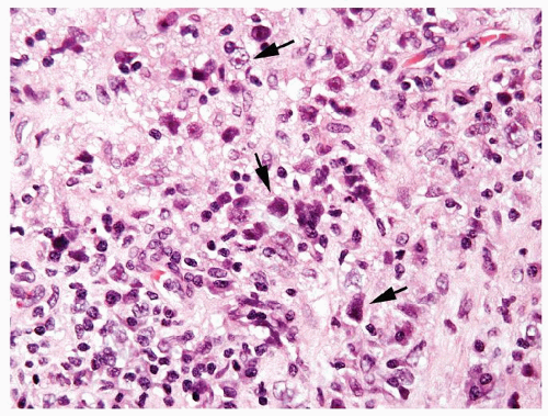
  4. The infiltrating neoplastic seminoma cells are monotonous round-to-polygonal cleared cells with ovoid nuclei containing one or more prominent nucleoli imparting a classic “fried egg” appearance. Mitotic activity is variable. In some cases, granulomatous infiltrate so prominent almost obscuring seminoma cells (Figs. 4.5.2 and 4.5.3)


  5. Seminiferous tubules containing IGCNU are almost invariably found in the surrounding testis


  1. Necrotizing and/or nonnecrotizing granulomatous inflammation composed of epithelioid histiocytes and multinucleated giant cells mainly affecting the testicular interstitium. The same interstitial distribution is also seen in sarcoidosis, yet without necrotizing granulomas (Figs. 4.5.5 and 4.5.6)


  2. Granulomas tend to be well formed


  3. Interstitial chronic inflammatory infiltrate composed mainly of lymphocytes


  4. Invasive seminoma cells absent


  5. IGCNU absent


Special studies


  • No need for organism stains


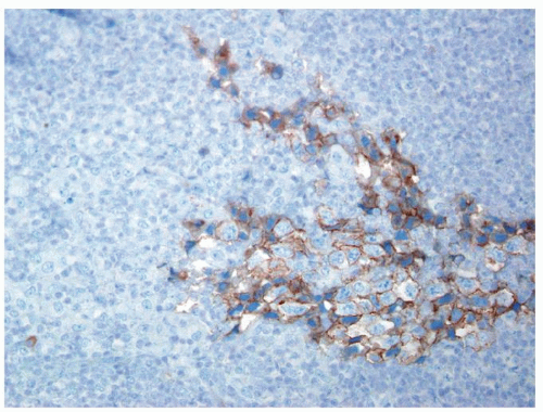
  • Invasive seminoma cells positive for PLAP, OCT4, CD117, and SALL4 (Fig. 4.5.4)


  • Histochemical stains for mycobacterial (acid-fast stains), fungal (periodic acid-Schiff and methenamine silver), and bacterial organisms aid in establishing the infectious etiology


  • Negative for PLAP, OCT4, CD117, and SALL4


Treatment


Stage dependent. Radical orchiectomy followed by active surveillance or adjuvant radiotherapy and/or platinum-based chemotherapy


Antibiotics to the specific infectious agent


Prognosis


Excellent prognosis with up to 90% relapse-free 5-y survival


Excellent


Figure 4.5.1 Focal nonnecrotizing interstitial granulomatous inflammation composed of histiocytes is seen in septae dividing sheets of seminoma cells.


Figure 4.5.2 Rare seminoma cells (arrow) associated with extensive nonnecrotizing interstitial granulomatous inflammation composed of epithelioid histiocytes admixed with lymphocytes, plasma cells, and multinucleated histiocytes.


Figure 4.5.3 Rare seminoma cells (arrows) associated with extensive nonnecrotizing interstitial granulomatous inflammation. Some seminoma cells are very hyperchromatic where it is difficult to see nuclear detail as these cells are fragile and can show crush artifact. Others have preserved prominent nucleoli.


Figure 4.5.4 Seminoma cells labeled with PLAP immunostain.


Figure 4.5.5 Nonnecrotizing granulomatous orchitis in sarcoidosis composed of well-formed granulomas. Note surrounding scarring and atrophic seminiferous tubules lacking IGCNU.


Figure 4.5.6 Mycobacterial granulomatous orchitis with discrete granuloma.


4.6 INTRATUBULAR GERM CELL NEOPLASIA UNCLASSIFIED (IGCNU) VS. SERTOLI CELL-ONLY PATTERN (GERM CELL APLASIA)


Intratubular Germ Cell Neoplasia Unclassified (IGCNU)


Sertoli Cell-Only Pattern (Germ Cell Aplasia)


Age


15-35 y


Pre- or postpuberty


Location


Usually unilateral


Usually bilateral except when in association with the cryptorchid testis


Symptoms


Asymptomatic


Infertility


Signs


None except for those of associated conditions such as cryptorchidism and infertility


Hypogonadism in some cases. Azoospermia, cryptorchidism. Variable levels of serum FSH, LH, and testosterone depending on the etiology


Etiology


Associated risk factors include cryptorchidism (4% risk) and infertility (IGCNU in 1%-2% of biopsies)


Associated with hypogonadotropic hypogonadism due to combined deficit of FSH and luteinizing hormone (LH) during infancy; chromosomal anomalies, including 45X, 48XYYY, 46XX, and structural anomalies of chromosome Y; cryptorchidism; iatrogenic; exposure to ionizing radiation or chemotherapy; and corticosteroids. 5%-10% of male infertility cases


Histology


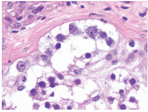
  1. Seminiferous tubules contain IGCNU cells, which are enlarged, atypical germ cells residing as isolated cells or as a single row along a usually thickened basement membrane (Figs.4.6.1, 4.6.2, 4.6.3)


  2. IGCNU cells have clear cytoplasm, irregular nuclear contours, coarse chromatin, and enlarged single or multiple enlarged variably sized nucleoli


  3. Seminiferous tubules containing IGCNU usually lack active spermatogenesis and contain mostly Sertoli cells. Can see contrast between Sertoli cells and IGCNU


  1. Seminiferous tubules contain only Sertoli cells


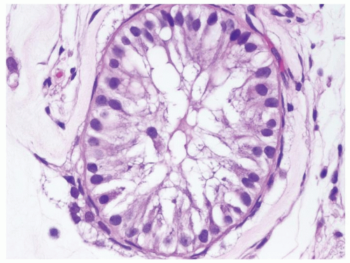
  2. Sertoli cells have a round-to-ovoid nucleus with a smooth outline with uniform chromatin and small uniform nucleoli (Figs.4.6.4, 4.6.5, 4.6.6)


  3. Uniform population of Sertoli cells


Special studies


  • IGCNU cells positive for PLAP and OCT4


  • IGCNU cells also positive for SALL4 and CD117 but not useful stains in this differential


  • Sertoli cells positive for inhibin


  • Negative for OCT4 and PLAP


  • Nonneoplastic germ cells may be positive for SALL4 and CD117 so not useful in this differential


  • Sertoli cells positive for inhibin


Treatment


Typically just close follow-up. In some countries, contralateral biopsy performed, and if positive, then sperm banking and bilateral low-dose radiation are offered


Directed toward inciting etiology. Micro-TESE (microdissection testicular sperm extraction) is attempted to achieve fertility


Prognosis


Up to 50% risk of developing invasive germ cell tumor within 5 y (up to 90% risk in 7 y). Risk of germ cell tumor is also increased in the contralateral testis


Fertility can be occasionally achieved


Figure 4.6.1 Seminiferous tubules contain IGCNU. Enlarged, atypical germ cells with cytoplasmic clearing in a single row along a thickened basement membrane are noted.


Figure 4.6.2 Seminiferous tubules contain IGCNU with enlarged hyperchromatic nuclei with coarse chromatin residing as a single row along a thickened basement membrane (arrows). Sertoli cell nuclei are smaller with more uniform chromatin and a single small central nucleolus (arrowhead).


Figure 4.6.3 IGCNU cells have clear cytoplasm, irregular nuclear contours, coarse chromatin, and enlarged single or multiple enlarged variably sized nucleoli.


Figure 4.6.4 Seminiferous tubules contain only Sertoli cells. Cells at low power often have a wispy appearance. Peritubular fibrosis is common.


Figure 4.6.5 Seminiferous tubules contain only Sertoli cells with small bland nuclei and abundant vacuolated cytoplasm.


Figure 4.6.6 Seminiferous tubules contain only Sertoli cells. Nuclei are round-to-ovoid with a smooth outline, uniform chromatin and small uniform nucleoli.


4.7 INTRATUBULAR GERM CELL NEOPLASIA UNCLASSIFIED (IGCNU) VS. PREPUBERTAL CRYPTORCHID TESTES


Intratubular Germ Cell Neoplasia Unclassified (IGCNU)


Prepubertal Cryptorchid Testes


Age


15-35 y


Congenital


Location


Usually unilateral


Usually unilateral but can rarely be bilateral


Symptoms


Asymptomatic


None


Signs


None except for those of associated conditions such as cryptorchidism and infertility


Congenital maldescent of one or both testes


Etiology


Associated risk factors include cryptorchidism (4% risk) and infertility (IGCNU in 1%-2% of biopsies)


Risk factors include intrauterine exposure to environmental chemicals with endocrine-disrupting properties; low birth weight; karyotypic abnormality (intersex syndrome); other genital tract malformation (i.e., hypospadias); and maternal smoking during pregnancy. Extremely rare to see IGCNU in prepubertal testes apart from an intersex disorder


Histology


  1. IGCNU cells are enlarged and have clear cytoplasm, irregular nuclear contours, coarse chromatin, and enlarged single or multiple nucleoli (Figs.4.7.1, 4.7.2, 4.7.3)


  2. Seminiferous tubules containing IGCNU usually lack active spermatogenesis and contain mostly Sertoli cells


  3. IGCNU cells are within seminiferous tubules as isolated cells or as a single row along a usually thickened basement membrane


  1. Spermatogonia have clear cytoplasm containing small size nuclei with round and regular nuclear contours, densely packed chromatin and no nucleoli. Occasionally, spermatogonia are otherwise identical, yet can be binucleated or have enlarged nuclei (giant spermatogonia). They are usually solitary in a given seminiferous tubule and can be dispersed throughout the testis (Figs.4.7.4, 4.7.5, 4.7.6)


  2. Prepubertal seminiferous tubules lack active spermatogenesis


  3. Sertoli cells are basally located as isolated cells along a usually thickened basement membrane


Special studies


  • OCT4 and PLAP positive


  • Other markers not as useful in this differential


  • PLAP and OCT4 negative in normal prepubertal testes


  • Delayed maturation in undervirilization syndromes [1] and in Down syndrome characterized by retention of fetal germ cell markers (PLAP, OCT4, CD117) but lacking typical seminoma-like morphology


Treatment


Typically just close follow-up. In some countries, contralateral biopsy performed, and if positive, then sperm banking and bilateral low-dose radiation are offered


Orchiopexy. Testicular biopsy to rule out IGCNU when cryptorchidism is associated with karyotypic abnormality or other male genital malformation


Prognosis


Up to 50% risk of developing invasive germ cell tumor within 5 y (up to 90% risk in 7 y). Risk of germ cell tumor is also increased in the contralateral testis


2%-4 % risk of developing IGCNU during adulthood. Increased risk of invasive germ cell tumor in the ipsilateral (4-7×) and contralateral testis (2-4×). Orchiopexy even before 2 y of age is not protective of subsequent neoplasia


Figure 4.7.1 Seminiferous tubules contain IGCNU. Enlarged, atypical germ cells are situated as a single row along a thickened basement membrane.


Figure 4.7.2 IGCNU cells have clear cytoplasm, irregular nuclear contours, coarse chromatin, and enlarged single or multiple enlarged variably sized nucleoli.


Figure 4.7.3 IGCNU cells with irregular nuclear contours and coarse chromatin.


Figure 4.7.4 Spermatogonia with clear cytoplasm containing small-size nuclei with round and regular nuclear contours are seen in this prepubertal testis.


Figure 4.7.5 Occasional spermatogonia in the prepubertal testes may increase in size but maintain regular nuclear contour. The lack of atypia distinguishes these giant spermatogonia from IGCNU.


Figure 4.7.6 Spermatogonia with clear cytoplasm containing small-size nuclei with round and regular nuclear contours are seen with occasional binucleated giant spermatogonia in this prepubertal testis. The lack of nuclear atypia and prominent nucleoli are helpful features.


4.8 INTRATUBULAR GERM CELL NEOPLASIA UNCLASSIFIED (IGCNU) VS. IGCNU WITH INTERTUBULAR SEMINOMA


Intratubular Germ Cell Neoplasia Unclassified (IGCNU)


IGCNU with Intertubular Seminoma


Age


15-35 y


Second to fourth decades


Location


Usually unilateral


Usually unilateral


Symptoms


Asymptomatic


Painless swelling of one testicle; one-third of cases complain of scrotal heaviness or dull pain. One-tenth of cases may present with symptoms due to metastatic disease such as abdominal/back pain, cough.


Signs


None except for those of associated conditions such as cryptorchidism and infertility


Usually palpable testicular nodule


Etiology


Associated risk factors include cryptorchidism (4% risk) and infertility (IGCNU in 1%-2% of biopsies)


Associated risk factors include cryptorchidism (10%), infertility, low socioeconomic status, and male genital malformations


Histology


  1. IGCNU cells are enlarged, atypical germ cells residing within seminiferous tubules as isolated cells or as a single row along a usually thickened basement membrane (see Sections 4.6 and 4.7) (Fig. 4.8.1)


  2. Lacks invasive seminoma cells


  3. Lacks prominent interstitial lymphoplasmacytic infiltrate


  1. Seminiferous tubules containing IGCNU are almost invariably present (Fig. 4.8.2)


  2. Invasive seminoma component can be subtle permeating the testicular stroma between intact seminiferous tubules. The invasive neoplastic cells are monotonous round-to-polygonal cleared cells with ovoid nuclei containing one or more prominent nucleoli imparting a classic “fried egg” appearance. Mitotic activity is variable (Figs. 4.8.3 and 4.8.4)


  3. Lymphoplasmacytic infiltrate at times with lymphoid follicles, and eosinophils are typically associated with the infiltrating seminoma. The latter is helpful in pointing to the occasionally subtle invasive intertubular seminoma component (Figs. 4.8.5 and 4.8.6)


Special studies


Positive for PLAP and OCT4


Intertubular infiltrating seminoma cells can be highlighted positive for PLAP and CD117, OCT4 and SALL4. Associated IGCNU positive for same markers


Treatment


Typically just close follow-up. In some countries, contralateral biopsy performed, and if positive, then sperm banking and bilateral low-dose radiation are offered


Early-stage disease typically requiring only radical orchiectomy followed by active surveillance


Prognosis


Up to 50% risk of developing invasive germ cell tumor within 5 y of diagnosis (up to 90% risk in 7 y). Risk of invasive germ cell tumor also increased in the contralateral testis


Excellent prognosis with over 90% relapse-free 5-y survival


Figure 4.8.1 Seminiferous tubules contain IGCNU.


Figure 4.8.2 Intertubular/interstitial invasive seminoma component adjacent to the seminiferous tubule with IGCNU.


Figure 4.8.3 Intertubular/interstitial invasive seminoma (arrows) adjacent to sclerotic seminiferous tubules.


Figure 4.8.4 Intertubular/interstitial invasive seminoma (arrows) permeating the testicular stroma between the seminiferous tubule with IGCNU.


Figure 4.8.5 Associated lymphoplasmacytic infiltrate is a clue raising the suspicion for the presence of an invasive component.


Figure 4.8.6 Same case as Figure 4.8.5 with intertubular invasive neoplastic seminoma cells in lymphoplasmacytic infiltrate.


4.9 SEMINOMA, CLASSIC TYPE VS. SPERMATOCYTIC SEMINOMA


Seminoma, Classic Type


Spermatocytic Seminoma


Age


Second to fourth decades. Can also be seen in older men in fifth to sixth decades


Older male, typically fifth to sixth decades. Uncommonly can be seen in younger men in second to fourth decades


Location


Usually unilateral. Can occur in extratesticular midline location


Usually unilateral. No primary extratesticular counterpart exists


Symptoms


Painless scrotal swelling. In one-third of cases, scrotal heaviness or dull pain. In one-tenth of cases, symptoms due to metastatic disease


Painless testicular mass


Signs


Usually palpable testicular nodule. Occasional elevation in serum human chorionic gonadotropin (beta hCG). 2% with gynecomastia


Usually palpable testicular nodule. Negative serum tumor markers


Etiology


Associated risk factors include cryptorchidism (10%), infertility, low socioeconomic status, and male genital malformations


No association with other risk factors typical of other testicular germ cell tumors


Histology


  1. Monotonous round-to-polygonal cleared cells with ovoid nuclei containing one or more prominent nucleoli (Figs. 4.9.1 and 4.9.2)


  2. Mitotic activity is variable


  3. Architecture varies from sheets, pseudoglandular/tubular, alveolar, cribriform, or intertubular (Fig. 4.9.4) between nonneoplastic seminiferous tubules


  4. Lymphoplasmacytic infiltrate in 85% of cases. May have lymphoid follicles. Eosinophils and histiocytes typically present in fibrovascular septae (Fig. 4.9.1).


  5. Nonnecrotizing granulomatous response seen in up to 50% of cases (see Section 4.5)


  6. Associated syncytiotrophoblastic giant cells in 10%-20% of cases (see Section 4.11)


  7. IGCNU cells are almost invariably associated (80% of cases) (Fig. 4.9.3)


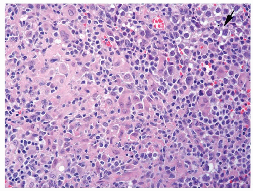
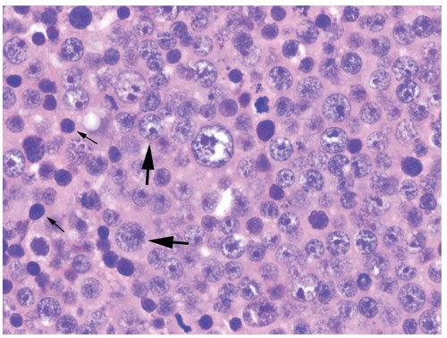
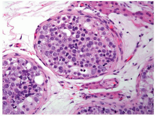
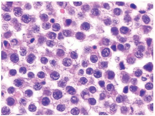
  1. Three basic cell types: Predominant is round-intermediate size with eosinophilic cytoplasm and round nuclei with characteristic lacy filamentous chromatin (spiremic pattern). Second type is smaller cells with dark nuclei and scant cytoplasm, while the third type is large mononucleated or multinucleated giant cells (Figs. 4.9.4 and 4.9.5)


  2. Mitotic activity can be brisk


  3. Typical architecture sheets of neoplastic cells with areas of stromal myxoid edema imparting a pseudoglandular pattern


  4. Lymphoplasmacytic infiltrate lacking


  5. No associated granulomatous response


  6. No associated syncytiotrophoblastic giant cells


  7. Lack IGCNU cells, yet may have intratubular spermatocytic seminoma (Fig. 4.9.6)


Special studies


  • OCT4 is negative


  • PLAP positive


  • CD117 positive


  • SALL4 positive


  • Gain of isochromosome 12p (i12p) seen in 80% of cases


  • OCT4 is negative


  • Focal PLAP positivity may be present


  • CD117 positivity in 50% of cases


  • SALL4 may also be positive


  • No gain of isochromosome 12p


Treatment


Radical orchiectomy followed by active surveillance or adjuvant radiotherapy and/or platinum-based chemotherapy


Radical orchiectomy. No adjuvant radiotherapy or chemotherapy


Prognosis


Stage dependent. Overall >90% relapse-free 5-y survival


With exception of extremely rare cases associated with sarcoma at presentation, benign behavior regardless of the presence of vascular or tunical invasion


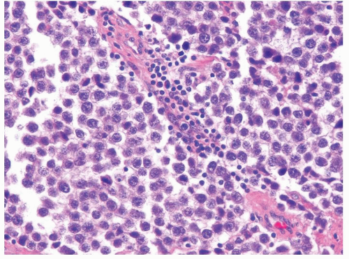
Figure 4.9.1 Classic seminoma composed of monotonous round-to-polygonal cleared cells with ovoid nuclei containing one or more prominent nucleoli with scattered interstitial lymphocytic infiltrate.


Figure 4.9.2 Monotonous round-to-polygonal cleared cells with ovoid nuclei typical of classic seminoma.


Figure 4.9.3 IGCNU is invariably present in seminiferous tubules associated with classic seminoma.


Figure 4.9.4 Spermatocytic seminoma with three cell types: (1) predominant round, intermediate-size cells with characteristic lacy filamentous chromatin (large arrows); (2) smaller cells with dark nuclei and scant cytoplasm (small arrows); and (3) large mononucleated or multinucleated giant cells (center). Note eosinophilic cytoplasm.


Figure 4.9.5 Spermatocytic seminoma is composed of three basic cell types. Lymphocytic host response is lacking.


Figure 4.9.6 Intratubular spermatocytic seminoma with the same three basic cell types filling seminiferous tubules.


4.10 SEMINOMA VS. EMBRYONAL CARCINOMA


Seminoma


Embryonal Carcinoma


Age


Second to fourth decades, yet can be older


Second to third decades


Location


Usually unilateral


Usually unilateral


Symptoms


Painless scrotal swelling. In one-third of cases, scrotal heaviness or dull pain. In 10% of cases, symptoms due to metastatic disease


Usually painless swelling but can present with testicular pain mimicking torsion. In 10% of cases, symptoms due to metastatic disease


Signs


Usually palpable testicular nodule. Occasional elevation in serum human chorionic gonadotropin (beta hCG) can be present. 2% with gynecomastia


Usually palpable testicular nodule


Etiology


Associated risk factors include cryptorchidism (10%), infertility, low socioeconomic status, and male genital malformations


Same as seminoma


Histology


  1. Monotonous round-to-polygonal cells with ovoid nuclei containing prominent nucleoli and clear cytoplasm (Figs. 4.10.1 and 4.10.2). Histologic variant with increased pleomorphism and mitotic rate in the past referred to as “anaplastic seminoma” or “high mitotic rate seminoma,” which mimics embryonal carcinoma (Fig. 4.10.3)


  2. Architecture varies from sheets, pseudoglandular/tubular, alveolar, cribriform, or intertubular (Fig. 4.10.4). Pagetoid spread to the rete testis can mimic embryonal carcinoma (Fig. 4.10.5)


  3. Cells are loosely cohesive


  4. Lymphoplasmacytic infiltrate in 85% of cases


  5. Nonnecrotizing granulomatous inflammation in up to 50% of cases


  6. Syncytiotrophoblasts can be associated in variable distribution


  7. IGCNU cells are almost invariably associated (80% of cases)


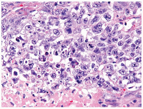
  1. Large cells with abundant granular amphophilic or eosinophilic cytoplasm and large vesicular irregular nuclei with one or more irregular nucleoli. Typically, more pleomorphic compared to seminoma (Fig. 4.10.6). Uncommonly can have focal clear cytoplasm (Fig. 4.10.7). Mitotic activity is brisk


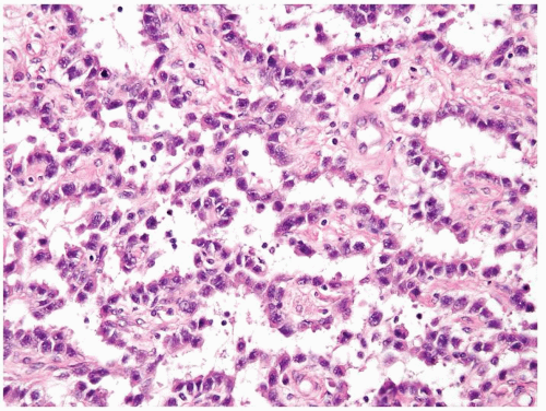
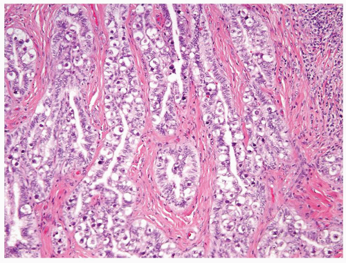
  2. Architecture varies from solid sheets to papillary structures with clefts and gland-like structures (Fig. 4.10.8)


  3. Cells are cohesive


  4. Lymphoplasmacytic infiltrate is infrequent (Fig. 4.10.9)


  5. Rare to see associated granulomatous inflammation


  6. Syncytiotrophoblasts not as common compared to seminoma


  7. IGCNU cells are almost invariably associated (80% of cases). Can also see intratubular embryonal carcinoma, which typically has very eosinophilic necrosis (Fig. 4.10.10)


Special studies


  • CD117 positive


  • CD30 negative


  • Typically keratin negative. However, occasionally, scattered isolated loosely cohesive keratin-positive cells that are still considered seminoma


  • CD117 negative


  • CD30 positive


  • Cytokeratin AE1/AE3 diffusely positive. In seminoma, if clusters of cohesive keratin-positive cells can be found on the corresponding H&E-stained slides to consist of more atypical cells, then admixed early embryonal carcinoma can be diagnosed


Treatment


Stage dependent. Radical orchiectomy followed by active surveillance or adjuvant radiotherapy and/or platinum-based chemotherapy


Stage dependent. Radical orchiectomy followed by active surveillance, retroperitoneal lymph node dissection (RPLND), or adjuvant platinum-based chemotherapy


Prognosis


Stage dependent with more than 90% relapse-free 5-y survival


Stage dependent with over 80% relapse-free 5-y survival


Figure 4.10.1 Classic seminoma is composed of loosely cohesive monotonous round-to-polygonal cells with clear cytoplasm.


Figure 4.10.2 Relatively uniform nuclei with central prominent nucleoli.


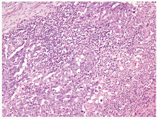
Figure 4.10.3 Seminoma with greater than usual pleomorphism. Cells are still loosely cohesive with interspersed lymphocytes.


Figure 4.10.4 Seminoma with pseudoalveolar pattern. Cells are still typical of seminoma.


Figure 4.10.5 Seminoma with pagetoid spread into the rete testis.


Figure 4.10.6 Embryonal carcinoma composed of large polygonal cells with abundant granular amphophilic cytoplasm and large vesicular irregular nuclei with one or more irregular nucleoli.


Figure 4.10.7 Embryonal carcinoma cohesive cells and focal area with clear cytoplasm.


Figure 4.10.8 Embryonal carcinoma with clefts and gland-like structures.


Figure 4.10.9 Unusual embryonal carcinoma with lymphocytic infiltrate. Cells are cohesive with clefts.


Figure 4.10.10 Intratubular embryonal carcinoma with eosinophilic necrosis.


4.11 SEMINOMA WITH SYNCYTIOTROPHOBLASTS VS. CHORIOCARCINOMA


Seminoma with Syncytiotrophoblasts


Choriocarcinoma


Age


Second to fourth decades, yet can be older


Second to third decades


Location


Usually unilateral


Usually unilateral


Symptoms


Usually, painless scrotal swelling. 10% present with symptoms due to metastatic disease such as abdominal/back pain, cough.


Usually present with symptoms due to widespread metastases including multiorgan hemorrhage (hemoptysis, hematemesis, cerebrovascular accident, etc.)


Signs


Usually palpable testicular nodule. Syncytiotrophoblasts lead to elevation in serum beta hCG usually in the levels of 100 mIU/mL. 2% with gynecomastia


Testicular mass may not be apparent. Very high levels of serum beta hCG usually in the levels of 100,000 mIU/mL. May have gynecomastia


Etiology


Same as other germ cell tumors (see Section 4.10)


Same as other germ cell tumors (see Section 4.10)


Histology


  1. Can be seen with pure seminoma or mixed germ cell tumor


  2. Composed of monotonous round-to-polygonal cells with clear cytoplasm and ovoid nuclei containing one or more prominent nucleoli


  3. Cells are loosely cohesive


  4. Seminoma admixed with syncytiotrophoblasts, which are multinucleated cells that contain abundant basophilic cytoplasm. No cytotrophoblasts (Fig. 4.11.1)


  5. Syncytiotrophoblasts scattered in midst of seminoma cells and tend to cluster around small blood vessels (Figs.4.11.2, 4.11.3, 4.11.4, 4.11.5, 4.11.6)


  6. Usually relatively few syncytiotrophoblasts, yet can be fairly numerous


  7. Lymphoplasmacytic infiltrate common


  8. Noncaseating granulomatous inflammation in up to 50% of cases


  9. Vascular invasion is uncommon


  10. IGCNU almost invariably associated (80% of cases)


  1. Can be pure but more often mixed with other germ cell tumor elements


  2. Cytotrophoblasts have pale cytoplasm surrounding an irregular, single nucleus with one or more nucleoli less prominent than in seminoma. Nuclei are usually more atypical than in seminoma (Figs.4.11.7, 4.11.8, 4.11.9)


  3. Cytotrophoblasts are cohesive


  4. Cytotrophoblasts are intimately admixed with syncytiotrophoblasts (Figs.4.11.7, 4.11.8, 4.11.9)


  5. Cytotrophoblasts and syncytiotrophoblasts often associated with hemorrhage and necrosis (Fig. 4.11.10)


  6. If in mixed germ cell tumor, usually the choriocarcinoma component is substantial, although uncommonly can be focal


  7. Lymphoplasmacytic infiltrate is infrequent


  8. Granulomatous reaction absent


  9. Vascular invasion is common


  10. IGCNU almost invariably present


Special studies


  • Seminoma cells positive for OCT4 and CD117. Syncytiotrophoblasts is negative


  • Seminoma cytokeratin AE1/AE3 negative or at most rare positive cells. Syncytiotrophoblasts positive for AE1/AE3


  • Syncytiotrophoblasts beta hCG positive


  • Cytotrophoblasts and syncytiotrophoblasts negative for OCT4 and CD117


  • Cytotrophoblasts and syncytiotrophoblasts positive for AE1/AE3


  • Syncytiotrophoblasts beta hCG positive


Treatment


Stage dependent. Radical orchiectomy followed by active surveillance or adjuvant radiotherapy and/or platinum-based chemotherapy


Stage dependent. Radical orchiectomy followed by retroperitoneal lymph node dissection (RPLND) or adjuvant platinum-based chemotherapy


Prognosis


Syncytiotrophoblasts does not impact prognosis in otherwise classic seminoma


Typically associated with advanced stage with poor prognosis. However, stage-for-stage prognosis is comparable to other nonseminomatous germ cell tumors


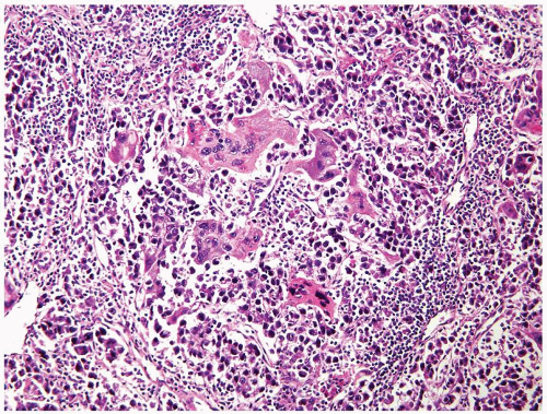
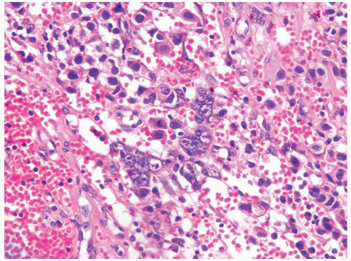
Figure 4.11.1 Seminoma admixed with syncytiotrophoblasts, which are multinucleated cells that contain abundant basophilic cytoplasm. No cytotrophoblasts are accompanied.


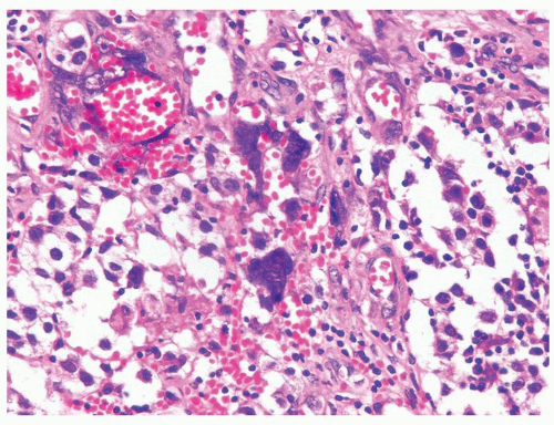
Figure 4.11.2 Syncytiotrophoblasts without associated cytotrophoblasts are seen adjacent to areas of hemorrhage in seminoma.


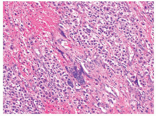
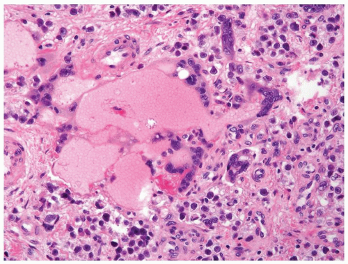
Figure 4.11.3 Syncytiotrophoblasts without associated cytotrophoblasts are seen surrounding vascular structures in seminoma.


Figure 4.11.4 Syncytiotrophoblasts without associated cytotrophoblasts are seen adjacent to areas of hemorrhage in seminoma.


Figure 4.11.5 Syncytiotrophoblasts without associated cytotrophoblasts are seen surrounding vascular structures in seminoma.


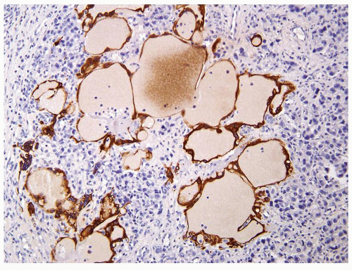
Figure 4.11.6 Same case as in Figure 4.11.5 with highlighting of syncytiotrophoblasts by HCG immunostains.


Figure 4.11.7 Cytotrophoblasts are intimately admixed with syncytiotrophoblasts in choriocarcinoma.


Figure 4.11.8 Cytotrophoblasts are intimately admixed with smaller syncytiotrophoblasts with amphophilic cytoplasm in choriocarcinoma. Cytotrophoblasts have paler cytoplasm surrounding an irregular, single nucleus with one or more nucleoli.


Figure 4.11.9 Choriocarcinoma with subtle syncytiotrophoblasts (arrows) enveloping cytotrophoblasts.


Figure 4.11.10 Choriocarcinoma with extensive hemorrhage and necrosis.


4.12 SEMINOMA VS. LARGE B-CELL LYMPHOMA


Seminoma


Large B-Cell Lymphoma


Age


Second to fourth decades but can occur in older overlapping with lymphoma


Seventh and eighth decades, most frequent testicular tumor in this age group


Location


Usually unilateral


Over one-third of cases are bilateral


Symptoms


Painless scrotal swelling. 10% present with symptoms due to metastatic disease


Painless swelling of the scrotum or inguinal region. No associated B-cell lymphoma symptoms


Signs


Usually a palpable testicular nodule


Usually a palpable testicular/paratesticular mass


Etiology


Associated risk factors include cryptorchidism (10%), infertility, low socioeconomic status, and male genital malformations


Diffuse large B-cell lymphoma is the most common subtype (80%) in adults. Usually represents secondary involvement of the testis from the lymph node primary. Rarely, primary testicular lymphoma without nodal involvement


Histology


  1. Typically a solid mass with relatively little intertubular growth that spares seminiferous tubules


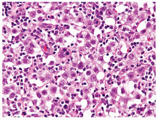
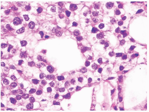
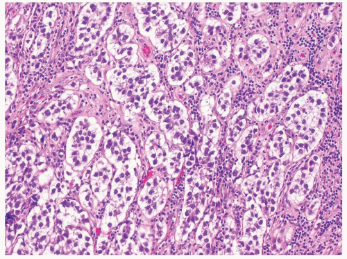
  2. Monotonous round-to-polygonal cleared cells with ovoid nuclei that are typically relatively uniform, although pleomorphism can be present (Figs. 4.12.1 and 4.12.2)


  3. Cytoplasm clear


  4. One or more prominent nucleoli imparting a classic “fried egg” appearance


  5. Architecture varies from sheets, pseudoglandular/tubular, alveolar, cribriform, or interstitial/intertubular


  6. Granulomatous inflammation in up to 50% of cases


  7. Syncytiotrophoblasts present in minority of cases


  8. IGCNU almost invariably present (80% of cases)


  9. Does not tend to spread into adjacent adipose tissue extensively and when it extends out of the testis has more of a solid growth


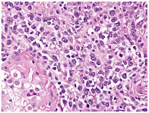
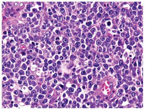
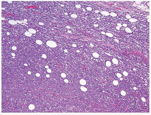
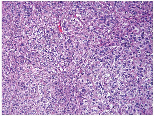
  1. Typically a solid mass with relatively little intertubular growth that spares seminiferous tubules (Figs.4.12.3, 4.12.4, 4.12.5, 4.12.6, 4.12.7)


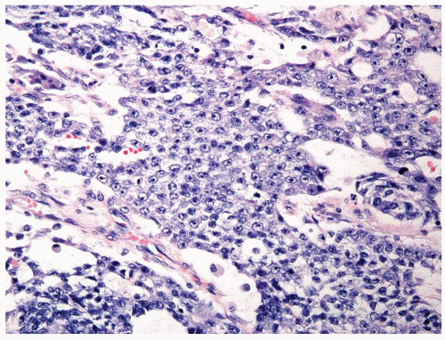
  2. Predominantly large cleaved and large noncleaved atypical lymphocytes. Nuclei more irregular than seminoma (Figs. 4.12.3, 4.12.5, 4.12.6, 4.12.7)


  3. Most cases with eosinophilic to amphophilic cytoplasm (Figs. 4.12.3 and 4.12.7)


  4. Multiple irregular nucleoli of varying sizes


  5. Diffuse sheets of cells


  6. Granulomatous inflammation typically not seen in testicular lymphomas


  7. Syncytiotrophoblasts not encountered


  8. IGCNU typically absent


  9. Can extensively involve paratesticular soft tissue with relative sparing of adipose tissue (Fig. 4.12.8)


Special studies


  • SALL4 positive


  • OCT4 positive


  • Negative for CD45 and CD20


  • SALL4 negative, except anaplastic large cell lymphoma can be positive


  • OCT4 not as useful in this differential as rare lymphomas positive


  • Positive for CD45 and CD20


Treatment


Stage dependent. Radical orchiectomy followed by active surveillance or adjuvant radiotherapy and/or platinum-based chemotherapy


In vast majority of cases, orchiectomy is performed to make the diagnosis. Subsequently treated by systemic chemotherapy


Prognosis


Stage dependent with more than 90% relapse-free 5-y survival


Stage dependent with 60% 5-y tumor-free survival for early stage and 20% for advanced stage. As most patients present with advanced stage, the overall survival is 50% at 5 y. High relapse rate mainly in extratesticular location


Figure 4.12.1 Classic seminoma composed of sheets of back-to-back monotonous neoplastic cells separated with associated lymphocytic infiltrate.


Figure 4.12.2 Classic seminoma with relatively uniform round-to-polygonal cleared cells with ovoid nuclei.


Figure 4.12.3 Large cell lymphoma with cells showing more pleomorphism than seminoma. Also the cells lack central prominent nucleoli in the majority of cells.


Figure 4.12.4 Lymphoma with relative preservation of testicular architecture.


Figure 4.12.5 Large cell lymphoma with less uniformity than seminoma. Also the cells lack central prominent nucleoli in the majority of cells.


Figure 4.12.6 Lymphoma with clear cytoplasm. Cells lack vesicular nuclei with prominent nucleoli.


Figure 4.12.7 Lymphoma mimicking seminoma with prominent nucleoli, yet cells have amphophilic cytoplasm.


Figure 4.12.8 Lymphoma spread outside of the testis with entrapment of adipocytes.


4.13 SEMINOMA VS. YOLK SAC TUMOR, SOLID PATTERN


Seminoma


Yolk Sac Tumor, Solid Pattern


Age


Second to fourth decades, yet can be older


Two peaks: Infants/early childhood (median age 1.5 y) and postpubertal (second and third decades)


Location


Usually unilateral


Usually unilateral


Symptoms


Painless scrotal swelling. 10% present with scrotal pain and 10% with symptoms due to metastatic disease


Painless scrotal mass. Occasionally acute pain. In children, symptoms of metastatic spread (i.e., hemoptysis) at presentation in up to 10%-20% of cases


Signs


Usually palpable testicular nodule


Usually palpable testicular nodule. Serum AFP elevation in 90% of cases


Etiology


Same as other germ cell tumors (see Section 4.10)


Same as other germ cell tumors (see Section 4.10)


Histology


  1. Monotonous round-to-polygonal cleared cells with ovoid nuclei containing one or more prominent nucleoli (Fig. 4.13.1). Mitotic and apoptotic activity is variable


  2. Cells are loosely cohesive


  3. Architecture varies from sheets of back-to-back monotonous neoplastic cells, pseudoglandular/tubular, alveolar, cribriform, or interstitial/intertubular (Figs. 4.13.2 and 4.13.3)


  4. Lacks intracytoplasmic hyaline globules


  5. Lymphoplasmacytic infiltrate typically present, yet absent/minimal in 15% of cases


  6. Noncaseating granulomatous reaction seen in up to 50% of cases


  7. Syncytiotrophoblasts can be associated in variable distribution


  8. IGCNU almost invariably associated (80% of cases)


  1. Polygonal, medium-sized cells with clear to eosinophilic cytoplasm, uniform vesicular nuclei, and prominent nucleoli. Mitotic activity can be high (Figs.4.13.4, 4.13.5, 4.13.6, 4.13.7, 4.13.8, 4.13.9)


  2. Cells are tightly cohesive


  3. Solid pattern mimic seminoma (Figs.4.13.4, 4.13.5, 4.13.6, 4.13.7 and 4.13.9). Other more common patterns include microcystic, macrocystic, glandular, papillary, myxomatous, polyvesicular vitelline, hepatoid, and endodermal sinus with Schiller-Duval bodies (Figs. 4.13.4 and 4.13.8)


  4. Often presence of intracytoplasmic hyaline globules


  5. Lymphoplasmacytic infiltrate typically absent


  6. Noncaseating granulomatous reaction absent


  7. Syncytiotrophoblasts less common than in seminoma but can be seen


  8. IGCNU almost invariably associated in adults while absent in children


Special studies


  • OCT4 positive


  • Glypican 3 and AFP negative


  • CD117 positive


  • Cytokeratin AE1/AE3 negative or rare cells positive


  • OCT4 negative


  • Glypican 3 in >80% of cases. AFP expressed to a lesser extent (Fig. 4.13.10)


  • CD117 can be positive in minority of cases


  • Strong cytokeratin AE1/AE3 and Cam 5.2


Treatment


Stage dependent. Radical orchiectomy followed by active surveillance or adjuvant radiotherapy and/or platinum-based chemotherapy


Stage dependent. In postpubertal patients, same therapy as other mixed germ cell tumors. With exception of cases with extratesticular spread at presentation, radical orchiectomy is followed by active surveillance in prepubertal cases


Prognosis


Stage dependent with more than 90% relapse-free 5-y survival


Stage dependent with more than 80% relapse-free 5-y survival in postpubertal patients. Prepubertal patients 95% relapse-free survival regardless of stage with a 100% relapse-free survival in stages I and II


Figure 4.13.1 Classic seminoma is composed of monotonous round-to-polygonal cleared cells with lymphocytic infiltrate.


Figure 4.13.2 Classic seminoma with pseudoglandular spaces with uniform, loosely cohesive seminoma cells.


Figure 4.13.3 Classic seminoma with nesting appearance.


Figure 4.13.4 Yolk sac tumor composed of sheets of cells (right) with a more typical microcystic component (left).


Figure 4.13.5 Same case as Figure 4.13.4 with cells mimicking seminoma, however lacking the seminoma cells’ uniform shape, vesicular nuclei, and large prominent eosinophilic nucleoli.


Figure 4.13.6 Solid yolk sac tumor.


Figure 4.13.7 Same case as Figure 4.13.6 with polygonal, medium-sized, cells with clear to eosinophilic cytoplasm and variably shaped nuclei.


Figure 4.13.8 Same case as Figures 4.13.6 and 4.13.7 with more typical myxoid features.


Figure 4.13.9 Solid yolk sac tumor, which is more cohesive than seminoma.


Figure 4.13.10 Same case as Figure 4.13.9 with diffuse positivity for AFP.


4.14 SEMINOMA WITH EXTENSIVE NECROSIS VS. TORSION


Seminoma with Extensive Necrosis


Torsion


Age


Second to fourth decades, can be older


Any age but more common during childhood (two peaks: Neonatal and adolescent)


Location


Usually unilateral


Usually unilateral


Symptoms


Painless scrotal swelling. One-third of cases complain of scrotal heaviness or dull pain. One-tenth of cases may present with symptoms due to metastatic disease such as abdominal/back pain, cough.


Sudden onset severe testicular pain (76%), scrotal swelling (65%), abdominal pain, nausea, and vomiting (22%)


Signs


Usually palpable testicular nodule. Occasional elevation in serum human chorionic gonadotropin (beta hCG). 2% with gynecomastia


No palpable nodule. Spherical testicular enlargement, redness, and inflammatory scrotal signs termed “acute scrotum”


Etiology


Associated risk factors include cryptorchidism (10%), infertility, low socioeconomic status, and male genital malformations


Complicated pregnancies (prolonged labor, preeclampsia, gestational diabetes) leading to fetal stress/mechanical factors that may play a role in the pathogenesis of perinatal testicular torsion. Recently, seasonal predilection (cold temperature and humidity) has been suggested as a risk factor in childhood and adolescent cases as a result of associated hyperactive cremasteric reflex


Histology


  1. Typically, does not involve the entire testis


  2. Occasional seminomas can undergo extensive necrosis, yet in the center of coagulative necrosis see ghosts of seminoma cells. At edge of necrosis, may see necrotic seminoma with more cellular preservation composed of monotonous round-to-polygonal cleared cells with ovoid nuclei containing one or more prominent nucleoli (Figs.4.14.1, 4.14.2, 4.14.3, 4.14.4, 4.14.5, 4.14.6)


  3. Nonneoplastic testis surrounding the necrotic seminoma remains viable and lacks evidence of ischemia or hemorrhage


  4. Atrophic seminiferous tubules containing IGCNU may be seen in the adjacent testis


  5. May see some lymphocytic infiltrate


  1. Torsion occurs as a result of testicular rotation around the cord leading to interruption of blood supply and hemorrhagic infarction, such that the entire testis is affected


  2. In advanced cases, complete necrosis with shadows of seminiferous tubules with basement membranes outlines can still be identified (Figs. 4.14.9 and 4.14.10)


  3. The entire testis shows similar ischemic changes


  4. No IGCNU


  5. When present, inflammation is predominantly acute and composed of polymorphonuclear leukocytes


Special studies


  • Positive PLAP, CD117, OCT4, and SALL4 may be maintained in necrotic seminoma (Figs. 4.14.7 and 4.14.8)


  • PLAP, CD117, OCT4, and SALL4 negative


Treatment


Remains stage dependent despite regression. Radical orchiectomy followed by active surveillance or adjuvant radiotherapy and/or platinum-based chemotherapy


By the time the testis is so necrotic that it mimics necrotic seminoma, the only treatment is orchiectomy and contralateral orchiopexy


Prognosis


Stage dependent with more than 90% relapse-free 5-y survival


Diagnosis and treatment over 6 h, which would correspond to an entirely necrotic testis, have no likelihood of testicular salvage

Only gold members can continue reading. Log In or Register to continue

Stay updated, free articles. Join our Telegram channel

Jul 9, 2016 | Posted by in PATHOLOGY & LABORATORY MEDICINE | Comments Off on Testis

Full access? Get Clinical Tree
Get Clinical Tree app for offline access