Synovial Sarcoma
Key Facts
Macroscopic Features
Large, well-circumscribed, pleural-based mass
May extend along pleural surface mimicking malignant mesothelioma
May show cystic and hemorrhagic changes
Microscopic Pathology
May be biphasic or monophasic
Biphasic tumors are composed of cleft-like and glandular structures with intraluminal eosinophilic proteinaceous material admixed with population of atypical spindle cells
Monophasic tumors are composed of monotonous atypical spindle cell proliferation with herringbone, storiform, or hemangiopericytic growth patterns
Variable mitotic activity (range: 2-10 mitoses per 10 high-power fields)
Spindle cell population is very monotonous and uniform
Absence of nuclear pleomorphism or prominent nucleoli
Glands lined by tall columnar cells with abundant cytoplasm in biphasic type
Focal (patchy) positivity for epithelial markers (cytokeratin and EMA)
Strong positivity for Bcl-2 and CD99
Shares many markers with malignant mesothelioma, including calretinin and HBME-1
Some cases may be positive for CD34
Characteristic chromosomal translocation, t(X;18) (SYT-SSX) seen in > 80% of cases
Distinctive translocation may be assayed by FISH, PCR, or DNA in situ hybridization
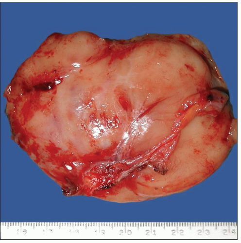 Gross appearance of primary synovial sarcoma of the pleura shows a well-circumscribed mass with a fleshy and soft appearance and a lobulated external surface. |
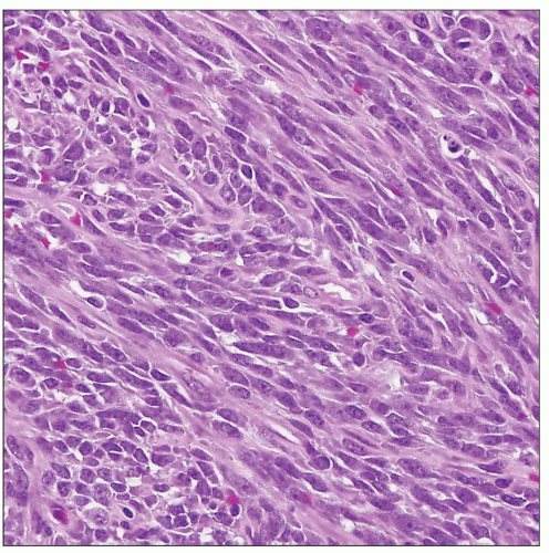 Typical histologic appearance of monophasic synovial sarcoma of the pleura shows monotonous fascicles of spindle cells almost completely devoid of connective tissue stroma. |
TERMINOLOGY
Abbreviations
Synovial sarcoma (SS)
Definitions
Pleural malignant neoplasm composed of nonmesothelial cells showing epithelial differentiation with distinctive spindle cell pattern of growth and chromosomal translocation
ETIOLOGY/PATHOGENESIS
Pathogenesis
Unknown
CLINICAL ISSUES
Presentation
Chest pain
Pleural effusion
Pleural-based mass on CT scan
Age range: 10-50 years (average: 25 years)
Treatment
Surgical excision, radiation therapy, chemotherapy
Prognosis
Similar prognosis to soft tissue counterpart
Aggressive behavior with local recurrence and metastases
MACROSCOPIC FEATURES
General Features
Large, well-circumscribed, pleural-based mass
May extend along pleural surface mimicking malignant mesothelioma
May infiltrate underlying structures
May show cystic and hemorrhagic changes
May attain large sizes (up to 16 cm in greatest dimension)
MICROSCOPIC PATHOLOGY
Histologic Features
May be biphasic or monophasic
Biphasic tumors are composed of cleft-like and glandular structures with intraluminal eosinophilic proteinaceous material admixed with population of atypical spindle cells
Monophasic tumors are composed of monotonous atypical spindle cell proliferation with herringbone, storiform, or hemangiopericytic growth patterns
Poorly differentiated variants are characterized by round epithelioid cells rather than spindle cells
Variable mitotic activity (range: 2-10 mitoses per 10 high-power fields)
May display hemorrhage and necrosis
Cytologic Features
Spindle cell population is very monotonous and uniform
Absence of nuclear pleomorphism or prominent nucleoli
Glands lined by tall columnar cells with abundant cytoplasm in biphasic type
Absence of fibrous stroma
Absence of marked anaplasia or multinucleated tumor cells
ANCILLARY TESTS
Immunohistochemistry
Cytogenetics
Characteristic chromosomal translocation, t(X;18) (SYT-SSX) seen in > 80% of cases
Distinctive translocation may be demonstrated by FISH, PCR, or DNA in situ hybridization
Distinctive translocation has not been recognized in other similar tumors
DIFFERENTIAL DIAGNOSIS
Biphasic Malignant Mesothelioma
More intimate admixture between glandular structures and spindle cell component seen in synovial sarcoma
Stay updated, free articles. Join our Telegram channel

Full access? Get Clinical Tree


