Superficial Vascular Lesions and Mimics of Vascular Lesions


Superficial Vascular Lesions and Mimics of Vascular Lesions


INTRODUCTION

This chapter includes reactive and neoplastic lesions arising in the superficial soft tissues in adults and children. The differential diagnosis is presented in Table 17.1.

Benign pediatric vascular lesions can be divided into the two broad categories of tumors (hemangiomas) and vascular malformations. Hemangiomas are characterized by rapid growth during infancy followed by gradual involution, whereas malformations are present at birth (although not necessarily clinically apparent), growing proportionally with the child and persisting without regression. Vascular malformations have subsequently been further subdivided according to the predominant type of vascular channel (capillary, venous, arterial, lymphatic, or a combination) and their hemodynamic features (low or high blood flow).


REACTIVE LESIONS

This group includes vascular proliferations that result from infectious processes, immunologic disturbances, or host responses to a variety of other etiologies, including trauma. Angiolymphoid hyperplasia with eosinophilia (epithelioid hemangioma) is included in this section, in part to highlight its contrasting features with Kimura disease, with which it has been confused in the past, and in part as a reflection of the belief that some examples have features consistent with a reactive process, although some examples may be neoplasms (epithelioid hemangioma).


BACILLARY ANGIOMATOSIS


Clinical Features

Bacillary (epithelioid) angiomatosis is a pseudoneoplastic vascular proliferation, first described in individuals infected with HIV.1 The disease has a predilection for, but is not restricted to, immunocompromised patients. The predominant pathogen is Bartonella (Rochalimaea) henselae, which


is also the etiologic agent of cat scratch disease, but other Bartonella species are associated.2 The organism is commonly harbored by domestic cats (which have a seroprevalence of about 5% to >60% depending on geographic area3) and transmitted to their owners. These organisms are susceptible to antibiotics, but untreated disease can prove fatal. Although cutaneous lesions are the most frequently recognized manifestation of bacillary angiomatosis (e-Fig. 17.1), a systemic infection typically ensues in the immunosuppressed host. In immunocompetent individuals, lesions are believed to remain localized to the inoculation site. Bacillary angiomatosis can masquerade as Kaposi sarcoma (KS) in resource-poor areas with a high prevalence of HIV-associated disease.4


TABLE 17.1 Differential Diagnosis of Superficial Vascular Lesions


Typical Clinical Features


Microscopic Features


Ancillary Investigations


Bacillary angiomatosis


Immunocompromised patients with cutaneous lesions


Lobulated proliferation of capillarysized vessels


Aggregates of neutrophils, leukocytoclastic debris


Warthin-Starry stain (pH 4.0), shows clumps of small curved rods of Bartonella henselae


Verruga Peruana (Oroya fever)


Skin lesions in persons in endemic areas (Peru)


Cutaneous vascular proliferation with plump endothelial cells and inflammation


Weak staining of Bartonella bacilliformis organisms with Giemsa


Papillary endothelial hyperplasia


Within thrombi anywhere in the body, often distal extremities and in hemorrhoids; essentially a variant of organizing thrombus


Fibrin cores coated by monolayers of endothelial cells, typically all within a vessel


Typically not required


Intravascular fasciitis


Distal extremity nodules in young adults


Intravascular myofibroblastic proliferation accompanied by lymphocytes and osteoclast-like giant cells


SMA+, calponin+, caldesmon−, desmin−


Vascular markers (CD34, CD31, ERG)−


Intravascular leiomyomatosis


In women with uterine leiomyomas that extend into veins


Essentially a leiomyoma in a vessel; perpendicularly oriented fascicles of brightly eosinophilic cells with blunt-ended nuclei without mitotic activity


SMA+, calponin+, caldesmon+, desmin+


Vascular markers (CD34, CD31, ERG)−


Angiolymphoid hyperplasia with eosinophilia (epithelioid hemangioma)


Adults (20-40 y), head-neck region, single or multiple smooth papules or plaques (superficial)


Some cases in bone


Lobular vascular proliferation surrounded by lymphoid cuff, often associated with damaged artery


Vessels lined by epithelioid endothelial cells, background eosinophils—benign


Typically not required; ZFP36-FOSB fusions detected in atypical cases suggests that at least some examples are neoplastic


Epithelioid hemangioendothelioma


Deep soft tissues, organs (lungs, liver)


Infiltrative lesion composed of single (not forming capillaries) epithelioid endothelioid cells in chondroid background, sometimes associated with a vessel—looks similar to lobular breast carcinoma in some cases—malignant


CD34+, CD31+ ERG+, variable keratin expression


Rearrangements in WWTR1-CAMTA1 and YAP-TFE3


Kimura disease


Asians, head and neck region


Associated lymphadenopathy and peripheral blood eosinophilia, elevation in the serum immunoglobulin E, sometimes nephrotic syndrome


Fibroinflammatory lesion with numerous eosinophils, sometimes eosinophilic microabscesses, vessels inconspicuous


Typically not required


Capillary hemangioma (infantile hemangioendothelioma, juvenile hemangioma, strawberry nevus)


Newborns and babies, female predominance, head and neck, grow rapidly and then usually regress


Cellular capillary proliferation with commensurate proliferation of supporting cells (smooth muscle, pericytes)


GLUT-1+


Pyogenic granuloma/lobular capillary hemangioma


Head and neck of adults, some associated with pregnancy, do not regress


Lobular capillary proliferation of capillaries, overlying epidermal collarette common


GLUT-1−


Cavernous hemangioma


Any age, usually upper half of body, do not regress


Gaping blood-filled spaces


GLUT-1−


Superficial arteriovenous tumor/malformation


Perioral region and extremities of adults, small red or purple papules, do not regress


Well-circumscribed masses of thick- and thin-walled vessels, in dermis (skin) or submucosa (lip)


Some vessels have a “transitional” appearance between arteries and veins


Typically not required


Microvenular hemangioma


Small solitary nodule or plaque involving the extremities of young adults


Permeative proliferation of small irregularly branching vessels which are well-formed but often have collapsed, somewhat inconspicuous lumina; vessels are thicker than


Typically not required


Glomeruloid hemangioma


Found in patients with POEMS syndrome—polyneuropathy, organomegaly (hepatosplenomegaly and lymphadenopathy), endocrinopathy (amenorrhea, gynecomastia, hypothyroidism, adrenal insufficiency, and glucose intolerance), monoclonal protein (marrow plasmacytosis, paraproteinemia), and skin lesions


Dilated, ectatic vessels with intraluminal capillary conglomerates, architecturally resembling renal glomeruli


Typically not required


Angioblastoma (acquired tufted angioma, progressive capillary hemangioma, angioblastoma of Nakagawa)


Children and young adults, upper half of body, some painful, do not regress


Tightly packed clusters of capillaries with a rounded or ovoid (“cannonball”) appearance in a plexiform pattern


GLUT-1−, CD34+, CD31+, podoplanin (D2-40)+, prox1+, LYVE1+, ERG+


Hobnail hemangioma (targetoid hemosiderotic hemangioma)


Solitary lesion, trunk or extremities of young adults


Early examples with central purple, violaceous or brownblack papule, bordered by an inner pale zone and a more peripherally situated ecchymotic ring (“targetoid”)


Ectatic thin-walled vascular channels situated in the superficial dermis and lined by plump, sometimes epithelioid, endothelium


Deeper vessels, located in the reticular dermis, have an angulated, irregular and slit-like appearance


Typically not required—endothelial cells CD34+, CD31+, podoplanin (D2-40)−, ERG+


Progressive lymphangioma (benign lymphangioendothelioma)


Head and neck of adults


Anastomosing vascular channels with a “collagen dissection” pattern


CD34+, CD31+, podoplanin (D2-40)+, ERG+


Angiokeratoma


Some sporadic; systemic type (angiokeratoma corpus diffusum) is associated with Fabry disease, fucosidosis, and deficiency of beta-galactosidase


Dilated vessels which are primarily restricted to the uppermost dermis and are bordered or enclosed by elongate rete ridges


Typically not required


Verrucous hemangioma


Reflects superficial extension (into epidermis) of an underlying deep hemangioma or vascular malformation


Dilated vessels in uppermost dermis enclosed by elongate rete ridges associated with underlying vascular lesion


Typically not required


Angioma serpiginosum


Lower extremities of young women


Superficial vascular ectasia


Typically not required


Cutaneous epithelioid angiomatous nodule


Blue papules, usually on the trunk of adults


Circumscribed, unilobular, mainly solid proliferations of large polygonal epithelioid endothelial cells with vesicular nuclei and conspicuous nucleoli and at least focal vasoformation


Variable staining with CD34, CD31, and FVII-related antigen


Massive localized lymphedema in the morbidly obese


Large soft tissue lesions (usually of thigh) with superficial component of longstanding duration in morbidly obese adults


Dermal fibrosis, expansion of the fibrous septa between fat lobules with increased numbers of stromal fibroblasts, lymphatic proliferation and lymphangiectasia


Typically not required MDM2, CDK4 both − if performed to exclude liposarcoma


Well-differentiated liposarcoma


Deep adipose tissue mass of thighs or retroperitoneum, only rarely involves the subcutaneous fat, no skin changes


Mature-appearing fat punctuated by enlarged hyperchromatic nuclei, occasionally lipoblasts


MDM2, CDK4+, nuclear labeling


MDM2 amplification on fluorescence in situ hybridization


Spindle cell hemangioma


Slow-growing, often multifocal small painless mass involving the dermis and/or subcutis, usually of hands and feet in young adults


Association with Milroy disease, Maffucci disease, Klippel-Trenaunay syndrome


Cavernous vascular spaces and a spindle cell proliferation vaguely reminiscent of Kaposi sarcoma


Typically not required; CD34+ and CD31+ in the endothelial cells of the dilated vessels and interleukin 8+ and vascular endothelial growth factor receptor+ in the spindle cells; IDH1 and IDH2 mutations in cases associated with Maffucci syndrome


Malignant endovascular papillary angioendothelioma, papillary intralymphatic angioendothelioma, Dabska tumor


Extremely rare, possibly more common in children but all ages, no favored site


Intercommunicating vascular channels, epithelioid to columnar endothelium, sometimes arranged in papillary (primitive glomeruloid) tufts with a central avascular hyaline core, intravascular lymphocytes may be prominent


CD34+, CD31+, podoplanin (D2-40)+ and vascular endothelial growth factor receptor-3+


Kaposiform hemangioendothelioma


Superficial or deep lesion of children (often) and adults (occasional)


In some infants, large retroperitoneal mass associated with a consumptive coagulopathy (Kasabach-Merritt syndrome), aggressive local behavior, association with lymphangiomatosis


Infiltrative yet lobular growth of well-formed capillaries admixed with short fascicles of spindle cells with slit-like vascular lumina


Podoplanin D2-40+ in the Kaposi sarcomalike proliferative capillaries but not in the surrounding dilated vessels


Endothelial cells in nodules are CD31+, CD34+, prox1+, LYVE1+, vascular endothelial growth factor receptor-3+, FLI1+, but GLUT-1−


Atypical vascular lesion


In (usually mammary) skin following radiation


A single or several small papules of patches in the irradiated site


Most benign, but a subset progresses to angiosarcoma, often after several recurrences


Small (<1 cm) resembling benign lymphangiomas, lymphangioendothelioma, or a lobular capillary proliferation


CD34+, CD31+, podoplanin (D2-40)+ for lesions that are lymphangioma-like or CD34+, CD31+, podoplanin (D2-40)− for capillary-like lesions


Retiform hemangioendothelioma


Rare neoplasms of young adults without gender preference involving skin and subcutaneous tissue, usually of lower extremities


Occasionally metastases to regional lymph nodes but not distant sites


Narrow arborizing vascular channels lined by plump hobnail endothelial cells with pattern reminiscent of rete testis, often with a striking lymphoid background


CD31+, CD34+, factor VIII-related antigen+, podoplanin−, vascular endothelial growth factor receptor 3−


Composite hemangioendothelioma


Longstanding dermal and subcutaneous nodules and plaques, usually in the extremities of young adults


Admixture of benign-appearing well-formed vessels, areas that resemble spindle cell hemangioma, areas similar to retiform hemangioendothelioma, and areas similar to epithelioid hemangioendothelioma


CD34+, CD31+, factor VIII-related antigen+, podoplanin (D2-40)−


Kaposi sarcoma


Lesions of immunosuppressed patients or in endemic areas


Tumors can involve skin or viscera


Spindle cells in fascicles and sheets, extravasated erythrocytes, siderophages, hyaline globules, lymphoplasmacytic infiltrate


Early lesions with more subtle features


CD34+, CD31+, human herpesvirus 8+, podoplanin (D2-40)+, vascular endothelial growth factor receptor 3+


Superficial angiosarcoma


Variable tumors (e.g., bruiselike lesions of face and scalp of elderly), associated with lymphedema, postirradiation


Wide range from hemangiomalike with collagen dissection to overtly malignant epithelioid neoplasms


Variable staining with CD34, CD31, ERG, podoplanin (D2-40), Fli-1, keratins


Human herpesvirus 8−, Prox1 (limited by lack of specificity)


Angiomatoid fibrous histiocytoma


Small lesions usually of proximal limb girdle, trunk, head and neck arising in first two decades, slight female predominance


About 15% recurrences, rare metastases to nodes, rarer systemic metastases


Multinodular sheets of ovoid cells


Some cases with focal nuclear pleomorphism and multinucleated giant cells, variable hemorrhage, some cases with large cysts lined by tumor cells, not endothelium, variable fibrous and lymphoid cuff


Desmin+, about 60%, variable calponin+, caldesmon+, SMA−, myogenin−


Negative vascular markers


EWSR1-CREB1, EWSR1-ATF1, FUS-ATF1


Aneurysmal fibrous histiocytoma


Trunk and extremities of adults, cutaneous


Overlying epidermal hyperplasia, dermal short spindle cells with focal storiform pattern, peripheral thickened collagen bundles and minimal extension into subcutis, variably sized cystic spaces filled with blood but lacking endothelial lining cells, abundant hemosiderin, multinucleated giant cells


CD34−, CD31−, human herpesvirus 8−, S100 protein−, desmin−, factor XIIIa+


Multinucleate cell angiohistiocytoma


Violaceous erythematous papules of acral regions in middle-aged to elderly women


Capillaries and small venules of subpapillary plexus and the middermis, in association with angulated multinucleate cells


Vessels: CD34+, CD31+, podoplanin (D2-40)−


Multinucleate cells: variable CD68 and FXIIIa


Pathologic Features

The lesions of bacillary angiomatosis consist of a lobulated proliferation of capillary-sized vessels which are often well formed (Fig. 17.1, e-Figs. 17.2 to 17.4). The endothelium lining the vessels may be plump with an “activated” or mildly atypical appearance. Aggregates of neutrophils, leukocytoclastic debris, and granular flocculent material are present both within the vascular lobules and the adjacent soft tissue (e-Figs. 17.5 and 17.6). The infectious agent is harbored within these foci and appears, with aid of the Warthin-Starry stain (pH 4.0), as clumps of small curved rods approximately 0.5 to 1.0 micron in length (Fig. 17.2, e-Figs. 17.7 and 17.8).


Ancillary Investigations

Organisms are recognized on Warthin-Starry staining but can also be detected by polymerase chain reaction.


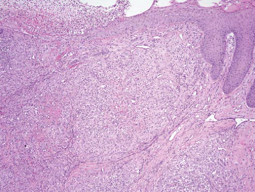
FIGURE 17.1 Bacillary angiomatosis. This lesion arose in the knee area a patient who had had a renal transplant. Beneath the ulcerated skin is a lobular but solid endothelial proliferation. The centers of the lobules contain eosinophilic necrotic material.


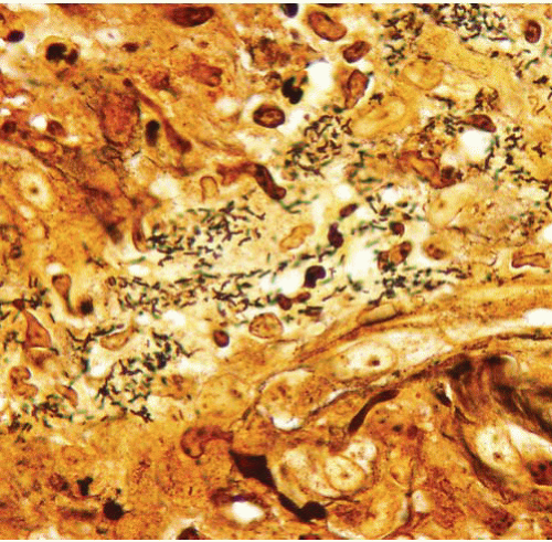
FIGURE 17.2 Bacillary angiomatosis, Warthin-Starry stain. Numerous Bartonella henselae organisms are apparent.


VERRUGA PERUANA (OROYA FEVER, CARRION DISEASE)


Clinical Features

The etiologic agent is Bartonella bacilliformis, a hemotropic, mobile, pleomorphic, gram-negative bacterium which has been shown to elaborate an angiogenesis factor in vitro.5 The disease is transmitted by Lutzomyia (Phlebotomus) verrucarum, a nocturnal sandfly. Humans are the only known reservoir. The disease is restricted to high altitude valleys of Ecuador, Colombia, and Peru.6 After an incubation period of approximately 3 weeks, individuals without tolerance develop a fever and an acute, severe, hemolytic anemia, the result of virtually 100% parasitization of circulating erythrocytes by the microorganism. Infiltration of the reticuloendothelial system results in hepatosplenomegaly and lymphadenopathy. In the preantibiotic era, the mortality at this stage was approximately 40%; the current figure is around 8%. A tissue phase, which signals improvement in the patient’s condition, appears approximately 2 months after the hematic phase and is characterized by the development of verruga peruana nodules (e-Fig. 17.9) which primarily involve the face and extremities.


Pathologic Features

The lesions can be subclassified histologically as miliary, when small and confined to the papillary dermis, nodular, when small to medium-sized and present in the reticular dermis or subcutis, or mular, when large and
involving both skin and subcutaneous tissue.7 All contain vasoformation elements, commonly admixed with inflammatory cells and typically arranged in a multinodular or lobulated pattern (Fig. 17.3, e-Figs. 17.10 to 17.13). Superficial examples may be polypoid and, when accompanied by well-formed vessels with patent lumina, may be suggestive of a pyogenic granuloma or bacillary angiomatosis. Lesions which arise in less distensible tissues may exhibit more solid “angioblastic” growth suggestive of a neoplastic process.7 All examples are unified by the presence of plump atypical appearing endothelial (verruga) cells. Depending on the stage of the lesions, lymphocytes and plasma cells or polymorphonuclear leukocytes with microabscess formation may be the predominant inflammatory constituents. Fibrin deposition, cellular necrosis, and fibrosclerosis may also be evident. The identification of Rocha-Lima inclusions, which may be fleeting, assists in confirming the diagnosis. These granular structures are best seen when tissues are fixed in buffered neutral formalin, embedded in glycol methacrylate, and stained with Giemsa.8 They also stain lightly, although less convincingly, with the Warthin-Starry and Periodic acid-Schiff-diastase stains.


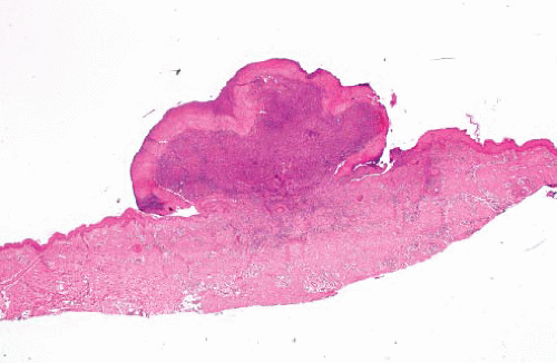
FIGURE 17.3 Verruga peruana. This lesion appears similar to those associated with bacillary angiomatosis; diagnosis requires correlation with the clinical history. (Image courtesy of Dr. Javier Arias-Stella.)


PAPILLARY ENDOTHELIAL HYPERPLASIA

This benign reactive process, which is an exuberant form of organizing vascular thrombus, was first described in 1923 by Pierre Masson as “vegetant intravascular hemangioendothelioma,” reflecting his belief that it was neoplastic.


Clinical Features

Papillary endothelial hyperplasia occurs at any age and involves virtually any vessel, including those of neoplastic processes and vascular malformations. However, in its “pure” form, it presents as a solitary,
superficial, firm, bluish or erythematous mass, involving the fingers, head and neck, or anorectal region. Physical examination reveals limited mobility of the process about its longitudinal axis. Astute gross examination by the surgeon or the pathologist may reveal confinement of the process to the lumen of a dilated vessel, usually a vein.


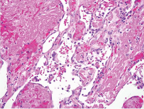
FIGURE 17.4 Papillary endothelial hyperplasia is an exaggerated form of organization of vascular thrombi. Note the fibrin cores coated by a monolayer of endothelial cells.


Pathologic Features

There is a proliferation of endothelial cells within vessels (hemorrhoidal; e-Figs. 17.14 and 17.15) forming papillary fronds associated with thrombus. The fronds contain a core of fibrin or hyalinized collagen, depending on the stage of the lesion, and are lined by a single layer of endothelial cells which may be plump or swollen but lack significant atypia and mitotic activity (Fig. 17.4, e-Figs. 17.16 and 17.17).9 Occasionally, due to the plane of sectioning or an actual event, the fronds appear to be “free-floating” within the vessel lumen. Extravascular extension may occasionally be seen, although it is usually not extensive. However, large, predominantly extravascular examples arising in hematomas have also been reported.


ANGIOLYMPHOID HYPERPLASIA WITH EOSINOPHILIA (EPITHELIOID HEMANGIOMA)


Clinical Features

These are lesions of adults (20 to 40 years) without gender predominance and unaccompanied by systemic symptoms.10 They are most common in the head and neck region and are characterized by single or multiple smooth papules or plaques of varying color.11 Most are unilateral.
Local recurrence has been noted in up to one-third of patients. Whether this is the result of a persisting underlying vascular anomaly (i.e., arteriovenous shunt) or an indication of inherent neoplastic potential is unresolved. Occasional multicentric lesions have been reported. There are no reported deaths due to this process. Optimal management is complete local excision.


Pathologic Features

Subcutaneous examples of angiolymphoid hyperplasia with eosinophilia are characterized by a prominent proliferation of capillary-sized vessels which, although well-formed, often have an “immature,” uncanalized appearance. Many are lined by plump epithelioid (histiocytoid12) endothelial cells (Fig. 17.5, e-Figs. 17.18 to 17.23). Mild cytologic atypia may be noted, but generally, the nuclei have small nucleoli, an abundance of euchromatin, and little heterochromatin clumping along the nuclear membranes. Lobulation and folds in the nucleus may occasionally be observed. The vascular proliferation is frequently centered about, and emanates radially from, a medium-sized artery or vein which shows histologic evidence of damage.10 The process is commonly well-marginated and bordered peripherally by lymphoid follicles (see e-Figs. 17.20 and 17.21). Eosinophils (see e-Fig. 17.18) without eosinophilic microabscesses and lymphocytes are usually present.

Dermal examples of angiolymphoid hyperplasia with eosinophilia11 are similar to subcutaneous lesions but less well marginated. They more frequently have a mature, canalized appearance, and are not generally associated with a medium-sized artery or vein. Lymphoid follicles are also less commonly observed in this superficial location.


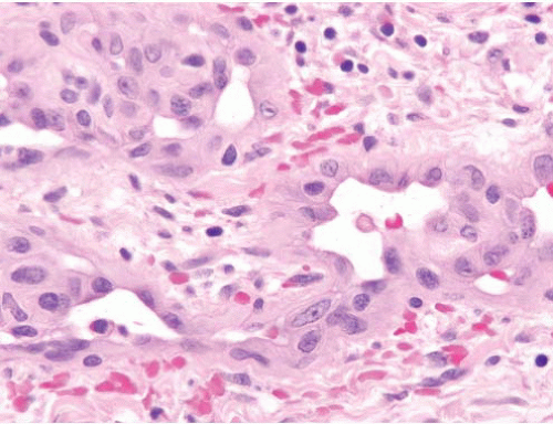
FIGURE 17.5 Angiolymphoid hyperplasia with eosinophilia/epithelioid hemangioma. This lesion shows well-formed vessels lined by plump epithelioid endothelial cells.


Ancillary Investigations

The endothelial cells are reactive for CD31 and CD34 but not GLUT1; the latter feature confirms that they differ from juvenile-type hemangiomas (see “Hemangiomas” section). Some atypical examples have been shown to display fusions of ZFP36-FOSB,13 but these tumors lack the WWTR1- CAMTA1 fusions that characterize epithelioid hemangioendothelioma.14


KIMURA DISEASE

This is not actually a vascular lesion but has been confused with epithelioid hemangioma/angiolymphoid hyperplasia with eosinophilia in the past and is thus listed here.


Clinical Features

Kimura disease appears almost exclusively in the Orient where it presents as a mass lesion, primarily in the head and neck region. Other reported sites of involvement include the groin, upper limb, axilla, and chest wall.15 The masses may be multifocal or bilateral. Regional lymphadenopathy and peripheral blood eosinophilia are commonly noted, and serum immunoglobulin G or immunoglobulin E can be elevated. Some cases have been associated with renal disease, and there is speculation concerning a link with immunoglobulin G4-related disease, but this remains poorly understood.16


Pathologic Features

The condition is characterized by a poorly marginated fibroinflammatory mass (Fig. 17.6) predominantly involving the subcutaneous tissue but also
occasionally extending into the dermis, underlying musculature, or adjacent structures such as the salivary glands. Although the microscopic features vary somewhat with the duration of the lesion, all stages feature fibrosis, a reactive vascular proliferation, and an inflammatory infiltrate dominated by eosinophils and lymphocytes (e-Figs. 17.24 to 17.26). Earlier examples typically exhibit a more pronounced inflammatory reaction while later stages are more fibrotic. Lymphoid follicles are a universal finding and are generally distributed through the process. Eosinophilic microabscesses are sometimes present (Fig. 17.7). In contrast to angiolymphoid hyperplasia with eosinophilia (epithelioid hemangioma), the vessels in Kimura disease are typically well canalized, lack epithelioid morphology, and, for the most part, tend to be a relatively minor or inconspicuous component of the process.


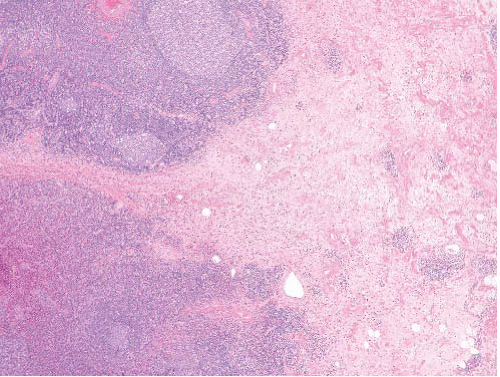
FIGURE 17.6 Kimura disease. Kimura disease is not a vascular lesion but, rather, a fibroinflammatory process. It is included here based on confusion between it and angiolymphoid hyperplasia with eosinophilia/epithelioid hemangioma in the past.


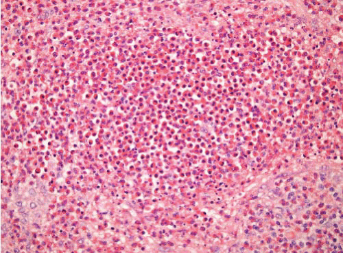
FIGURE 17.7 Kimura disease. Eosinophilic microabscesses are a feature of Kimura disease but not of angiolymphoid hyperplasia with eosinophilia/epithelioid hemangioma.


Ancillary Investigations

Human herpesvirus 8 and Epstein-Barr virus are negative.


HEMANGIOMAS

In general, the following morphologic features in a vascular tumor support a benign diagnosis: (1) zonated/lobular growth, (2) bland morphology with minimal mitotic activity, (3) arrangement of the endothelium in a monolayer, and (4) the presence of a “mature” vascular pattern with appropriate supporting elements or evidence of intralesional maturation. However, as is commonly the case in surgical pathology in general, no single feature can be relied on with absolute certainty. For example, angiosarcomas may contain deceptively bland foci closely reminiscent of hemangioma, and
pediatric vascular tumors (infantile or juvenile hemangiomas) are often cellular with prominent mitotic activity.

Hemangiomas of all types often contain additional supporting elements that include pericytes, smooth muscle, and interstitial fibroblast-like cells. These components are frequently more pronounced in pediatric tumors and can be highlighted with special histochemical stains (i.e., reticulin, Movat, pentachrome, etc.), as well as by immunohistochemical techniques.


CAPILLARY HEMANGIOMA


Clinical Features

Capillary hemangiomas are the most common tumor of infancy, seen in about 10% of term and 20% of preterm babies.17 There is a female preponderance with a female to male ratio up to 3:1. In children, capillary hemangiomas are typically cellular and have thus been referred to as infantile hemangioendothelioma, juvenile hemangioma, strawberry nevus, and, perhaps most appropriately, cellular capillary hemangioma.

They may, on rare occasion, be associated with right-sided aortic arch coarctation, sternal clefting, midline abdominal raphe, and sacral and genitourinary tract defects,18 or with anomalies of the brain, cerebral vasculature, eyes, aorta, and chest wall in the neurocutaneous disorder called PHACE syndrome.19 The PHACE acronym refers to posterior fossa anomalies, hemangioma, arterial lesions, cardiac abnormalities/aortic coarctation, and abnormalities of the eye, although the vascular lesions in these syndromes may in fact be malformations.

Sporadic capillary hemangiomas most frequently involve the skin or subcutis of the head and neck region, although a wide variety of other sites may also be involved. Rarely, they may grow to sufficient size to damage adjacent structures and even less frequently, they may have life-threatening or fatal consequences. The vast capillary surface area associated with large lesions can cause platelet trapping, thrombocytopenia, and a consumptive coagulopathy (Kasabach-Merritt syndrome), which can also occur in a variety of large vascular lesions of diverse histologic type.

Capillary hemangiomas typically proliferate in the first few months of life and then regression and, in many instances, completely involute. Approximately half of untreated patients with superficial lesions are left with normal skin by 5 years of life and from 70% to 90% of patients are “cured” by 7 years of age.20 These results do not appear to be influenced by gender, duration of the proliferative phase, size, or site of the lesions. The rare examples with life-threatening or severely disfiguring consequences due to massive size or strategic location have been managed by embolization, laser, corticosteroid therapy, interferon alpha-2A or -2B, or vincristine.

Superficial capillary hemangioma (strawberry nevus or hemangioma) needs to be differentiated from vascular stains. The term nevus flammeus has been used in the past to encompass a wide variety of congenital
macular stains, including both small, transient lesions (e.g., nevus simplex) and the larger, persistent, port wine stains. Common sites of involvement include the glabella, eyelids, and the nape of the neck. Those located in the center of the face commonly disappear by 1 year of life, whereas nape lesions fade more slowly and often incompletely with faint vestiges persisting into adulthood. Port wine stains (nevus venosus), on the other hand, occur far less frequently. They consist of larger macules with irregular borders which tend to enlarge in proportion to the growth of the child and generally do not fade. They are typically flat in infancy and childhood but may become elevated and nodular later in life.

Lateral facial port wine stains in the distribution of the trigeminal nerve with abrupt termination at the midline are a component of the Sturge-Weber (leptomeningeal nevus flammeus or encephalotrigeminal angiomatosis) syndrome. Port wine stains are also associated with the Klippel-Trenaunay (osteohypertrophic nevus flammeus) and Parkes Weber syndromes, which are further discussed in Chapter 18.


Pathologic Features

During the proliferative phase, capillary hemangiomas are characterized histologically by a mixture of endothelial cells, pericytes, and fibroblastlike cells (e-Fig. 17.27). The endothelial cell nuclei can have prominent nucleoli, and mitotic activity may be pronounced. Many of the vessels may lack apparent lumina, although tubular or cord-like angiogenic growth can be highlighted with a reticulin stain. On low-power examination, the lesions often have a marginated or circumscribed appearance, and the proliferating vessels are typically arranged in a lobular pattern (Fig. 17.8).
Maturation is first noted at the periphery of these lobules and is characterized by the presence of flattened or less active appearing endothelium lining vessels with patent lumina. Over time, a greater percentage of the capillaries assume this morphology. The vascular lobules also become more accentuated due to an ingrowth of fibrous and adipose connective tissue.


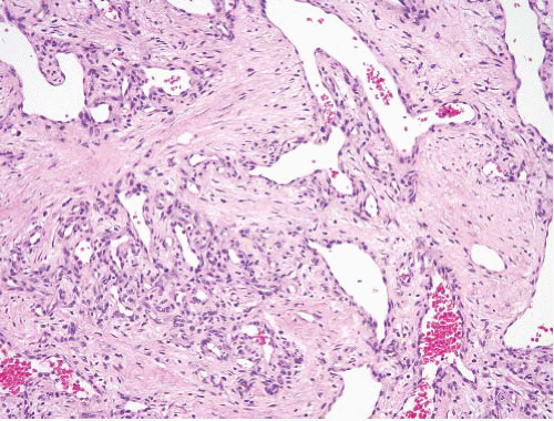
FIGURE 17.8 Capillary hemangioma. This lesion from a small child has a lobular growth pattern, showing larger capillaries branching into smaller ones. The smallest are supported by cells other than endothelial cells (such as myoid cells and pericytes).

Unlike capillary hemangiomas, congenital macular stains lack endothelial proliferation. In fact, when biopsied early in life, they typically reveal minimal or no apparent histologic abnormality. Persistent, laterally located lesions of the port wine stain type, however, eventually develop pronounced capillary ectasia. Macular stains are regarded as telangiectasias rather than true hemangiomas (and do not regress).


Ancillary Investigations

GLUT1 is usually strongly and diffusely expressed in the lesional endothelial cells of most infantile hemangiomas (which are likely to regress) during all stages of their natural history but usually not in other vascular tumors or malformations (which will not regress). GLUT1 also labels a variety of nonvascular neoplasms. The endothelial cells in capillary hemangiomas express CD34 and CD31 but not podoplanin (D2-40).21


PYOGENIC GRANULOMA/LOBULAR CAPILLARY HEMANGIOMA

The term pyogenic granuloma is a misnomer. An infectious etiology has long been disproved and current observers favor a lobular variant of capillary hemangioma, but there are some important distinctions from classic (pediatric) capillary hemangioma.


Clinical Features

The lesions may involve mucosal, cutaneous, subcutaneous, or even intravascular sites. Patients under the age of 18 are predominantly males, whereas those between 19 and 40 are mostly females, with equal sex incidence over 40. About a third have a history of trauma. Local recurrence develops in about 10% of cases treated by simple excision. The most commons sites are gingiva and fingers, followed lips, face, and tongue. Tumors of the oral and nasal mucosa (but not other sites) show a marked predilection for women in their reproductive years.22

Approximately 1% of pregnant women develop pyogenic granulomas in the oral mucosa, especially the interdental regions of the gingiva. These typically appear in the first trimester and regress postpartum. Similar lesions may develop with oral contraceptive use. These associations suggests hormonal sensitivity although ER and PgR are negative.23

Pyogenic granulomas are solitary or rarely multiple. Disseminated lesions can occur spontaneously or be associated with an underlying malignancy. Superficial pyogenic granulomata, involving the mucous membranes or skin,
may be sessile, polypoid, or pedunculated. They frequently have a rapid onset but stabilize at a maximum size of several millimeters to a few centimeters after several weeks or months. The lesions range in color from bluish-purple to bright red. Their epithelial surface may be attenuated or ulcerated.

Some pyogenic granulomas are intravascular (e-Figs. 17.28 to 17.30).24 These lesions present in the third decade over a period of 2-8 weeks and have a predilection for small to medium-sized veins of the neck and upper extremities. They have polypoid intraluminal architecture. Apart from the intravascular location, their histology is typical of pyogenic granuloma.


Pathologic Features

On histologic examination, pyogenic granuloma is characterized by a relatively well-marginated proliferation of small capillary-sized vessels arranged in a multilobular pattern (Fig. 17.9, e-Figs. 17.31 and 17.32). Larger feeder vessels may be seen in association with the vascular lobules. The process is set in a fibromyxoid stroma which, in the absence of surface ulceration, has minimal inflammation. Both endothelial and stromal elements are mitotically active. Polypoid or pedunculated lesions often have a collarette of hyperplastic epithelium (see Fig. 17.9). Deep-seated have less edema and vascular dilatation.


Ancillary Investigations

The endothelial cells express CD34 and CD31 but not GLUT1.


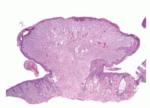
FIGURE 17.9 Pyogenic granuloma (lobular capillary hemangioma). The histologic appearances of these lesions are similar to those of capillary hemangiomas found in infants and small children, but in contrast to those of infants, these are not as likely to regress. Note again the lobular configurations of the proliferating vessels. This example has a collarette of hyperplastic skin.


CAVERNOUS HEMANGIOMA


Clinical Features

Cavernous hemangiomas are common in both children and adults and have a predilection for the upper half of the body. Roughly equal numbers of males and females are affected. Unlike capillary hemangiomas, cavernous hemangiomas do not regress. Examples occurring in deep soft tissue and organs (liver, bone, intestines, lung) are also well-known. Rarely, large deep-seated examples may be associated with the Kasabach-Merritt consumptive coagulopathy syndrome.

Only gold members can continue reading. Log In or Register to continue

Stay updated, free articles. Join our Telegram channel

Jun 18, 2016 | Posted by in PATHOLOGY & LABORATORY MEDICINE | Comments Off on Superficial Vascular Lesions and Mimics of Vascular Lesions

Full access? Get Clinical Tree

Get Clinical Tree app for offline access