Superficial Aspiration Technique
Rose Anton, MD
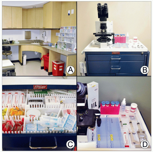 (A) Fine-needle aspiration (FNA) may be performed in a physician’s office, clinic, radiology, or at the bedside. A room dedicated for performing FNAs is optimal for the storage of needed supplies, patient comfort, and overall convenience. (B) A mobile cart is useful to maneuver required supplies to other locations, such as patient rooms. (C) Supplies can be discreetly stored in several drawers of the cart. (D) The essential components of any superficial FNA are shown in this photograph. |
BEFORE PROCEDURE
Examination and Patient Consent
Obtain history and examine site to be aspirated
Fully discussing procedure and answering all questions ameliorates apprehension and often obviates use of anesthetic
Risk of bleeding is minimized by use of fine needle and application of pressure after needle removal
Risk of infection is minimized by using sterile technique
SUPPLIES FOR PROCEDURE
Required
Needles, usually 25-gauge 5/8 inch
Consider larger gauge (21) for fat pad aspirates
May use 1.5 inch for deeper lesions
Syringe, usually 10 mL with Luer-Lok tip
Betadine &/or alcohol
Sterile gloves
Sterile gauze
Slides (plus slides optimal for possible immunocytochemical applications)
Consider frosted slides for aspirates containing fatty tissue
Alcohol spray fixative
Cell preservative solution
For flow cytometry (RMPI) or cell block (RMPI or CytoLyt)
Culture tubes (routine, fungus, and TB)
Slide tray
Marker for labeling of slide with patient identifiers
Diff-Quik stain for immediate adequacy assessment
Microscope
Optional
Topical or local anesthetic
Handle or “gun” to contain syringe
Bandage (usually not required with small-gauge needles but may be necessary to protect clothing, etc.)
Stay updated, free articles. Join our Telegram channel

Full access? Get Clinical Tree


