Stomach and Proximal Duodenum: Inflammatory and Miscellaneous Disorders


Stomach and Proximal Duodenum: Inflammatory and Miscellaneous Disorders


CLASSIFICATION OF GASTRITIS AND GASTROPATHY

Though no classification of gastritis satisfies everyone, the overall goal of any classification is to help clear thinking and be clinically useful. Inevitably much of the early thinking regarding gastritis was centered on “peptic ulcer disease” (PUD). Ignorance regarding the role of both Helicobacter and medications gave rise to theories that were to some extent flawed, yet they still dominate traditional teaching. Gastritis was considered physiologic and intestinal metaplasia an aging phenomenon. We also need to recall that

1. Gastritis originally meant “redness”—which now is usually associated with a gastropathy rather than gastritis; conversely, most histologic gastritis has a normal endoscopic appearance.

2. Many disorders that are characterized by abnormal endoscopy also have a typical biopsy appearance. From a classification viewpoint are these best considered from an endoscopic or histologic viewpoint? Most classifications can only be viewed from one vantage point.

3. From a clinical viewpoint, “ulcers” have played a major role in gastric disease because of the symptoms with which they or their complications are associated (pain, bleeding, perforation, and obstruction/stenosis). However, the term “peptic ulcer disease” has been in common parlance for decades, with the implication that this is associated with acid, the “proof” being that symptoms are markedly ameliorated with therapy, whether antacids, H2-receptor antagonists, or proton pump inhibitors (PPIs). In the early 1980s, it was ultimately shown that some ulcer disease, especially in the duodenum, was related to Helicobacter pylori, so that its eradication virtually guaranteed that duodenal ulcer, the archetypal peptic ulcer, would not recur. Thus PUD changed from being primarily acid related to primarily bacterial, or a combination of both.

4. Nonsteroidal anti-inflammatory drugs (NSAIDs), aspirin (acetylsalicylic acid—ASA), and other medications now play a huge role in gastric pathology. While the introduction of NSAIDs around 1970 was a major step forward therapeutically, it came at a price that included numerous gastrointestinal (GI) side effects. Prior to this time, ASA had been “the” analgesic and antipyretic of choice. Bayer introduced ASA in the market around 1900, and within a decade or two this “wonderdrug” was present in virtually every household in the more developed countries, and used widely for numerous ailments—colds, coughs, headaches, migraines, and all arthritides. Yet the erosive, ulcerative, and bleeding diathesis associated with this drug was not widely appreciated. In retrospect, from about 1900 on, many “peptic ulcers” may well have been as much ASA associated as Helicobacter associated, and this association even creeps, almost inadvertently, into case reports back in the 1950s.1 So while we typically think of “peptic ulcer disease” historically as unrecognized Helicobacter infection, ASA was very likely a major contributor. This continued until acetaminophen/paracetamol/Tylenol came into the market in to the 1960s. Further, it is now well recognized that, especially in the very young2, 3 and elderly,4 not only that NSAIDs are likely “the” culprit irrespective of the presence of H. pylori, but that the risk of complications such as bleeding (and therefore the erosions and ulcers that bleed) can be largely prevented using PPIs. Thus, historically, the disease we consider to be “peptic ulcer disease” may have been as much NSAID/ASA associated as Helicobacter associated, especially in the presence of abundant acid.

5. Historically, alcohol, which not only has a social role in many societies but is also an analgesic in large doses, has been around much longer than any other gastric damaging agent except for Helicobacter, and produces histologic changes similar to NSAIDs (i.e., a chemical/reactive gastropathy). From around 1900 on when aspirin became available, the big three, became Helicobacter, alcohol, and ASA, and from 1970 on, NSAIDs was added to these.

6. The notion of “peptic ulcer disease” and “no acid— no ulcer” is therefore likely true in that in the major causes of gastroduodenal erosions and ulcers, namely, Helicobacter and NSAIDs/ASA, and other medications or chemicals, especially alcohol, the presence of acid facilitated the development of injury caused by these agents. Although the nature of the interaction of these common causes of peptic ulcer is still unclear, it would make most sense if, when antral-predominant H. pylori is present, that the risk of NSAID/ASA and alcohol-induced damage was increased, but that when the organism spread proximally, resulting in a decrease in acid output, that there may well be a degree of protection from NSAID/ASA, and possibly alcohol-associated damage (Table 13-1).


Current Classification of Gastritis

Until the early 1970s, chronic gastritis was classified into three main varieties (superficial, atrophic, and hypertrophic) as suggested by Schindler in 19395 (Table 13-2). Wood as well as Schindler later concluded that chronic hypertrophic gastritis is a variation of normal mucosal function.6, 7 Thus, chronic gastritis was classified as superficial or atrophic.

Whitehead’s classification was the first to understand the importance of noting location, and grading
the depth and degree of inflammation and the presence or absence of both intestinal and pseudopyloric metaplasia, separating them from atrophic changes8 (Table 13-2). This really formed the basis of all subsequent morphologic classifications of gastritis. In 1973, Strickland and Mackay classified gastritis based on detecting parietal cell (PC) antibodies, clarifying the etiology of autoimmune gastritis (AIG) (type A) despite the fact that these can develop in Helicobacter infected patients. It is associated with atrophic changes in body and fundic (oxyntic) mucosa. Antral predominant gastritis was type B. Glass and Pitchumon added type AB into Strickland-Mackay classification to encompass cases that did not fit type A or type B, essentially pangastritis.


Table 13-1 ABC Classification of Gastritis


Autoimmune


Pernicious anemia


Bacterial


Bugs including post-Rx effects:


Helicobacter pylori


Enterococcus


Syphilis


Chemical


Bile reflux


Drug-associated/Iatrogenic


NSAIDs/ASA


Anti-platelet medications


Chemotherapy/GVHD


Iron


Alcohol


Eosinophilic


Eosinophilic gastritis/gastroenteritis


Food allergies, medications


Focal


Crohn’s disease


Granulomatous


Tuberculosis


Sarcoid


Crohn’s disease


Foreign body


Helicobacter pylori


Hypertrophic (big folds)


“Ménétrier’s disease”


Lymphocytic gastritis


Eosinophilic gastritis


Gastric varices


Gastritis cystica profunda


Lymphoma (MALT)


Gastric adenocarcinoma


Helicobacter pylori gastritis (lymphocytic), CMV


Zollinger-Ellison syndrome


Multiple polyps/polyposis


Idiopathic


Juvenile (pediatric)


Follicular with H. pylori, CMV


Lymphocytic


Helicobacter pylori, celiac disease


Chronic erosive (varioliform) gastritis


Multifocal intestinal metaplasia with/without atrophic front


In atrophic gastritis, isolated


Modified from Wyatt JI, Dixon MF. Chronic gastritis—a pathogenetic approach. J Pathol. 1988;154(2):113-124.


With the rediscovery of H. pylori (originally Campylobacter pylori) by Warren and Marshall in the early 1980s,9 it became clear that H. pylori is a principal component of most gastritides. In 1988, two classification systems emerged. That by Wyatt and Dixon incorporated reactive gastropathy (then called chemical gastritis/gastropathy) as the “C” of the ABC classification system, A being autoimmune and B being bacterial (=Helicobacter but, at that time, C. pylori).10 The same year, Correa proposed classifying gastritis based on clinical and etiopathogenetic information. He classified chronic gastritis into superficial gastritis, diffuse antral gastritis (DAG), usually Helicobacter associated and related to duodenal ulcer disease, diffuse corporal atrophic gastritis (autoimmune), and multifocal atrophic gastritis (MAG—considered to be “environmental”). MAG was related to intestinal-type adenocarcinoma and gastric ulcer, and intestinal metaplasia in the antrum and body.11 Diffuse corporal atrophic gastritis was often related to AIG and pernicious anemia, with inflammation and atrophy in the corpus and relative sparing of the antrum11, 12 (Fig. 13-1).

The Sydney system is the basis of most contemporary classifications of gastritis. Proposed by a group of European pathologists and clinicians (World Congress of Gastroenterology, Sydney, August 1990),12 it recommended incorporating the topography of gastric mucosal changes with the immunology and microbiology of the disease. The classification depends on separate assessment of the antrum and corpus by taking a minimum of two biopsies from the anterior and posterior walls of the respective gastric compartments as well as any specific lesions identified. An important feature is a standard three-tier grade of mild, moderate, and severe applicable to a selected number of morphologic variables. As a broad guideline, each successive grade represents an increment in severity of about one-third. Graded variables included inflammation (acute and chronic), atrophy, metaplasia, and density of H. pylori. The Sydney system also expanded previous classifications by adding a variety of other “special forms” of gastritides (collagenous, eosinophilic, granulomatous, lymphocytic, etc.).

The Sydney classification was updated in 1994,13 which expanded the section on specific entities (special forms) and includes a 4-point visual analog (equivalent to none, mild, moderate, and severe) to aid with morphologic grading of inflammation and atrophy.13 Gastric atrophy is loss of normal glands, often with replacement by an epithelium that could be either native or metaplastic (Table 13-3). The score is an average from each region’s biopsies. Antral atrophy was the average score for atrophy from all antral biopsies and corpus atrophy (the average score for atrophy from all corpus biopsies).13 The updated Sydney classification depends on the separate assessment of the
antrum and corpus. It needs a minimum of two biopsies from the lesser and greater curvature of the respective gastric compartments as well as the incisura and any specific lesions identified (Fig. 13-2).13 On all occasions accurate grading depends on correctly oriented full-thickness mucosal biopsies. In practice, other than for academic studies, grading is rarely required.


Table 13-2 Gastritis Classification “Historical Prospective”


YEAR, AUTHOR


CLASSIFICATION


1942, Schindler


Gastritis


Superficial


Atrophic


Hypertrophic (later dropped as Schindler and others concluded hypertrophy is normal function variation)


1972, Whitehead et al.8


Mucosa type


Gastritis grade


Metaplasia


Pyloric


Superficial


Body


Quiescent


Pseudopyloric


Cardiac


Active


Transitional


Atrophic (used synonymous with deep inflammation).


Intestinal


Indeterminate


Mild (quiescent or active)


Moderate (quiescent or active)


Severe (quiescent or active)


1973, Strickland and Mackay828


Type A or autoimmune gastric atrophy of pernicious anemia


Type B—nonautoimmune (pyloro-cardial extension type in connection with PUD (Kimura, 1972181)


1976, Glass and Pitchumoni modification of Strickland and Mackay829


Type A (autoimmune)


Type B (antrum)


Type AB (cases that did not fit Type A or Type B)


1988, Correa11


Morphologic


Mechanistic


Synonyms


Not atrophic


Superficial


Initial stage of other types?


Simple?


Diffuse antral (DAG)


Hypersecretory?


Antral


Psychosomatic?


Type B


Infectious? (C. pylori)


Atrophic


Postgastrectomy


Reflux


Chemical


Diffuse corporal


Autoimmune


Type A


Multifocal


Dietary?


Environmental


Type B


Type AB


Pangastritis


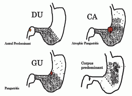
Figure 13-1. Prototypes of gastritis pattern predict disease outcome. In practice, all tend to have some degree of both antral and corpus inflammation. Top left: Duodenal ulcer (DU) patients have antral predominant inflammation with little corpus inflammation. Bottom left: Pangastritis is seen in gastric ulcer (GU) patients. Corpus mucosa is inflamed and often extends into the specialized mucosa but still tends to be antral predominant. Top right: Pangastritis with atrophy is seen in patients with the intestinal type of gastric adenocarcinoma (CA). Bottom right: Corpus-predominant gastritis is usually seen in AIG or end-stage Helicobacter infection.


GASTRITIS

Gastritis (in its broadest sense) and its complications account for millions of doctors’ office visits each year. Symptoms are often associated with acute changes or complications described as mild upper abdominal discomfort, indigestion, heartburn, coated tongue, foul breath, and bad taste to more ominous symptoms such as loss of appetite, nausea, vomiting blood or coffee-ground material, diarrhea, and dark stools. Most patients with chronic gastritis have no symptoms. Even so, these symptoms are not specific and include broad differentials such as H. pylori infection, other infections, bile reflux, inflammatory
bowel disease (IBD), and side effects of medications (Table 13-4). As treatment depends on the cause, it is important to know the cause for appropriate management. Occasionally, it may be necessary to list possible etiologies for gastric inflammation, rather that reporting “nonspecific chronic inflammation”—which is an unnecessarily complex term as all inflammation is “nonspecific,” so these words can always be omitted from reports without deleterious effect. If it is specific, the cause (e.g., Helicobacter) should be stated.


Table 13-3 Gastritis Classification “Sydney System”


GASTRITIS TYPE


ETIOLOGY


SYNONYMS


1996, Updated Sydney13


Non-atrophic


Helicobacter pylori


? Other factors


Superficial


Diffuse antral,


Chronic atrophic


Interstitial—follicular


Hypersecretory


Type B


Atrophic


Autoimmune


Autoimmunity


Type A


Diffuse corporal


Pernicious-anaemia associated


Multifocal


Helicobacter pylori


Dietary


? Environmental


Type B


Type AB


Environmental


Special forms


Metaplastic


Chemical


Chemical irritation:


Bile


NSAIDs/Antiplatelet


Other medications


Reactive, Reflux


NSAID


Type C


Radiation


Radiation injury


Lymphocytic


Idiopathic? Immune mechanism


Gluten


Drug (ticlopidine)


Varioliform (endoscopic)


Celiac disease-associated


Noninfectious granulomatous


? H. pylori


Crohn’s disease


Sarcoidosis


Wegener’s granulomatosis and other vasculitides


Idiopathic


Isolated granulomatous


Eosinophilic


Food sensitivity, drugs, Churg-Straus


Allergic


Other infectious gastritis


Bacteria (other than H. pylori)


Viruses


Fungi


Parasites


Phlegmonous


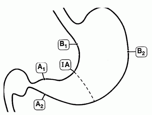
Figure 13-2. The updated Sydney biopsy protocol requires a minimum of two biopsies from the lesser and greater curvature of the respective gastric compartments as well as the incisura and any specific lesions identified. This identifies all of the patterns of gastritis illustrated in Figure 13-1, as well as estimating the extent of atrophy present, which often starts at the incisura/angulus (IA), affects the antrum (A1, A2), and then extends proximally to the oxyntic zone (B1, B2), so that, as antral inflammation extends proximally, biopsy site B1 is first affected, and B2 is the last site affected.


Distinctive (Specific) Types of Gastropathies

Gastropathies are biopsies in which epithelial (noninflammatory) changes predominate. The mucosa is often mucin depleted, causing it to appear red endoscopically (invariably interpreted by endoscopists as “gastritis” rather than areas of redness). They include biopsies with primary epithelial reactive changes (such as chemical/reflux (bile) gastropathy, chemotherapy effect) and a smaller subset of biopsies with predominant vascular pathology (such as gastric antral
vascular ectasia [GAVE], portal hypertension gastropathy, Dieulafoy, and hemorrhagic/shock) (Table 13-4). Graft versus host disease (GVHD) is usually normal endoscopically.


Table 13-4 Classification by Predominant Histologic Change


CATEGORY


SUBCATEGORY


Gastropathy (predominantly noninflammatory)


Predominant epithelial changes


  1. Medications/reflux (bile) gastropathy


  2. Alcoholic gastropathy


  3. Caustic-induced injury


  4. GVHD


  5. Radiation/chemotherapy


  6. Ischemia


Predominant vascular pathology


  1. GAVE


  2. Portal hypertension


  3. Hemorrhagic/shock gastropathy


Gastritis (predominant inflammatory)


Infectious


  1. Bacterial


    1. Helicobacter pylori bacterial infection (autoimmune gastritis, lymphocytic gastritis, granulomatous gastritis, carditis)


    2. NHPH infections—“H. heilmanii


    Other bacteria—TB, syphilis, phlegmonous and emphysematous gastritis


  2. Viral (CMV, Herpes)


  3. Fungal (Candida, histoplasma, mucormycosis, Aspergillosis)


  4. Parasitic (Cryptosporidium, Anisadikosis, other parasites, and nematodes)


Noninfectious


  1. IBD (endoscopic, histologic features, clinical implication)


  2. Eosinophilic gastroenteritis


Part of systemic involvement


  1. GVHD


  2. Vasculitis (including Churg-Strauss)


Endoscopic gastropathies


Distinctive macroscopic (endoscopic) appearance with appropriate histology


  1. Erosive and hemorrhagic


    Varioliform gastritis


    Watermelon stomach (GAVE)


    Portal gastropathy


    Hemorrhagic gastritis/gastropathy


  2. Nonerosive


    Nodular gastritis, children


    Atrophic front, adults


  3. Distinctive hypertrophic gastropathy


Reactive (Predominant Epithelial) Changes

Reactive (chemical/reflux-associated) gastropathy is a reaction to noninfectious irritants. This can be due to protracted exposure to bile and pancreatic juice (especially postgastric surgery14). The most infamous of irritants are NSAIDs, which include over-the-counter drugs such as aspirin and ibuprofen, and many prescription medicines. Other medications— such as bisphosphonates used for osteopenia, iron pills and irritants in food such as capsaicin in peppers and chilies and alcohol—can all cause this lesion.15 These irritants usually cause no clinical problems when taken for the short term, although endoscopic damage can be seen even with short-term use. However, regular (or excessive) use can lead to a more severe gastropathy as well as erosions and ulcers. With the increasing use of aspirin and other NSAIDs, and decreasing prevalence of Helicobacter, chemical/reactive gastropathy is increasingly seen in gastric biopsies, and may co-exist. Anti-platelet mediations also cause similar injury.

Pathogenesis: Aspirin is the best-studied NSAID, the mechanism of injury is inhibition of prostaglandin synthesis by inhibiting cyclooxygenase (COX) 1 and 2.16 Aspirin also changes the ability of the mucosa to maintain a pH gradient causing gastric acid back-diffusion with resultant mucosal injury.16 Further, its anticoagulant properties increase the risk of bleeding once erosions or ulcers are present. Conversely, some other NSAIDs have antiplatelet properties but do not possess this therapeutic anticoagulant effect. NSAIDs produce mucosal injury by both local and systemic effects.16 Newer NSAIDs are predominantly COX-2 inhibitors, which make them less likely to cause gastric injury and the risk of gastric (or duodenal) injury is reduced, but not abolished. A variety of antiplatelet medications are increasingly being implicated causing similar injury.

Histology: The histology of reactive gastropathies has both an acute and a chronic phase, although
in practice it is often reported without qualifying it as acute or chronic. In some patients both are present together.

Reactive gastropathy. The morphologic changes that accompany ingestion of medications such as NSAIDs have been known for decades, 15 but they are now more commonly recognized.

In the acute phase, as in any reparative process, the main changes are

1. Mucin depletion—the amount of supranuclear mucin is markedly reduced or absent, so that at low power the cells appear more basophilic—often the most apparent low-power indication that this change is present

2. A reduction of the normal cell size so that the cells are frequently low columnar to cuboidal

3. A corresponding increase in nuclear size, and also an increase in hyperchromatism; nuclei that are normally compressed at the cell base markedly increase in size, and in conjunction with the smaller cell size cause a marked increase in nuclear-cytoplasmic ratio.

4. Because this appears to be a reparative, partly restitutional process, the number of cells and nuclei appears reduced; this results in nuclei being distinct and separated, one of the best indicators that this is not dysplasia. However, especially following erosions, the reactive changes can be more marked so that the nuclei become more open and vesicular with distinct nucleoli, and there may also be concomitant increase in nuclear hyperchromatism (Fig. 13-3).

5. Changes are usually most marked in the mucous neck region, and tend to decrease superficially. Interestingly, some of these changes appear on the surface, especially the mucin depletion, but most of the other changes are maximal in the mucous neck region suggesting that these are all a reaction to injury and not dysplasia. These changes can be easily missed in the fundic glands as the foveolae are shorter and changes can be mistaken for biopsy artifacts. Occasionally, the changes may extend deeper to involve the entire length of the oxyntic or antral glands (Fig. 13-3E).

6. Erosions or ulcers may be present. When this occurs, careful examination of the erosion or ulcer base should be carried out for the presence of crystals or foreign material representing medications. Iron encrustation can readily be confirmed using Perl’s stain (Fig. 13-3). When erosions or ulcers occur, the immediately adjacent epithelium may be
restituting, and appears attenuated as seen in any restitutional processes.


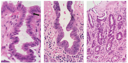
Figure 13-3. Reactive changes in gastric mucosa. A: Pit in which there is total mucin loss but nuclei are separated from each other. This is most marked superficially where the epithelial cells are more cuboidal and attenuated. Hints of mucin secretion are reappearing superficially (arrow) at the apex of the cell—an indication of maturation. Note the lack of any inflammation in the lamina propria in this biopsy. B: Similar features but there is more attenuation of epithelium superficially, and in the generative zone at the bottom nuclei are becoming stratified. The hyperchromatism associated with most dysplasias is absent. A modest chronic inflammatory infiltrate is present in the lamina propria but this disappears superficially. C: Chemical (NSAID) erosion. The attenuated epithelium is visible superficially with diffuse mucin depletion. Foveolar hyperplasia (corkscrewed pits) are visible, as is the normal architecture. At the surface the hyalinized zone is typical of NSAID damage. The lamina propria is largely empty indicating that this cannot be a Helicobacter-associated erosion.


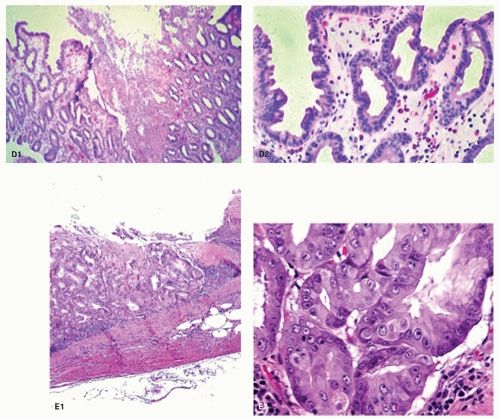
Figure 13-3. (Continued) D: D1. An erosion with almost a pseudomembranous appearance. D2. The adjacent mucosa has typical reactive changes and scattered eosinophils predominate. E1: Further NSAID erosion with the superficial hyalinized band that approaches the muscularis mucosae and E2: Very reactive nuclei, again most marked at the bases of the pits, nuclei remain separated but here have a prominent nucleolus. More superficially nuclei are even more widely separated indicating restitution. Note also that these nuclear changes do not correspond to intestinal, foveolar, or pyloric dysplasia.

7. Occasionally there is focal edema in the lamina propria, which may also be devoid of inflammatory cells or have a predominantly acute or eosinophilic (sometimes both) infiltrate. A sparse chronic inflammatory infiltrate can also be seen, but in most biopsies chronic inflammation is usually conspicuous by its absence or minimal presence (Fig. 13-3), indicating that Helicobacter infection is not the etiology of the changes present.

In the chronic phase, other changes become apparent. Sometimes the acute and chronic phases coexist, sometimes only the chronic changes persist, and it is presumed that they followed the acute changes (Fig. 13-4).

1. Foveolar hyperplasia can develop that results in tortuosity of pits in the mucous neck region.

2. Proliferation of smooth muscle in the lamina propria above that normally seen

3. A degree of vasodilatation of capillaries with congestion and edema17, 18

4. If erosions have occurred, a degree of lamina propria fibrosis ensues.

Iron toxicity, especially in children, may result in gastric mucosal necrosis, sometimes with extension into the submucosa.19 The encrustation is often visible (Fig. 13-5).

Biopsies show reactive mucosal changes (mucin depletion, foveolar hyperplasia, and smooth muscle hyperplasia) without the severe inflammatory component seen in infectious gastritis (Fig. 13-5).


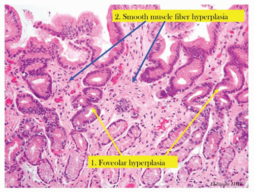
Figure 13-4. Chronic reactive gastropathy. The pits have foveolar hyperplasia, being elongated and have a corkscrew configuration (yellow arrows). There is hyperplasia of the smooth muscle fibers (blue arrow) that are normally found in the stomach. Note the lack of inflammation.

Foveolar hyperplasia appears to be a result of excessive cell exfoliation from the surface epithelium over a period of time and, accordingly, is likely to be seen in all types of active gastritis.17 Further, if the insult is ongoing, superimposed changes of acute reactive gastropathy may also be present, and this may include erosions. These histopathologic changes are not seen in all patients and when present are usually patchy (postgastrectomy states usually being more diffuse and therefore the exception). Pathology is more likely to be seen in biopsies obtained from incisura angularis.20 If biopsies are taken, then those from areas of endoscopic abnormality are preferred.

Gastric glands may be distorted and dilated, with an absence or paucity of plasma cells. In some areas there is no gland distortion, just simple thinning of the mucosa.21 Other features include stomal erosions,22 lipid islands, and intramucosal cysts21 (Fig. 13-6). Sometimes the cysts become large enough to be visible grossly and extend into the submucosa. These cysts have been labeled with a variety of names, including gastritis cystica polyposa and gastritis cystica profunda.23 Adenocarcinoma in the postoperative stomach has been reported in association with these cysts, but this association appears to be coincidental, especially because the cysts are so commonly found microscopically in the postoperative stomach.21, 24


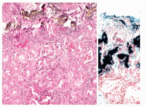
Figure 13-5. Reactive gastropathy. Iron medication may result in gastric mucosal necrosis (H&E stain) with iron encrustation (Perl’s stain—right)

Toxic gastropathy. Changes can be seen characterized by vacuolated cells in the specialized mucosa. The cause is not always apparent but may be prominent in uremic patients, the vacuolation tending to occur in chief cells rather than PCs (Fig. 13-7).


Reactive Changes with Erosions in Helicobacter—One or Two Diseases?

It should be appreciated that while “reactive gastropathy” is usually applied to changes with minimal chronic inflammation, identical epithelial and lamina propria changes can be seen in other etiologies such as Helicobacter infections, especially if acute inflammation is present. However, in the presence of Helicobacter gastritis with relatively little acute inflammation, erosions are almost certainly not related to the underlying infection, and the possibility that the patient has medication-related erosions or ulcers superimposed on Helicobacter gastritis should be considered as the distinction can be made in many instances.25 Helicobacter-type associated erosions invariably occur on a background of severe chronic active gastritis, so if this is not present they should always be viewed with suspicion, and a second etiology considered. Further, the nature of the erosion (see Fig. 13-3) can distinguish the two on biopsy, with a dense hyalinized band in the superficial mucosa being indicative of NSAID type-associated injury.26 Indeed, in patients with both diseases it is likely that a medication caused the damage.25, 26

Caveat: Severe (disproportionate) reactive changes resembling those seen in, for example, NSAID gastropathy in patients with only a modest chronic Helicobacter gastritis may well be related to medications rather than the concurrent Helicobacter infection. This is discussed subsequently.


Distinction of Reactive Changes from Dysplasia

It is imperative to distinguish reactive changes from dysplasia as they resolve when the acute insult is withdrawn. The most helpful feature is that at the surface there is invariably maturation in the form of
small mucin droplets at the surface. While “bottom-up” dysplasia (dysplasia maximal in the pit bases) does occur, it is quite rare, so the diagnosis of dysplasia should only be made if the diagnosis is absolutely clear, and ideally conforms to one of the usual forms of foveolar dysplasia (see following chapter). The adage that dysplasia should never be diagnosed in the presence of overlying or adjacent ulcers, erosions, or restituting epithelium unless absolutely clear is a good one. Making a diagnosis of dysplasia under these circumstances is fraught with danger. Unless there is absolutely no diagnostic uncertainty, it is usually best to rebiopsy the area following antisecretory therapy (e.g., PPIs) to ensure that the changes persist when the erosions have healed. Fortunately, even if dysplasia is diagnosed and graded, most can be visualized and treated endoscopically (Fig. 13-8).


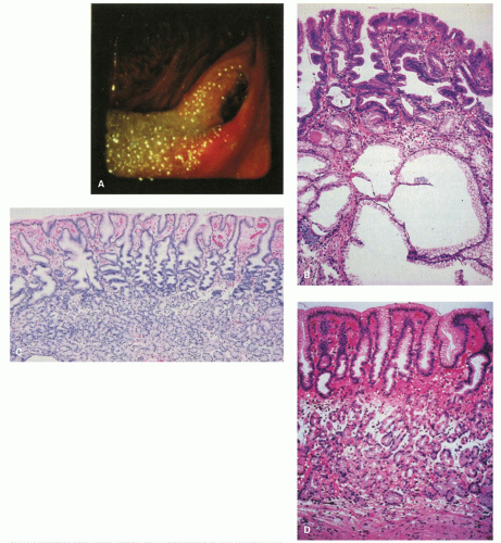
Figure 13-6. The postoperative stomach. A: Endoscopic view of a Billroth II stoma, which is typically red. Bile-stained fluid is refluxing into the gastric remnant. B: Biopsy specimen from the stoma of a Billroth II anastomosis. There is marked foveolar hyperplasia (corkscrew pattern), with minimal or no increase in the number of inflammatory cells. The epithelium in the surface and pits is dark and mucin depleted. Large intramucosal cysts are present. C: Fundic gland mucosa from the gastric body after Billroth II anastomosis. There is mild interfoveolar edema and marked foveolar hyperplasia with the corkscrew pattern, but an intact gland zone without increased numbers of inflammatory cells. D: Biopsy specimen from the greater curvature of the midbody region after Billroth II anastomosis. Many biopsy specimens in such patients simply show a thin fundic gland mucosa with a shallow epithelial gland zone, especially when the antrum has been removed as the gastrin drive for growth is lost. This specimen also shows subepithelial hemorrhage and edema in the interface between the pits and glands. It is not possible to exclude endoscope trauma as the cause of this finding.

Reactive gastropathy may be confused with dysplasia and may be one reason why some have reported large numbers of cases of dysplasia in the postoperative stomach.21 We suspect that the vast majority of these changes represent “regenerative atypia” rather than dysplasia. Highly reactive cytologic changes are seen in other conditions, such as in the mucosa
adjacent to alcohol- and NSAID-induced erosions27 in some patients without erosions on NSAIDs17 and in the mucosa at or near-healed gastric ulcer sites. Though, at times, it can be challenging, atypical reparative changes can be distinguished from dysplasia (intraepithelial neoplasia or dysplasia) as discussed in the previous section.


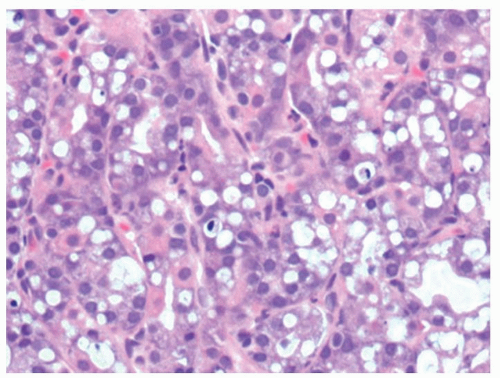
Figure 13-7. Vacuolated cells that are prominent in toxic states, in this patient the association was uremia.

Reactive changes in intestinal metaplasia. It should also be recognized that gastric intestinal metaplasia, whether incomplete (residual foveolar epithelium admixed with goblet cells) or complete (goblet and absorptive cells with or without Paneth cells), can be subject to surface injury and reactive changes. However, the same principles apply regarding using surface maturation as an indicator of reactive changes and not diagnosing it in the presence of ulcers, erosions, or restituting epithelium. These can also be recognized by the presence of metaplasia in the adjacent mucosa. However, complete intestinal metaplasia starts with intestinal nuclei that are already considerably larger than native gastric mucosa. Nuclei in incomplete intestinal metaplasia are more open and vesicular with distinct small nucleoli. Reactive changes enhance all of these features, so this needs to be taken into account. All forms of reactive mucosa have both mucin depletion and enlarged pleomorphic nuclei occupying most of the cell, and may be accompanied by erosions or ulcer. The tip-off is the presence of (a) restituting mucosa (low cuboidal or columnar) with nuclei that are usually more widely separated than in normal mucosa, especially superficially, and (b) usually a degree of maturation superficially, to the degree that a diagnosis of dysplasia should be made very cautiously in the presence of active restitution. It is worthwhile to remember that in the bases of these pits, nuclei can overlap and be stratified and hyperchromatic causing confusion with adenoma/dysplasia.


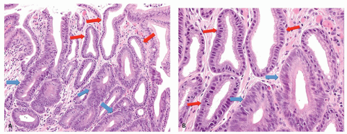
Figure 13-8. A: Reactive changes versus dysplasia. Typical reactive changes with mucin depletion, but widely spaced nuclei and superficially attenuated epithelium (red arrows). These contrast with the closely packed stratified nuclei in the dysplastic crypts (blue arrows). B: Detail of (A). Reactive changes (red arrows) versus low-grade dysplasia (blue arrows).

Reporting reactive gastropathy: Minor degrees of superficial mucin depletion are relatively common, and it is unclear how much surface mucin depletion is required to report the changes, or indeed whether they can be seen physiologically. As a guide we do not report reactive changes unless the mucin droplet in the superficial epithelial cells (usually about 75%-80% of the cell) is <50%, but there are no data to support this. However, when reported we usually indicate the most common causes.

Reporting chronic changes: Usually mild chronic reactive changes alone, such as isolated foveolar hyperplasia, are not reported unless marked, as they
tend to refer to events that happened at some point in the past, and it is unclear how long these changes take to reverse. If accompanied by acute changes of damage, then “reactive changes” covers both acute and chronic changes without the need to specify.

Clinical Implications: Of the millions of patients who every day ingest NSAIDs/ASA, only about 2% per year develop a GI complication severe enough to require medical attention, usually a bleeding gastric ulcer. Yet even 2% of a million is 20,000 events. It is possible that these patients represent a subset of individuals with a predisposition (increased sensitivity) to greater loss of their physiologic mucosal defense mechanisms. The risk of GI bleeding with NSAID use increases with age, duration of use, comorbidities, anticoagulant use (including aspirin that may also cause the damage itself), and a history of bleeding ulcers. However, it also causes bleeding in infants.28 A subset of patients (about one-third in one series20) may suffer a modest mucosal injury that results in one of the characteristic chronic changes of reactive gastropathy. However, in a series looking at the protective use of PPIs on naproxen 500 mg b.i.d.,4 within a week 25% developed antral ulcers, 12.5% duodenal ulcers, and 9.4% ulcers in multiple locations (one developed ulcers in the antrum and body; two in the antrum and duodenum). Of those taking PPIs, only 11.8% developed ulcers, all of which were antral. Anecdotally we have seen inflammatory masses in the cardia and proximal duodenal that seem likely NSAID related. They may take weeks/months to resolve or persist for months.

The gastric mucosa of the majority of users may therefore never develop changes that can be detected by endoscopic or histopathologic examination. In practice, however, most NSAID users have been taking them for long periods of time, and it is less clear how well the stomach is able to adapt to chronic NSAID ingestion. Overall, only a small subset of chronic NSAID users have biopsies with all features we commonly associate with chemical gastropathy; most may only have foveolar hyperplasia.20 In addition, such changes may occasionally occur in persons with no history of chemical injury. Concurrent H. pylori infection makes a firm diagnosis of chemical gastropathy extremely arduous.20


Figure 13-9. A: Biopsy specimen of a subepithelial hemorrhage in a patient with alcoholism. There is diffuse subepithelial hemorrhage across the full span of the fundic gland mucosa, but there is no inflammation present. B: Mucosal edema with an empty appearance of the interpit regions throughout the span of the biopsy (arrows). This is from a biopsy specimen adjacent to an area of subepithelial hemorrhage in a patient with alcoholism.

Alcoholic gastropathy. The term alcoholic gastritis (or gastropathy) is commonly used in a clinical or endoscopic context to explain abdominal pain or gastric lesions in alcoholic patients. Gastric hemorrhages, erosions, or both are found in 20% or less of actively drinking alcoholics with GI bleeding27 (Fig. 13-9A). In humans, there are few data concerning the histologic basis of gastric erosions or subepithelial (lamina propria, rarely with submucosal) hemorrhages. However, in 1954 (pre-Helicobacter days), servicemen had their gastric mucosa examined after acute alcoholic ingestion.29 In most, a variety of lesions were noted: patchy hyperemia, erosions, petechiae, and “exudate.” Biopsy specimens showed mainly superficial gastritis with prominent neutrophils.29 Some specimens exhibited edema of the foveolar region. More recently, actively drinking alcoholics had biopsy specimens taken from either subepithelial (lamina propria) hemorrhages or erosions, with specimens for comparison from adjacent sites.27 The subepithelial hemorrhage specimens revealed foveolar region hemorrhage in target lesions and sometimes striking edema in the adjacent mucosa (Fig. 13-9B). The erosions were verified histologically in 70% and commonly exhibited a pseudomembranous appearance. In both the erosions and the hemorrhages, the associated inflammatory change
was mild and was similar in severity in the lesions and the adjacent mucosa. In actively drinking alcoholic patients, the gastric mucosa may, in addition, exhibit the features of congestive gastropathy if there is portal hypertension. This is discussed in the next section.

Caustic-induced injury. Accidental or suicidal ingestion of acids or alkalis (commonly in the form of household cleaners) may cause a wide range of oral, esophageal, and gastric lesions.30, 31 The gastric antrum is especially vulnerable, with lesions ranging from superficial erosions to gangrene. A late complication in some cases is the development of gastric antral strictures.31

Graft versus host disease. is discussed in detail in Chapter 3. GVHD is seen in severely immunosuppressed patients after allogeneic bone marrow transplant where the donor T cells attack host cells leading to cell necrosis. Upper endoscopic examination in the context of suspected or proven GVHD is done if upper GI symptoms are prominent. The severity of change in the upper gastrointestinal tract (UGT) frequently do not parallel the colonic changes (see Chapter 3). The endoscopic spectrum ranges from normal to subtle swelling to erosions, ulcers, and mucosal sloughing. In addition to mild reactive changes, variable degrees of epithelial injury are seen in a background of few inflammatory cells. In general, the histopathology is that of epithelial injury/death at variance with the amount of inflammation present. In the acute phase, epithelial injury can be seen as increased apoptosis (occasionally more numerous in the neck area), attenuated regenerative-appearing epithelial cells in less injured glands, granular eosinophilic debris intermixed occasionally with nuclear debris within dilated glands, sloughed mucosa, and total destruction of gastric glands in severely injured glands. The histopathology in chronic cases can include crypt loss, inflammatory polyps in severe disease, and architectural distortion with crypt branching and atrophy.32, 33, 34 Telangiectatic vessels suggestive of gastric vascular ectasia have been identified.34 Biopsy specimens are commonly taken to also rule out infections such as cytomegalovirus (CMV). Nonetheless, similar histopathology can be seen in CMV and human immunodeficiency virus (HIV) infection, transplant recipients, and in primary immunodeficiency.34


Figure 13-10. A: With chemotherapy, gastric injury is not uniform. There is architectural distortion and individual crypts are in various stages of repair. B: Individual crypts vary from some that are very attenuated and undergoing restitution (blue arrow) to others in which more typical regenerative changes can be found (red arrows). These are irregularly admixed with more normal-appearing pits.

Chemotherapy and radiation. can cause both gastritis and stomach ulcers. The pathology is similar to that seen in chemical/reactive gastropathy with glandular atypia and increased apoptosis (apoptotic gastropathy), and there may be abnormal mitosis.35 A typical feature, identifiable at low power, is that adjacent pits with relatively normal epithelium, and pits with attenuated mucosa, and all stages between, can be immediately adjacent to each other (Fig. 13-10). Gastric injury induced in short-term exposure is often temporary. The acute response to massive irradiation occurs largely in the antrum and the prepyloric region. Not uncommonly, gastric erosions or discrete ulcers are encountered in patients with malignancy who have received abdominal irradiation and are on chemotherapy. Biopsy specimens and cultures may be obtained to rule out recurrent or metastatic disease and opportunistic infection. With larger doses, the damage may be irreversible with destruction of acidproducing glands.

Ischemia. Ischemic disease of the stomach is extremely rare. See Chapter 2 for a more detailed discussion. Atheromatous embolization of cholesterol,36 therapeutic
embolization to help control bleeding, accidental entry of selective intra-arterial radiotherapy (SIRT) beads, and vasculitis37, 38 or hypovolemic states are reported causes of erosive gastritis and gastric ulcers. There have also been isolated reports of patients with chronic gastric ulcers and erosions that healed after intestinal revascularization.39 The reported histology in these cases may lack the classic features of ischemia. In severe disease, epithelial and glandular cells are shed in the lumens of pits. Although this sounds innocuous, the mucous-producing cells can take on the appearances of signet ring cells, mimicking signet ring carcinoma (Fig. 13-11), analogous to similar lesions seen in pseudomembranous colitis. Another potential mimic of signet ring cells are the normal mucous-producing cells that appear in the oxyntic mucosa as it approaches the antrum. The polarity of these cells may appear abnormal, but these are terminally differentiated cells with no proliferative activity (Fig. 13-11E,F), while parietal cells can be shed into the lumen in oxyntic mucosa and raise the question of parietal cell carcinoma because of apparent disorderly sheets of cells (Fig. 13-12). The gastroduodenal subepithelial hemorrhages and erosions reported in some children with Henoch-Schonlein purpura might be due to vasculitis-induced mucosal ischemia.40


Figure 13-10. (Continued) C: Overview of second biopsy with more severe changes. The admixture of pits of different stages of degeneration and repair, some benign columnar and others lined by restituting epithelium. D: Severe chemotherapy changes with most glands being lined by restituting epithelium although focally they are more columnar.


Predominantly Vascular Changes

Gastric antral vascular ectasia. is an uncommon cause of chronic GI bleeding with occult iron deficiency anemia. It is characterized by telangiectatic

capillaries with fibrin thrombi and marked fibromuscular hyperplasia of the lamina propria41 (Fig. 13-13). Though it has been primarily described in the gastric antrum, proximal involvement has been reported.42

Only gold members can continue reading. Log In or Register to continue

Stay updated, free articles. Join our Telegram channel

Jun 8, 2016 | Posted by in PATHOLOGY & LABORATORY MEDICINE | Comments Off on Stomach and Proximal Duodenum: Inflammatory and Miscellaneous Disorders

Full access? Get Clinical Tree

Get Clinical Tree app for offline access