Solitary Fibrous Tumor/Fibrosarcoma
Key Facts
Clinical Issues
Most common mesenchymal spindle cell tumor of anterior mediastinum
Cough
Chest pain
Dyspnea
Hypoglycemia
May be discovered incidentally on routine imaging studies
Majority of cases reported in mediastinum behaved more aggressively than those at other locations
Microscopic Pathology
Fascicles of bland-appearing spindle cells with small dark nuclei generally devoid of mitotic activity
Usually shows short storiform pattern resembling “fibrohistiocytic” tumors
Small to medium-sized branching vessels with open lumens and “staghorn” appearance
Hypercellular areas with increased number of nuclei are seen alongside hypocellular areas
Haphazard distribution of spindle cells separated by keloidal-like strands of collagen
Large dilated vessels are seen surrounded by dense collagenous tissue
Advanced cases may show extensive areas of sclerosis and collagenization
Tumors cells are strongly positive for CD34, Bcl-2, vimentin, and CD99
Tumor cells are generally negative for cytokeratins, SMA, desmin, S100 protein, and other differentiation markers
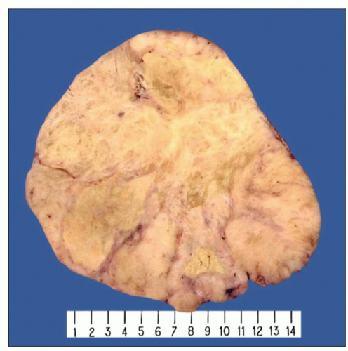 Gross appearance of solitary fibrous tumor of the mediastinum shows a well-circumscribed mass with a lobulated, tan-white homogeneous cut surface and focal areas of necrosis. |
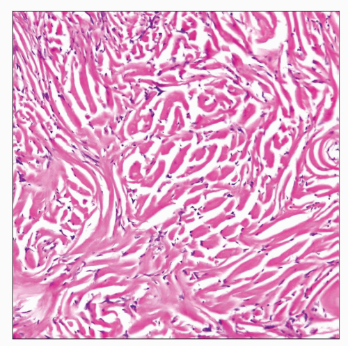 Characteristic histologic appearance of solitary fibrous tumor shows parallel linear arrays of rope-like collagen strands flanked by small, bland-appearing spindle cells. |
TERMINOLOGY
Abbreviations
Solitary fibrous tumor (SFT)
Synonyms
Localized fibrous tumor, malignant solitary fibrous tumor, mediastinal fibroma, mediastinal fibrosarcoma, hemangiopericytoma, fibrous mesothelioma
Definitions
Neoplastic, tumor-forming proliferation of dendritic fibroblasts
CLINICAL ISSUES
Site
Most common mesenchymal spindle cell tumor of anterior mediastinum
May also occur in posterior and middle mediastinal compartments
Presentation
Cough
Chest pain
Dyspnea
Hypoglycemia
Asymptomatic
May be discovered incidentally on routine imaging studies
Treatment
Surgical excision
Prognosis
Majority of cases reported in mediastinum behaved more aggressively than those at other locations
Large and poorly circumscribed tumors may recur, metastasize, and kill patient
Histology may not show strict correlation with prognosis
In general, cytologic atypia, mitotic activity, necrosis, and invasion are associated with aggressive behavior
MACROSCOPIC FEATURES
General Features
Most tumors are well circumscribed, firm, and lobulated
Larger tumors are usually infiltrative and adherent to lung and pleura
May be attached to thymus by a short fibrous pedicle
Tan-gray, whorled appearance on cut surface
Larger tumors may show areas of hemorrhage and necrosis
Gross cystic changes can also be seen on cut surface
Size
4-25 cm
MICROSCOPIC PATHOLOGY
Histologic Features
Tumors are characterized by large variegation of histologic growth patterns that may resemble other types of spindle cell tumors
Fascicular pattern
Fascicles of bland-appearing spindle cells with small, dark nuclei generally devoid of mitotic activity
Fascicles may resemble those seen in smooth muscle or neural tumors
Storiform pattern
Usually shows short storiform pattern resembling “fibrohistiocytic” tumors
Storiform pattern is usually only focal and admixed with other areas
Hemangiopericytic pattern
Small to medium-sized branching vessels with open lumens and “staghorn” appearance
Tumors with these features were commonly labeled hemangiopericytoma in previous years
Alternating hypo- and hypercellular areas
Hypercellular areas with increased number of nuclei are seen alongside hypocellular areas
This growth pattern may resemble malignant peripheral nerve sheath tumors or synovial sarcoma
Neural-like growth pattern
Fascicles composed of wavy nuclei, areas with focal palisading of nuclei, and areas resembling Verocay bodies
This growth pattern can be easily confused for schwannian neoplasms
“Herringbone” pattern
Short fascicles of spindle cells are seen emanating from central area reminiscent of a herring bone
This growth pattern can simulate fibrosarcoma, malignant peripheral nerve sheath tumor, and synovial sarcoma
“Patternless” pattern
Haphazard distribution of spindle cells separated by keloidal-like strands of collagen
Thin, rope-like strands of keloidal collagen, usually showing parallel distribution, are characteristic of these tumors
Dense hypercellular pattern
Densely packed sheets of spindle cells with little intervening stroma
This growth pattern closely resembles monophasic synovial sarcoma
Epithelioid growth pattern
Sheets or nests of round, epithelioid cells with round nuclei
This pattern may be confused for melanoma and metastatic carcinoma
Angiofibromatous pattern
Large dilated vessels are seen surrounded by dense collagenous tissue
Advanced cases may show extensive areas of sclerosis and collagenization
Unusual stromal changes
Stromal calcification, metaplastic bone formation, cartilaginous differentiation
Extensive degeneration of collagen simulating tumor necrosis
Cystic degeneration of the stroma
Hemorrhage and necrosis
Stromal myxoid changes may be prominent and extensive
Irregular stellate deposits of dense collagen (so-called amianthoid fibers)
Cytologic Features
Spindle cells are usually small and bland appearing with evenly dispersed chromatin and absent or inconspicuous nucleoli
Mitotic activity is generally very low (< 3 per 10 HPF)
Giant cells of osteoclast-type or multinucleated tumor cells may be seen
Malignant cases may show marked nuclear pleomorphism and high mitotic activity
Cells may rarely adopt round, epithelioid appearance with abundant rim of eosinophilic cytoplasm
Ancillary Techniques
Immunohistochemistry
Tumors cells are strongly positive for CD34, Bcl-2, and vimentin
Tumor cells are positive for CD99
Tumor cells are generally negative for cytokeratins, SMA, desmin, S100 protein, and other differentiation markers
Electron microscopy
Limited role in diagnosis
Ultrastructural features of fibroblastic cells with dendritic cytoplasmic prolongations
Cytogenetics
No known recurrent cytogenetic abnormalities
Molecular pathology
So far plays no role for diagnosis
No distinctive alterations have been demonstrated to date
DIFFERENTIAL DIAGNOSIS
Hemangiopericytoma
Older term used to designate tumors with distinctive branching pattern of vessels
Term “hemangiopericytoma” has been replaced by “solitary fibrous tumor” as most such cases represent examples of SFT cases
Hemangiopericytoma and SFT are currently regarded as synonymous terms in soft tissue locations
Synovial Sarcoma
High cellularity with marked cytologic atypia and variable mitotic activity
Monotonous spindle cell population with very scant connective tissue stroma
Tumor cells show focal cytokeratin and EMA positivity
Show distinctive t(X;18) translocation
May be very difficult to distinguish from SFT in some cases, requiring molecular studies for confirmation
Peripheral Nerve Sheath Tumors
Fascicular proliferation composed of spindle cells with wavy nuclei
Association with neurofibromatosis and with nerve trunks
S100 protein positivity in spindle cells
Complex, interdigitating slender dendritic prolongations seen on ultrastructural examination
Spindle Cell Thymoma
Fascicular spindle cell proliferation separated by broad fibrous bands into lobules
Usually shows minor component of scattered, small T lymphocytes
May display rosette-like structures or microcystic growth pattern
Spindle cells are positive for cytokeratins and negative for CD34
Sarcomatoid Malignant Mesothelioma
Atypical spindle cell proliferation with marked nuclear pleomorphism and high mitotic activity
Focal positivity of spindle cells for keratin and calretinin
Diffuse growth pattern with spread along pleural surface
Occupational history of previous asbestos exposure
Pleomorphic High-Grade Sarcoma
Histologic appearance can be indistinguishable from that of malignant solitary fibrous tumors
Lacks well-preserved areas showing features of solitary fibrous tumors
Lacks cells positive for CD34 and Bcl-2
DIAGNOSTIC CHECKLIST
Clinically Relevant Pathologic Features
Well-circumscribed mass in anterior mediastinum
History of hypoglycemia or clubbing of fingers
Pathologic Interpretation Pearls
Bland-appearing spindle cell population with variegation of histologic growth patterns within same tumor
Prominent hemangiopericytomatous growth pattern admixed with other growth patterns
Prominent vascularity of stroma with perivascular hyalinization and sclerosis
Striking, “rope-like” linear deposition of keloidal collagen flanking the spindle cells
SELECTED REFERENCES
1. Eguchi T et al: A solitary fibrous tumor arising from the thymus. Interact Cardiovasc Thorac Surg. 11(3):362-3, 2010
2. Suehisa H et al: Solitary fibrous tumor of the mediastinum. Gen Thorac Cardiovasc Surg. 58(4):205-8, 2010
Stay updated, free articles. Join our Telegram channel

Full access? Get Clinical Tree


