Small Round Cell Tumors


Small Round Cell Tumors


INTRODUCTION

Small round cell tumors of soft tissue are a group of malignant neoplasms composed of monotonous undifferentiated cells with high nuclearcytoplasmic ratio, which occur more frequently, although not exclusively, in childhood. They often share similar morphology and sometimes show immunophenotypic overlap. Many of them have characteristic genetic aberrations, including distinct chromosomal translocations that produce novel fusion genes resulting in disordered cellular function. Molecular genetic detection of the translocation (usually by fluorescence in situ hybridization [FISH]) or fusion gene transcripts (using reverse transcriptionpolymerase chain reaction [RT-PCR]) is therefore an essential part of the diagnostic workup of many of these tumors. Genetic studies also provide important prognostic information and have an increasing influence on clinical management.

The differential diagnosis includes poorly differentiated synovial sarcoma, soft tissue lymphoma (especially lymphoblastic lymphoma), leukemic deposits, and small cell carcinoma. These can be identified by their clinical setting and by the use of appropriate immunohistochemical panels. The differential diagnosis is summarized in Tables 14.1 and 14.2.


ALVEOLAR RHABDOMYOSARCOMA

Rhabdomyosarcomas are primitive malignant neoplasms that show incomplete differentiation toward skeletal muscle and are the most common soft tissue sarcomas in children. They constitute about 7% of childhood cancers, although are much rare in older adults. The most common subtype is embryonal rhabdomyosarcoma (ERMS) (see Chapter 10), which typically affects children younger than 10 years and shows a better response to established treatment regimens compared with alveolar rhabdomyosarcoma (ARMS). ARMS affects a slightly older population and accounts for approximately 31% of rhabdomyosarcomas.1 It is an aggressive neoplasm that has been shown to have a 5-year event-free survival rate of 45% compared to 70% in ERMS, and distinguishing the two is crucial for treatment and prognostication. ARMS is consistently associated


with characteristic translocations that result in chimeric transcription factors, which are hypothesized to generate novel transcriptional programs in an unknown target cell.2


TABLE 14.1 Differential Diagnosis of Small Round Cell Tumors


Typical Clinical Features


Microscopic Features


Ancillary Investigations


Alveolar rhabdomyosarcoma


Adolescents and young adults


Rapidly growing mass


Extremities, head and neck, trunk


Sheets and nests of cells with round nuclei of small to intermediate size and scant cytoplasm, separated by fibrous septa


Central necrosis and peripheral cellular preservation causing classic “alveolar” morphology


Solid variant lacks alveolar pattern


Rhabdomyoblasts in some cases


Desmin+, nuclear myogenin, and MyoD1+


Fluorescence in situ hybridization for FOXO1 rearrangements and RT-PCR for PAX3/7-FOXO1 fusion transcripts


Ewing sarcoma/primitive neuroectodermal tumor


Children and young adults


Extraskeletally in extremity deep soft tissue, paravertebral sites, retroperitoneum, thoracopulmonary region


Sheets of uniform cells with round nuclei, fine chromatin and scant cytoplasm


Sometimes rosette formation


Many tumors CD99+ (supporting diagnosis)


Recurrent chromosomal translocations resulting in EWS-ETS family fusion oncoproteins, detectable by reverse transcriptionpolymerase chain reaction


EWSR1 rearrangements detectable by fluorescence in situ hybridization


Minimal or absent intercellular reticulin


CIC-DUX4-associated small round cell sarcoma


Young adult males, but there is a wide age distribution (from first to seventh decade)


50% arise in limbs


Sheets of small- to mediumsized round to oval cells, often prominent nucleoli, high mitotic activity and necrosis


Sometime spindling


Associated with a higher degree of morphologic heterogeneity than classical Ewing sarcoma


Variable membranous CD99 (varying from diffuse to focal and weak)


WT1+, FLI variable


Very rarely express focal cytokeratin, EMA, desmin and S100 protein


CIC-DUX4 fusion (fluorescence in situ hybridization for CIC rearrangement)


BCOR-CCNB3-associated round cell sarcoma


Predominantly in bone; smaller numbers in deep soft tissues


Adolescents and young adults


Male preponderance


Relatively wide histomorphologic spectrum; most predominantly showing undifferentiated, small round cell Ewing sarcoma-like morphology


Smaller numbers show spindle cell morphology


CCNB3 immunohistochemostry


Also bcl-2+ and sometimes CD99 and CD117+


BCOR fluorescence in situ hybridization assay


Poorly differentiated synovial sarcoma


Young adults


Any site, but most common in extremity deep soft tissue, especially around knee and adjacent to joints and tendons


Sheets of rounded to ovoid cells


More spindled cells may be seen focally


Mast cells


CK, EMA, CD99, S100 protein all focally+, bcl-2+, TLE1+ (nuclear), CD34 and CD117−. Nested reticulin pattern t(X;18)(p11;q11), SS18-SSX fusion genes


Extraskeletal mesenchymal chondrosarcoma


Young adults


Head and neck, extremities


Exclude metastasis from extraosseous site


Biphasic morphology


Hypercellular sheets of primitive small cells with round, ovoid, or spindled nuclei


Interspersed islands of well-differentiated hyaline cartilage


Hemangiopericytic vascular pattern


Small cell component CD99+


Cartilaginous component S100 protein+


Small cell osteosarcoma


All ages, but most frequently in second decade


Sheets of small, uniform, round to focally short spindled cells


Foci of malignant osteoid, and sometimes malignant cartilage


Variable focal S100 protein+


CD99−


Desmoplastic small round cell tumor


Adolescents and young adults, particularly males


Abdomen or pelvis


Often widespread serosal involvement


Sheets of tightly packed monotonous cells with small nuclei and usually minimal cytoplasm


Striking desmoplastic stromal reaction which divides the tumor into sharply demarcated islands


Necrosis common


Nuclear expression of WT1 (carboxy terminal)


Divergent differentiation with coexpression of desmin, cytokeratins and neural markers t(11;22)(p13;q12) and EWSR1-WT1 fusion gene


Neuroblastoma


Infants and children in first 2 y of life


Follow distribution of sympathetic ganglia, with majority arising in retroperitoneum (of which about half arise in adrenal gland)


Sheets and lobules of small- to intermediate-sized cells, with hyperchromatic round nuclei


Pale neurofibrillary matrix resembling neuropil


Cells may show rosette formation


May show small amounts of ganglionic differentiation


NSE+


Neurofilament+, NB84+


CD99−


Amplification of MYCN oncogene in approximately 25% of cases


Ganglioneuroblastoma


Retroperitoneum, mediastinum


Neuroblastoma-like areas of primitive neuroblasts with areas of ganglioneuromatous stroma, subclassified according to arrangement and proportion of these areas


Ganglion cells NSE+, CD56+, NF+


Spindle Schwann cells S100 protein+


Olfactory neuroblastoma


Adults with bimodal peak (second and sixth decades)


Roof of nasal fossa, maxillary and ethmoid sinuses, nasopharynx


Lobules and nests of uniform small cells with round vesicular nuclei and scanty cytoplasm, often within neurofibrillary background


Rosettes may be present


NSE+, NFP+ variably, synaptophysin+, chromogranin+


S100 protein+ in sustentacularlike cells that surround islands of tumor


CD99−


Ectomesenchymoma


Children, M > F


Intracranial, nasal cavity, genitourinary tract, and soft tissue


Neuroectodermal elements (usually ganglioneuroma or ganglioneuroblastoma) and mesenchymal component, usually rhabdomyosarcoma


Appropriate immunoreactivity for each component


Small cell malignant peripheral nerve sheath tumor


Deep soft tissue of trunk and limbs


Uniform, small, round cells with scanty cytoplasm


Sometimes foci of uniform, short, spindle-shaped tumor cells arranged in whorl-like patterns


S100 protein+ focally, occasional GFAP+


CD99−


Small cell carcinoma


Older adults


Usually lung, but can occur in many other primary sites


Sheets of small to medium round to fusiform cells with hyperchromatic nuclei


Nuclear molding, necrosis and frequent mitoses


CK+ (dot), many TTF1+, chromogranin+, synaptophysin+, CK20−, NF−


Merkel cell carcinoma


Older adults


Face and extremities


Tumor in dermis or subcutis


Monotonous round cells, multiple nucleoli


Low molecular weight CK+, CK20+ in perinuclear dotlike distributions


NSE, neurofilament+


TTF1−


Melanoma


Primary dermal lesion may be apparent


May have junctional component


Often in nested pattern


Melanin pigment


S100 protein+, HMB45+, melan-A+


Lymphoma


May have lymphadenopathy


Cells have dispersed morphology, and tumor can have a more heterogeneous appearance with a mixed population of other inflammatory cells


CD45+, CD20, CD3+, TdT+ (lymphoblastic lymphoma)


Granulocytic sarcoma


Any site, including skin and lymph nodes


Patient may have underlying acute myeloid leukemia


Extramedullary tumor comprising immature myeloid (granulocytic or monocytic) cells


Lysozyme, CD43+, variably myeloperoxidase, CD117, CD13, CD33+


Round cell liposarcoma


Deep soft tissues of extremities of young adults


Often metastasizes to other soft tissue sites as well as to lungs


Monotonous small uniform rounded cells with minimal myxoid matrix, with some lipoblasts


Rich network of delicate vessels becomes inconspicuous in round cell liposarcoma


S100 protein+ in some cases


t(12;16)(q13;p11) or t(12;22) (q13;q12) with FUS-DDIT3 or EWSR1-DDIT3 fusion genes


Endometrial stromal sarcoma


Females


Can arise in a focus of endometriosis or present as metastasis in abdomen or elsewhere


Short closely packed spindle cells, variable focal myoid differentiation


Thick-walled vessels


CD10+, SMA+, occasional des+, CK+


Nuclear beta-catenin+ in 50% ER+, PgR + JAZF1-JJAZ1, JAZF1-PHF1, and EPC1-PHF1 fusion genes


NUT midline carcinoma


Mediastinum, head and neck


Occur over a wide age range


Roughly equal sex distribution


Relatively uniform, undifferentiated round or epithelioid cells in sheets, or sometimes nests within desmoplastic stroma


Variable squamous differentiation


Most are variably positive for p63, cytokeratins including CK7, and antibodies to the NUT protein


>50% show variable CD34+ CD99−; muscle, neural, melanocytic and hematolymphoid marker


Characterized by chromosomal rearrangements involving the gene encoding the nuclear protein of the testis (NUT) at 15q14


Most commonly due to t(15;19), fusing NUT with the BRD4 gene on chromosome 19, but in approximately a third of cases NUT is fused to a different partner gene


TABLE 14.2 Immunohistochemistry of Selected Small Round Cell Tumors


Alveolar RMS


ES


Desmoplastic SRCT


Neuroblastoma


PDSS


Small Cell Carcinoma


Lymphoma


Desmin


+


+a


+ dot


Myogenin


+


CD99


+a


+


±


±


CD56


+


+


+


+


+


±


NFP


±


+


+


±


Chromogranin


+a


+


±


Synaptophysin


±


+


±


CK


+a


+ dot


+


+ dot


EMA


+


Bcl-2


±


±


+


±


CD45


+


Others


MyoD1


FLI1


WT1


NB84


TLE1


TTF1


TdT (lymphoblastic)


a Rarely.


RMS, rhabdomyosarcoma; ES, Ewing sarcoma; SRCT, small round cell tumor; PDSS, poorly differentiated synovial sarcoma.


Clinical Features

Although the age distribution is wide, ARMS predominantly affects adolescents and young adults and is rare in adults over 45 years. It typically presents as a rapidly growing mass in an extremity. Other sites include the head and neck and the trunk. ARMS often metastasizes early and can present primarily with widespread nodal or bony metastases. Tumors metastatic to marrow can present with leukoerythroblastic anemia. Among patients with nonmetastatic ARMS, those with tumors harboring PAX3-FOXO1 fusion transcripts had a significantly poorer outcome compared with those with nonalveolar rhabdomyosarcoma or those harboring PAX7-FOXO1 fusions.3 Gene expression profiling can identify molecular risk groups, with outcome related to gene signatures.4,5


Pathologic Features

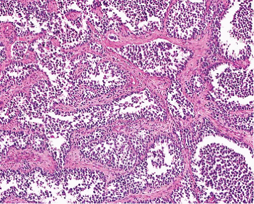
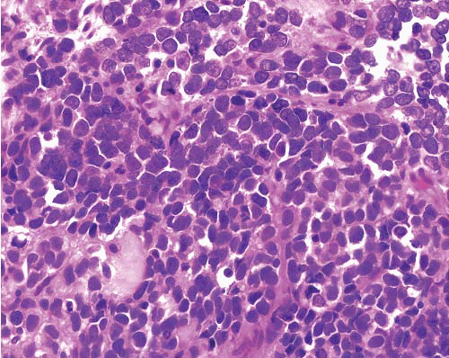
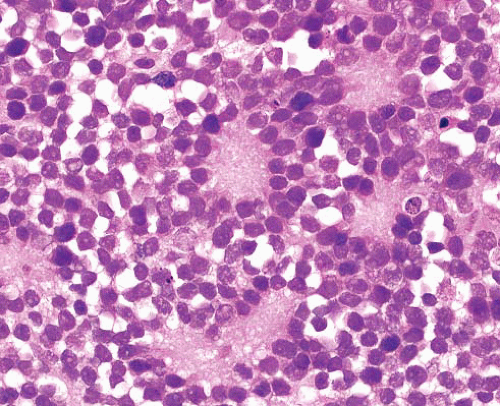
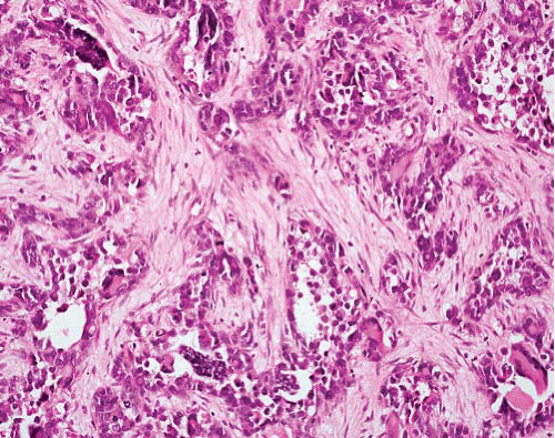
ARMS comprises nests and sheets of relatively small to intermediate cells with round to ovoid hyperchromatic nuclei and scanty cytoplasm (Figs. 14.1 and 14.2, e-Fig. 14.1), arranged in nests divided by thick fibrous septa. There is often central necrosis with cellular discohesion (e-Fig. 14.2) and peripheral preservation of cells creating an appearance resembling pulmonary alveoli. Tumor giant cells are fairly common, with abundant eosinophilic cytoplasm and nuclei arranged in peripheral
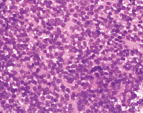
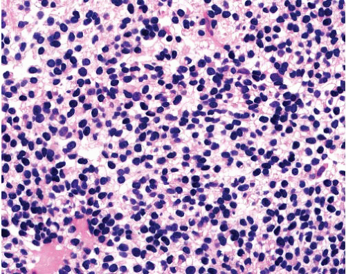
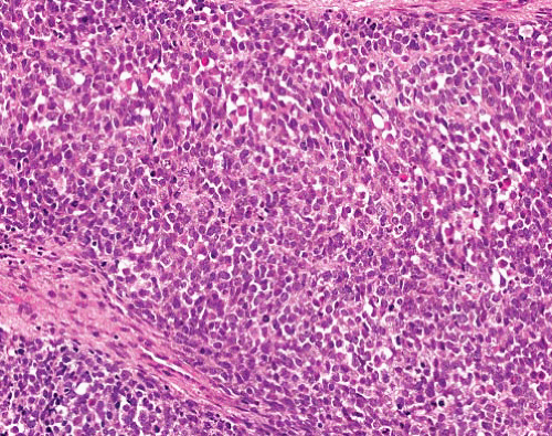
or “wreath-like” distributions (Fig. 14.2, e-Fig. 14.2). The solid variant has the same cellular features but lacks the characteristic architecture (Fig. 14.3, e-Figs. 14.3 to 14.8); however, small nests with typical appearances of ARMS may be found on thorough examination. In addition to the small cells, rhabdomyoblasts can be seen in some cases (Fig. 14.4,
e-Fig. 14.7), although not as commonly as in ERMS. Rarely, tumors may show clear cell morphology due to cytoplasmic glycogen6 (e-Fig. 14.9). These can mimic lipoblasts but lack the characteristic nuclear scalloping of the latter. Although the tumor cells are generally fairly uniform in appearance, there can be focal pleomorphism (e-Figs. 14.10 and 14.11). Cellular spindling is usually lacking in contrast to that seen in ERMS, but some tumors show mixed alveolar and embryonal histology, with foci of spindled cells in focally myxoid stroma. The presence of any alveolar pattern, however, usually indicates categorization as ARMS. Therapy can result in differentiation toward skeletal muscle, the extent of which relates to outcome.


FIGURE 14.1 Alveolar rhabdomyosarcoma. Central necrosis with peripheral preservation of cells creates an appearance resembling pulmonary alveoli. Thick fibrous septa traverse the tumor.


FIGURE 14.2 Alveolar rhabdomyosarcoma. The tumor nests contain multinucleated wreath-like cells.


FIGURE 14.3 Alveolar rhabdomyosarcoma, solid variant. There are sheets of small- to intermediate-sized cells with round hyperchromatic nuclei.


FIGURE 14.4 Alveolar rhabdomyosarcoma. Some cells have discernible eosinophilic cytoplasm reflecting early rhabdomyoblastic differentiation.


Ancillary Investigations

Desmin positivity is strong and frequently present in the majority of cells (e-Figs. 14.12 and 14.13). Myogenin and MyoD1 are intranuclear myogenic transcriptional regulatory proteins that are expressed at early stages of skeletal muscle differentiation and show strong nuclear positivity at least focally and often in a large proportion of nuclei (e-Figs. 14.14 and 14.15). Myogenin is more sensitive, and the proportion of cells with strong nuclear myogenin expression has been shown to be greater in ARMS than ERMS.7,8 Additionally, there may be focal SMA expression, and tumors can variably express neuroendocrine markers, particularly CD56 (e-Fig. 14.16). Immunostaining for myoglobin can give nonspecific positivity in a variety of other tumor types, and we no longer use this routinely. ALK is immunohistochemically positive in about half of ARMS.9


ARMS has two characteristic translocations that juxtapose the PAX3 or PAX7 genes on chromosomes 2 and 1, respectively, with the FOXO1 gene on chromosome 13. The resultant fusion genes encode PAX3-FOXO1 and PAX7-FOXO1 chimeric proteins, which serve as potent transcriptional activators. The fusion transcripts can be detected by RT-PCR and the genetic breakpoints by FISH. Also, it appears that immunostaining for PAX5 can identify translocation-positive variants of either type.10 Approximately 20% of ARMS do not have a detectable PAX-FOXO1 fusion, and these tend to be associated with a solid alveolar architecture.11 Gene expression profiling can distinguish fusion-positive and fusion-negative tumors.4,5


EWING SARCOMA/PRIMITIVE NEUROECTODERMAL TUMOR

Ewing sarcoma (ES) and primitive neuroectodermal tumor (PNET), or Ewing family tumors, are a group of small round cell sarcomas that show common histologic and molecular findings but varying degrees of neuroectodermal differentiation. They are characterized by recurrent balanced translocations involving the EWSR1 gene on chromosome 22 and the ETS family of transcription factors, which lead to the formation of novel fusion oncogenes that contribute to pathogenesis. There is no known normal counterpart cell, but these tumors are considered as neuroectodermal with capacity for divergent differentiation detectable at various levels.7,8 By convention, the term PNET is used for tumors with evidence of neuroectodermal differentiation by light or electron microscopy or immunohistochemistry, while ES is used for those lacking this. However, the tumors share overlapping features, and it is unclear that their distinction has clinical impact.12,13


Clinical Features

Most patients with ES are adolescents or young adults younger than 30 years,10 and ES/PNET is the second most common pediatric sarcoma, after rhabdomyosarcoma. It can occur anywhere in the skeletal system but especially in long bones. Extraskeletally, it may arise at virtually every site, but common regions include extremity deep soft tissue, the paravertebral area, retroperitoneum, and thoracopulmonary region (Askin tumor). With modern chemotherapy, patients with localized disease can achieve 90% 5-year survival, but this falls to about 30% in patients who have metastatic disease at presentation.


Pathologic Features

Macroscopically, the tumors tend to be solid and friable with frequent hemorrhage and necrosis, and occasional cyst formation. Histologically, most ES/PNET are composed of sheets of uniform cells with round or ovoid vesicular nuclei with smooth nuclear contours, fine chromatin,
small nucleoli, little cytoplasm (Fig. 14.5, e-Figs. 14.17 to 14.21) and indistinct cell borders. The nuclei appear evenly spaced with some overlapping (which is less in thin sections) but no molding. The cytoplasm often shows clearing (Fig. 14.6), owing to the presence of glycogen, which can be demonstrated with periodic acid Schiff stain with and
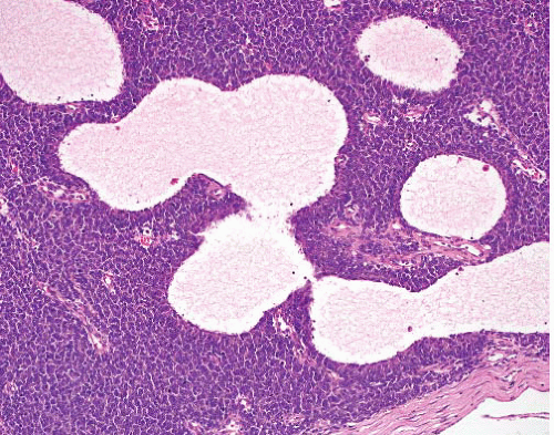
without diastase (e-Figs. 14.22 and 14.23) and may cause indentation of tumor nuclei. In some tumors, the cells are larger with moderately pleomorphic nuclei (e-Fig. 14.24) and often conspicuous nucleoli. These are referred to by some authors as large cell or atypical ES (e-Fig. 14.25).14 Rarely, the cells focally assume a more spindled shape. Rosette formation can be present (e-Fig. 14.26), most commonly as circular distributions of cells with a central fibrillary core termed Homer-Wright rosettes (Fig. 14.7). Flexner-Wintersteiner rosettes, which contain a well-defined central lumen, are rarely seen. Fibrous septa may divide the cells into lobules. Tumors can also have a pseudoalveolar pattern (e-Fig. 14.27), and cystic change sometimes occurs (Fig. 14.8). Necrosis is common (and increased after therapy) with preservation of tumor islands around blood vessels.


FIGURE 14.5 Ewing sarcoma/primitive neuroectodermal tumor. The tumor is composed of sheets of monotonous cells with round vesicular nuclei, finely distributed chromatin, and scant cytoplasm.


FIGURE 14.6 Ewing sarcoma/primitive neuroectodermal tumor. Cytoplasmic clearing is due to the presence of glycogen.


FIGURE 14.7 Ewing sarcoma/primitive neuroectodermal tumor. Rosettes can be prominent. These are of Homer-Wright type, with cells arranged around central fibrillary cores.

Several rare morphologic variants of ES/PNET have been described. “Adamantinoma-like” ES has nests of moderately pleomorphic cells in a nested growth pattern with peripheral palisading and a prominent host desmoplastic response12,13 and typically shows pancytokeratin expression.13 Sclerosing ES is characterized by an abundant eosinophilic hyalinized matrix,13 and spindle cell sarcoma-like ES has a greater degree of spindling than is typically seen in the usual ES, as well as a branching vasculature.15 Divergent differentiation is sometimes seen, including desmin expression that can lead to misinterpretation as rhabdomyosarcoma. These cases are particularly problematic for diagnosis and often require expert consultation.


FIGURE 14.8 Ewing sarcoma/primitive neuroectodermal tumor. Microcystic change in the stroma.


Ancillary Investigations

Ewing family tumors can show a variable immunoprofile, and as no specific markers exist, interpretation should be in the context of an immunopanel. Diffuse membranous positivity for CD99, a cell surface glycoprotein product of the MIC2 gene, is present in over 90% of cases (Fig. 14.9, e-Figs. 14.28 and 14.29). This is supportive of a diagnosis of ES/PNET but is not specific, as it is found in a variety of similar neoplasms (including poorly differentiated synovial sarcoma, ARMS, and lymphoblastic lymphoma, although this is often with a more nonspecific cytoplasmic staining pattern). About 70% show nuclear positivity for FLI1. ES/PNET can rarely express multiple lineage markers, including S100 protein (e-Fig. 14.30), cytokeratins (e-Fig. 14.31), CD117, and occasionally desmin.16 However, CD56, TLE1, and myogenin are negative. PNET has variable expression of neural or neuroectodermal markers such as NSE, neurofilament (e-Fig. 14.32), chromogranin, synaptophysin, and CD56. Although there is a wide range of available antibodies, it has been reported that at least three of a panel composed of CD99, FLI-1, HNK1, and caveolin-1 show immunoreactivity in all cases with caveolin-1 being positive in CD99-negative cases.17 Ewing family tumors with EWSR1-ERG rearrangement also typically show strong nuclear ERG immunoreactivity, while weak staining may be seen in a minority of non-EWR1-ERG cases.18 ERG immunoexpression is not specific, however, and tumors expressing ERG should not be confused with other neoplasms that express ERG, such as vascular neoplasms, prostatic adenocarcinomas,

Only gold members can continue reading. Log In or Register to continue

Stay updated, free articles. Join our Telegram channel

Jun 18, 2016 | Posted by in PATHOLOGY & LABORATORY MEDICINE | Comments Off on Small Round Cell Tumors

Full access? Get Clinical Tree

Get Clinical Tree app for offline access