Small Cell Carcinoma
Michael J. Thrall, MD
Key Facts
Clinical Issues
Rapidly growing and metastasizing lung mass
Very poor prognosis with life expectancy of months
Cytopathology
Molding and crush artifact
Large nuclei with “salt and pepper” chromatin
Very scant cytoplasm
Frequent mitosis and apoptosis
Necrotic diathesis
Ancillary Tests
Neuroendocrine markers: CD56 and synaptophysin are most useful
Top Differential Diagnoses
Large cell neuroendocrine carcinoma
Lymphoma
Metastatic small cell carcinoma
Non-small cell carcinoma
Reserve cell hyperplasia
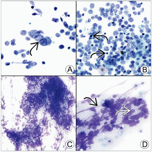 (A) Small cell carcinoma often shows mutual compression of nuclei in adjacent cells known as molding  , seen best on Pap stain. (B) This Pap-stained example of small cell carcinoma shows numerous cells without recognizable cytoplasm as well as frequent apoptotic bodies , seen best on Pap stain. (B) This Pap-stained example of small cell carcinoma shows numerous cells without recognizable cytoplasm as well as frequent apoptotic bodies  . (C) The typical low-power appearance on Diff-Quik stain is that of a markedly hypercellular smear with high nuclear:cytoplasmic ratio cells, raising the differential of lymphoma. (D) On high power, rosette-like structures . (C) The typical low-power appearance on Diff-Quik stain is that of a markedly hypercellular smear with high nuclear:cytoplasmic ratio cells, raising the differential of lymphoma. (D) On high power, rosette-like structures  can occasionally be seen, as well as nuclear streaks known as crush artifact can occasionally be seen, as well as nuclear streaks known as crush artifact  , highlighted by Diff-Quik stain. , highlighted by Diff-Quik stain. |
TERMINOLOGY
Synonyms


