Small Bowel Mucosal Disease


Small Bowel Mucosal Disease


SMALL BOWEL MUCOSAL DISEASE

A diverse array of disorders can be diagnosed with small bowel biopsy. However, in our experience, the evaluation of nonneoplastic small bowel biopsies is largely centered on “ruling out” celiac disease in the duodenum and Crohn’s in the terminal ileum; the latter is dealt with in the chapter on inflammatory bowel disease. The pathologist is commonly confronted with biopsy specimens only from the duodenum and unfortunately less commonly with biopsies from the both proximal and distal duodenum and proximal jejunum. These are the major topics of this chapter. Surgically resected specimens from patients with disorders such as ischemia or Crohn’s disease are discussed in Chapters 2 and 18. “Peptic” duodenitis is considered together with gastritis in Chapter 13.

The approach used here in the interpretation of small bowel biopsy specimens is based on that described initially by Rubin et al.1 and subsequently updated. The basic approach is to categorize lesions as specific (diagnostic) or nonspecific (nondiagnostic). In addition, the severity of mucosal injury is subjectively expressed as the degree of abnormality in villous architecture. In order to use this approach, it is essential to recognize those factors apart from disease that may alter the villous architecture. These are discussed in the following section.


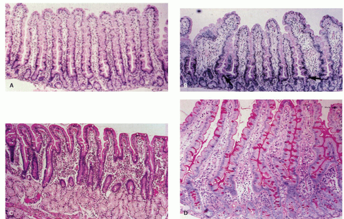
Figure 17-1. Normal small bowel biopsy. A: Rarely are this many villi seen in a row with such perfect orientation. B: More typical appearance of a well-oriented small bowel biopsy specimen. There is some variation in villous height. The arrow points to the location of the crypt-villus junction; the villus-crypt height ratio is about 4 to 1. The bent villi at the edges of the section are broader because they are tangentially cut. C: Biopsy specimen from the duodenal bulb. The villi are normally shorter here; the crypts overlie the mucussecreting Brunner’s glands. D: Biopsy specimen stained with the PAS stain. The normal columnar cells are well visualized, and have basally oriented nuclei and a prominent brush border that is stained red-purple. The intestinal goblet cells are also well visualized and are characterized by red-purple-staining globular cytoplasm.


NORMAL APPEARANCES AND ARTIFACTS


Normal Villous Architecture

Regional variations in mucosal biopsy appearances. In our experience, a great many of nonneoplastic small bowel biopsies are histologically normal (Fig. 17-1). In order to arrive at this “diagnosis” however, the pathologist must be cognizant of normal
anatomic variation, especially since most small bowel biopsies come from either the proximal duodenum or terminal ileum. The duodenal bulb is constantly assaulted by damaging peptic juices that often leads to Brunner gland hyperplasia, gastric surface cell metaplasia, and irregular villous architecture (see Chapter 13). The terminal ileum has numerous Peyer’s patches, which cause distortion and shortening of the villi (Fig. 17-2). Due to technical reasons, it is sometimes difficult to enter the terminal ileum resulting in biopsies that tend to be small, sometimes crushed, and frequently poorly oriented. In addition, the lamina propria is more cellular in this region. Hence, it behooves pathologists to let their endoscopists know that biopsies of the distal duodenum, proximal jejunum, or both are best for trying to diagnose malabsorptive diseases. Some villi appear more scalloped at their sides and shorter, whereas others appear more elongated and less pleated. In general, a villus to crypt ratio of 3:1 is felt to be normal.


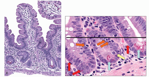
Figure 17-1. (Continued) E: Higher magnification of a normal intestinal villus showing the columnar-celled enterocytes with their basally oriented cytoplasm. F: Section of the lower half of the intestinal mucosa illustrating the intestinal crypts. Note the Paneth cells, with their characteristic red-staining granules at the base of the crypts (trichrome stain). It is important to remember that biopsy specimens fixed in Bouin’s solution rarely show Paneth cell granules, as these are dissolved by the fixative. The lamina propria between the intestinal crypts contains mononuclear cells. Plasma cells are more concentrated here than in the villous tips.


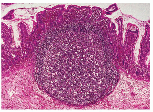
Figure 17-2. Normal small bowel mucosa overlying a hypertrophied lymphoid follicle. Note the marked blunting of the intestinal villi above the lymphoid follicle.

Duodenal biopsies: Suction biopsy has largely been replaced by direct-vision endoscopic biopsies from slightly more proximal locations in the duodenum.2, 3, 4 For adequate evaluation, a large biopsy forceps should be used, if available (see Chapter 1), and at least four biopsies specimens (at least 2 from bulb, 2 from second part) be taken to ensure that false-positive readings are kept to a minimum and that focal lesions are detected.5

Even with perfect orientation, one rarely encounters numerous normal villi in a row, all perpendicular to the lumen. Villi bend in different directions, and a number of crypts come in at different angles to join the bases (vestibules) of villi (see Fig. 17-1). Therefore, a diagnosis of “normal” requires the examination of a number of sections. In order to be secure in calling a biopsy specimen normal, three or four villi in a row should be observed.

In infants, the crypts appear to be more crowded than in adults. There is often an impression of more goblet cells than one usually sees in adults in relation to enterocytes, and there may be slightly more
branching of villi. Additionally, very shallow biopsy specimens may result in pseudoblunting (illustrated in Chapter 1). The muscularis mucosae may be absent; thus, villi appear to be stretched, separated from each other, and blunted. The clues that this appearance is an artifact and not a villous lesion are an absence of muscularis mucosae and a normal surface epithelium. In adults, villi from the duodenum are longer than ileal villi, have more indentations along their sides, and fewer goblet cells. The two can readily be distinguished by light microscopy. However, the increased numbers of goblet cells in the terminal ileum occur only over the last portion (that is the segment often biopsied), so that following ileal resections villi may look much more like proximal than distal villi.

Geographic variation. Biopsy specimens from asymptomatic natives of tropical areas or from individuals who go to live in those areas for prolonged periods may be “abnormal” by nontropical standards.6, 7 The abnormalities are usually mild, but occasionally can be more severe. Whether this results from endemic gastrointestinal (GI) infections, nutritional deficiencies, or other environmental factors is unclear.

Tangential artifact. This is the single most common cause of errors in small bowel biopsy interpretation. “Mild chronic inflammation” is commonly overdiagnosed for two major reasons:

1. Tangential orientation results in an illusion of shorter villi that are presumed to be accompanied by an increased number of lamina propria cells. The most common examples of tangential artifact in the small bowel are shown in Figure 17-1. Multiple layers of cross-sectioned crypts may occupy much of the mucosal thickness, and the villi appear blunted because they have not been sectioned perpendicular to the normal plane. In some villous tips, even in perfectly oriented sections, there will be multilayered nuclei in the columnar absorptive cells because a given villus was bent and sectioned tangentially. This should not be confused with increased numbers of intraepithelial lymphocytes.

2. The normal small bowel is inflamed, and some feel it necessary to state this in their reports as if it was pathological rather than physiological, leading to all biopsies being reported as mild (nonspecific) chronic inflammation. This can mislead the uninformed gastroenterologists to believe that biopsies are abnormal.

Traumatic artifact. Trauma is magnified by improper technique in attempting to flatten the freshly obtained specimen in the course of orientation. Some degree of traumatic artifact may be present at the edges of biopsy specimens, but the central portions should be intact if the tissue is oriented properly.

Pathologists are often confronted with biopsy samples from the small intestine that are traumatized and suboptimal in size and orientation. If they are handled properly in the laboratory from that point on, it is usually possible to exclude diseases associated with severe lesions such as celiac sprue. However, in such circumstances, it should be realized, milder, and variably severe abnormalities (including mild lesions of celiac disease) cannot be reliably excluded. In practice, despite best efforts and intentions, many biopsies tend to be poorly oriented and overreliance on the inability to assess the height of villi may result in reports that are clinically unhelpful and make the pathologist look inept. In such situations, absence of other diagnostically important features should be evaluated that include intraepithelial lymphocytosis and surface epithelial changes to exclude the diagnosis of celiac sprue. The limitations of a biopsy when encountered should however be stated in the pathology report, otherwise the clinician will fail to realize the kinds of material he or she is sending to the pathologist.


Evaluation of a Small Bowel Biopsy Specimen

In evaluating jejunal or multiple duodenal biopsy specimens, one should perform the initial screening at a low (2 or 4× objective) magnification to get an impression of whether or not obvious abnormalities are present. The overall grading of villous architectural change is usually done at this magnification. Next, a higher magnification is used to look for specific (diagnostic) features and to assess the mucosal components systematically in a number of adjoining well-oriented villi. The luminal aspects of villi and intervillous regions are examined for evidence of parasites, specifically Giardia lamblia and Cryptosporidium. A common abnormality in the surface epithelium is a cuboidal basophilic appearance. The surface epithelium is examined for loss of nuclear polarity, increased intraepithelial lymphocytes and neutrophils, and thickened subepithelial collagen band. Next, the lamina propria cell density and type are reviewed. The presence of plasma cells should be verified. They are most concentrated between the crypts. Isolated neutrophils and eosinophils are normally present, but any more than this is abnormal. The crypt zone is assessed mainly for the number of mitotic figures and for their location within the crypts, and to ensure that Paneth cells are present in normal numbers, while
the nuclear size is worth examining in crypt bases in case severe megaloblastic changes are present.

The average number of mitoses is one or two per well-oriented crypt. The number of mitotic figures is increased in many small bowel disorders, and this increase is usually paralleled by their upward displacement, so they appear halfway up or higher in the crypt-villus columns. A perceived decrease in the height of villi is commonly correlated with an increased number of mitoses in the crypts and is a useful check if villi have been artificially shortened. If a biopsy specimen is found to be abnormal but nonspecific, or normal on preliminary review, additional time needs to be spent ensuring that diagnostic features are not patchy. This mainly involves excluding the presence of parasites and granulomas; however, this may also occur with conditions like celiac sprue and Crohn’s disease.

If submucosa is contained in the specimen, the vessels should be examined with a view to discovering any telltale presence of abnormalities, such as amyloidosis or vasculitis.


The Abnormal Small Bowel Biopsy Specimen

The classification of abnormal small bowel biopsy appearances is given in Table 17-1. The term nonspecific refers to a histologic pattern, which may be common to diverse etiologic or pathogenetic mechanisms and requires either other diagnostic maneuvers or therapeutic trials. A specific or diagnostic histologic pattern is one in which the appearance of the mucosa is pathognomonic for a disease or has a distinctive appearance that narrows the differential diagnosis to a small group of disorders (e.g., granulomas). Regardless of whether a biopsy lesion shows nonspecific or diagnostic features, it is very useful to classify the severity of the lesion in order to transmit this information simply in reports. Severity is classified in relation to degrees of abnormality of the villous architecture. The grades are expressed as mild, moderate, or severe (“flat”) lesions. The Marsh (Marsh-Oberhuber) system is often used to grade the villous abnormalities in celiac disease.8 Marsh 1 is a lesion that has normal villous architecture but increased intraepithelial lymphocytes. Marsh 2 is a lesion that has normal villi but crypt hyperplasia (this is hard to identify in routine practice and hence is rarely mentioned). Marsh 3a refers to mildly blunted villi, while Marsh 3b has moderate blunting, and Marsh 3c is completely flat. The term variably severe refers to abnormalities in villous architecture that are patchily distributed and of variable severity.


Table 17-1 Overview Classification of Small Bowel Biopsy


ABNORMALITIESa


Severe (flat) lesionb


Nonspecific histology


Specific/distinctive histology


Variably severe lesionsc


Nonspecific histology


Specific/distinctive histology


Normal villous architecture


Specific/distinctive histology


a Based on the usual practice of reviewing three or four biopsy specimens from a given region in the small bowel, usually the middle to distal duodenum.

b A diffuse pattern of severe change with absent villi.

c Abnormalities in villous architecture that are patchily distributed and of variable severity. They range from normal to severe, but the usual pattern is a mild or moderate abnormality of villous architecture.


The mild lesion (Marsh type 1). The main abnormalities here are in the surface epithelium and the lamina propria. Villous height remains within the normal range (Marsh type 1 lesion). The surface epithelium commonly exhibits a disordered nuclear polarity and an increase in intraepithelial lymphocytes (Fig. 17-3). There may be an apparent increase in the number of mitotic figures, and these figures may extend higher in the crypts. The lamina propria may or may not exhibit an apparent increase in mononuclear cells. Sometimes, biopsy specimens with a nonspecific, mild lesion also have slightly branched or fused-appearing villi. However, in many cases there is no villous abnormality, and the main abnormalities consist of the surface epithelial and lamina propria changes. If one looks hard enough, it is often possible to find a single villus in a normal biopsy specimen that could be interpreted as exhibiting a mild change. It should be stressed that designation of a mild abnormality in a biopsy specimen generally refers to changes in numerous villi. The security in making this diagnosis is enhanced when several biopsy specimens from the region sampled exhibit this change.

Uncommonly, there is only an apparent increase in the number of mononuclear cells in the lamina propria, with no surface epithelial abnormalities, namely, loss of nuclear polarity and increased numbers of intraepithelial lymphocytes. In general, we are loath to label such an appearance as a mild lesion unless accompanying surface epithelial changes are present. The reason is that there is considerable normal variation in villous height and shape (see Fig. 17-1B) with differing appearances of cell density in the lamina propria.


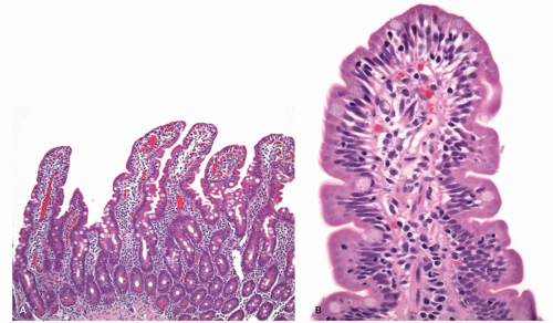
Figure 17-3. Normal villous architecture with increased intraepithelial lymphocytes (Marsh type 1). A: In this low-power view, the villous to crypt ratio is normal. B: Surface epithelium illustrates the loss of nuclear polarity and increased intraepithelial lymphocytes.

The moderate lesion (Marsh type 3a and 3b). The main difference between this grade and the mild lesion is that there is an unequivocal shortening and broadening of villi, with more exaggerated surface epithelial and lamina propria chronic inflammation (Fig. 17-4). The surface epithelium may exhibit both increased numbers of intraepithelial lymphocytes and a cuboidal appearance of the enterocytes (Fig. 17-5).

The severe flat lesion (Marsh type 3c). Here, the villi are virtually absent (see Figs. 17-6, 17-7, 17-8). The most common disorder accounting for this appearance is celiac sprue. However, rarer disorders with a more diagnostic appearance, such as Whipple’s disease and extensive infiltration by Mycobacterium avium-intracellulare, may rarely result in a flat appearance, although more usually villi are simply expanded. In other cases, the appearance of the severe flat lesion is described as “subtotal villous atrophy.” This appearance is best appreciated at low- or medium-power magnification (Fig. 17-8) (also known as Marsh type 3 lesion). Conceptually, this description is erroneous, since there is actually
increased cell turnover in the most common disorder associated with this lesion (i.e., celiac sprue). Biopsy specimens that are completely flat, many sometimes exhibit rudimentary villi, or villi of decreased height, all of which are encompassed by the term “partial villous atrophy.”


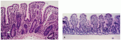
Figure 17-4. Moderate mucosal lesions (Marsh type 3b). A,B: Villi are shorter and crypts are elongated (hyperplastic), with a villus-crypt ratio of approximately 1:1. The villi are broader because of markedly increased numbers of inflammatory cells.

The Thin, Flat Mucosa This appearance is seen most typically in familial enteropathy (microvillus inclusion disease) and in conditions associated with marked reductions in cell turnover (radiomimetic injury) such as irradiation, chemotherapy, and severe folate and vitamin B12 deficiencies. These conditions are discussed and illustrated subsequently in this chapter. One can still use the general description of moderate or severe, but qualifiers are required. There are exceptions to the usual appearances: The mucosa may be very thin, the usual striking increase in lamina propria inflammatory cells may be minimal or absent, and the number of mitotic figures may be reduced or at least not increased, as they usually are in moderately or severely abnormal biopsy specimens.

Alternative terminology. Many pathologists and clinicians use variations on the term villous atrophy to describe small bowel mucosal abnormalities. Subtotal villous atrophy is used as a synonym for severe flat mucosa. Total villous atrophy would seem preferable in this regard, except that purists point out those little ridges of rudimentary villi (Fig. 17-8) may be found in the severe flat lesion of celiac sprue. Partial villous atrophy (sometimes subgraded further according to severity) is used to describe what is referred to here as mild or moderate lesions, or villous blunting (in some ways preferable). The problem with “atrophy” is that it often is taken as implying celiac disease, irrespective of any qualifiers the pathologist makes in the report.

We will not debate the pros and cons of the mild, moderate, and severe mucosal lesion classification versus the villous atrophy classifications. Some move between the two or combine them. Two qualifiers are necessary, however. First, villous atrophy should not be construed as referring to cell turnover, but only to the fact that the villi are shorter than usual. Second, as stressed in Chapter 1, the pathologist should ensure that the clinicians understand what they mean by the terms they use.

Although the term “villous atrophy” in celiac disease has been deeply ingrained in the literature and practice such that its use immediately evokes a mental diagnosis of celiac disease, some prefer to use a hybrid terminology, which is practical and omits all references to atrophy. Unless an intraepithelial lymphocytosis is present, the likelihood of the underlying disease being celiac disease is virtually zero and always needs to be mentioned. The hybrid system goes as follows:

1. “Intraepithelial lymphocytosis in the presence of normal shaped villi (see comment)” for Marsh 1 lesions.

2. “Mild/moderate/severe shortening of villi/villous blunting (see comment)” for anything where villi are present but not of normal height, that is, Marsh 3a and b lesions. More specific lesions (e.g. intraepitheilial lymphocytosis) are added when present.

3. “Severe shortening of villi/villous blunting/flat mucosa (see comment)” when no villi are present for Marsh 3c lesions. Again, other features are noted.

The comment should describe other pertinent findings and address the clinical issues. For example, in the first scenario of Marsh 1 lesions, it should be mentioned that there are numerous causes of this lesion such as ingestion of nonsteroidal anti-inflammatory drugs (NSAIDs), postinfectious states, bacterial overgrowth, an upregulated immune system (e.g. rheumatoid arthritis, thyroiditis), and immune deficient states (unlikely if normal numbers of plasma cells are present). However, the most important is celiac disease, which should be confirmed or excluded by appropriate serologic tests. One should recognize that a proportion of patients have disease largely localized to the bulb. The latter also requires gastric biopsies for Helicobacter to ensure that any disease in the bulb is not Helicobacter related.

For the other remaining scenarios, one should always include a statement whether an intraepithelial lymphocytosis is present or not. If it is present, then celiac disease is the most likely diagnosis, and should be confirmed using a serologic test (typically tissue transglutaminase [TTG], and then if positive confirmed by antiendomysial antibodies). These should always be accompanied by serum IgA levels, as the commonly used test is based on evaluation of IgA antibodies, and will be false negative if the patient is IgA deficient. In which case, tests based on IgG antibodies are specifically requested. In about 2% of adults and 10% of children, the duodenal bulb may be the preferred biopsy site with maximal disease, so routine biopsies from this site are encouraged (see subsequent discussion).


DISORDERS ASSOCIATED WITH ABNORMAL SMALL BOWEL BIOPSY SPECIMENS

Table 17-2 lists diseases associated with a severe, flat small bowel lesion in which the histologic features are nonspecific. The most common disease in this category is celiac sprue. Other conditions in this category may
be identified by their response to therapy. A few rare entities have no known treatment. In these disorders, multiple biopsy specimens from the duodenum have a diffuse, flat appearance. However, in some patients with these conditions, lesions of variable severity may be seen. A prime example is tropical sprue.


Table 17-2 Severe (Flat) Lesions: Nonspecific Histologya


DISEASEb


TREATMENT RESPONSE


Symptomatic celiac sprue


Gluten-free diet


Refractory celiac sprue


Partial only, with gluten-free diet


Unclassified sprue


Unknown


Other protein injury


Soy, milk, wheat, or other protein withdrawal


Medications olmesartan drug


Change/withdraw


Mycophenolate


Change drug


Infectious gastroenteritis of childhoold (some)


Spontaneous


Tropical sprue (some)


Antibiotic + folic acid


Childhood kwashiorkor


Adequate protein diet


Familial enteropathy


Unknown


aThree or four biopsy specimens in a region (e.g., distal duodenum) all exhibit a uniformly diffuse and severe abnormality.

b A single biopsy specimen from patients with the entities given in Table 17-4 may occasionally exhibit a severe flat lesion.


Table 17-3 presents small bowel biopsy lesions that are flat but in which the histologic changes may be diagnostic. Table 17-4 gives disorders that are associated with variably severe lesions in villous architecture that are nonspecific. The changes are usually mildly to moderately severe, but sometimes patchily distributed severe lesions are also present. Table 17-5 lists disorders with variably severe lesions but in which there may be specific (diagnostic) features. In some of the conditions, the diagnostic features are not invariably present because of focal involvement. Finally, Table 17-6 lists disorders in which the villous architecture is normal but other features, either in the mucosa or in the submucosa, yield specific clues to the diagnosis.


Table 17-3 Severe Flat Lesions: Specific/Distinctive Histology


DISEASE


DIAGNOSTIC HISTOLOGY


Collagenous sprue


Broad band of collagen below surface epithelium; may be patchy


Some common variable immunodeficiency (late-onset immunodeficiency)


Reduced or absent lamina propria plasma cells; nodular lymphoid hyperplasia; most have variably severe lesions


Autoimmune enteropathy/IPEX syndrome


Lack of goblet cells, Paneth cells and/or endocrine cells


Microvillous inclusion disease


Intracellular DPAS positive/CD10 positive inclusions


DISORDERS ASSOCIATED WITH A SEVERE FLAT MUCOSA: NONSPECIFIC AND SPECIFIC/DISTINCTIVE HISTOLOGY

This category includes lesions which, on multiple biopsy specimens from the distal duodenum, show a diffuse, flat change. In some of these disorders, lesions of variable severity may be seen. Prime examples are tropical sprue and late-onset immunodeficiency. In the latter, some individuals overlap between specific (diagnostic) and nonspecific categories (Tables 17-3, 17-4 and 17-5).


Celiac Sprue (Syn. Celiac Disease, Glutensensitive Enteropathy, Nontropical Sprue)

Celiac sprue is a small intestinal disease that is characterized by damage to the small intestinal mucosa and malabsorption of a variety of nutrients. The intestinal mucosal injury is due to an immunologic reaction to gluten, a group of proteins found largely in wheat, rye, and barley. Historically, Aretaeus in the first century AD is credited with earliest description of celiac disease, although it was Samuel Jones Gee in 1888 who first described the clinical findings of celiac sprue.9 Paulley is credited with providing the histopathologic correlates of the disease in 1954, although earlier Beneke (1910), Justi (1913), and Manson-Bahr (1924) had also made significant contributions in recognizing small bowel inflammation and villous atrophy as important components of the disease.10 The prevalence of the disorder has been estimated at approximately 1% in North America and Western Europe, being even higher in Ireland and Northern Europe.11

Pathogenesis. The grain proteins injurious to the small intestine consist of the gliadin fraction of gluten, which is present in wheat, barley, and rye. Oats, rice, and millets are considered safe in celiac disease as their proteins bear little similarity to gluten containing grains, although a small percentage of celiac
patients may harbor a response to avenin present in oats, besides some contamination with gluten that can be seen in commercial products.12 Gliadins can be categorized into four major fractions by gel electrophoresis, namely, alpha, beta, gamma, and omega gliadins. Early reports suggested that only the alphagliadins activated celiac sprue.13 However, subsequent studies indicate that all other components of gliadin may also activate disease.14, 15, 16 As one can imagine, defining and possibly eliminating the precise amino acid sequences of gliadin that are responsible for disease activation are topics of intense research.


Table 17-4 Variably Severe Lesions: Nonspecific Histologya


DISEASEb


TREATMENT RESPONSE


Subclinical celiac sprue (some dermatitis herpetiformis and some asymptomatic relatives)


Gluten-free diet


Medications


Withdraw the medication


NSAIDs


Mycophenolate mofetil (MMF)


Olmesartan


Helicobacter gastritis and reactive duodenitis


Treat Helicobacter (See Case 2 at end of chapter and Chapter 13.)


Infectious gastroenteritis


Spontaneous cure


Stasis syndromes (contaminated bowel syndromes)


Antibiotics


Tropical sprue


Antibiotics + folic acid


Some immunodeficiency syndromes


Some common variable immunodeficiency


Partial improvement if concomitant parasitic infestation or bacterial overgrowth is treated


Some acquired immunodeficiency syndrome (AIDS)


None unless a treatable opportunist is present


Graft versus host disease


Treatment for GVHD


Geographic variation


Environmental change


Acrodermatitis enteropathica


Oral zinc


Zollinger-Ellison syndrome


Antisecretory drugs or surgery


Idiopathic


Often resolve spontaneously


aRefers to a spectrum ranging from normal to completely flat in a group of three or four biopsy specimens from a given region. The usual finding is mild to moderate lesion.

b Except for tropical sprue and common variable hypogammaglobulinemia, the mucosa may be completely normal in some patients.


Table 17-5 Variably Severe Lesions—Specific/Distinctive Histologya


DISEASE


DIAGNOSTIC HISTOLOGY


Whipple’s disease


Typical macrophages filling lamina propria


Eosinophilic gastroenteritis


Masses of eosinophils; other causes must be excluded (e.g., helminths)


Crohn’s disease (some)


Noncaseating granulomas; most often Crohn’s disease, but clinical correlations are required


Primary intestinal lymphoma


Malignant lymphoid infiltrates


Immunoproliferative small intestinal disease (IPSID)


Massive infiltrates of mainly plasma cells, much more than in celiac sprue


Infections


Parasitic


Giardia, Cryptosporidium, microsporidiosis, Isospora belli, Strongyloides, hookworm, Schistosoma, Capillaria


Viral


Cytomegalovirus


Fungal


Candida, Histoplasma


Mycobacterial


Macrophages containing acid-fast organisms of Mycobacterium avium-intracellulare


Lymphangiectasia (primary or secondary)


Dilated lymphatics in lamina propria and submucosa


Macroglobulinemia


Hyaline deposits in lamina propria


Some common variable immunodeficiency


Decreased or absent plasma cells; nodular lymphoid hyperplasia


Autoimmune enteropathy/IPEX syndrome


Lack of goblet cells, Paneth cells and/or endocrine cells


Tufting enteropathy


Epithelial tufts at the tips of villi


Impaired epithelial cell replacement (severe vitamin B12 or folate deficiency, chemotherapy, irradiation)


Severe epithelial macrocytosis, and reduced mitoses suggest these entities more than any villous architectural change


aThe common pattern is mild or moderate abnormality in villous architecture. The key is to look for diagnostic features.


Patients with celiac disease have a genetic predisposition to the disorder, as virtually all patients are positive for HLA DQ2 or DQ8.17 These two HLA types are expressed in 30% to 35% of the population where celiac
sprue is prevalent, but only 2% to 5% goes on to develop the disease. There is a strong familial association, with as many as 15% of asymptomatic family members having some small bowel abnormalities on biopsy, especially after a gluten challenge.18, 19, 20, 21 Celiac sprue has also been found in identical twins, but only in 70% to 75% of cases,22, 23 suggesting that additional environmental factors are important for disease expression.18, 24, 25 Such environmental factors may include viral infection. Kagnoff et al. proposed a possible role for adenovirus serotype 12 (Ad 12) in the pathogenesis of celiac sprue, based on the observation that there is a homology of amino acid sequences between a portion of alpha-gliadin and a protein (E 1b) produced by Ad 12.26 To try to confirm this, the authors looked for evidence of previous infection with Ad 12 in patients with either treated or untreated celiac sprue and in controls. They found an increased association between antibodies to Ad 12 and celiac disease in the patients compared to the controls and concluded that this finding could not be accounted for by random viral infection.27 Unfortunately, PCR studies to try and confirm the presence of Ad 12 DNA in celiac patient biopsies have been negative.28


Table 17-6 Normal Villous Architecture: Specific/Distinctive Histology


DISEASE


DIAGNOSTIC HISTOLOGY


Abetalipoproteinemia


Fasting absorptive cells are clear and translucent because they are packed with fat


X-linked immunodeficiency


Virtual absence of lamina propria plasma cells


Amyloidosis


Congo red positive


Material in capillaries of the lamina propria and vessels of the submucosa


Lipid storage diseases


Vacuolated ganglion cells and macrophages


Chronic granulomatous disease


Pigmented, vacuolated macrophages in lamina propria


There is abundant evidence that immune mechanisms play a central role in celiac sprue.29, 30, 31, 32, 33 A number of antibodies have been found in celiac sprue patients, such as antigliadin, antireticulin, antiendomysial antibodies, and TTG.29, 30, 31, 32, 33, 34 TTG appears to play a central role in disease pathogenesis (Fig. 17-5).34, 35 How the gluten enters the lamina propria remains unclear. Various mechanisms postulated include entry through a paracellular pathway due to defective tight junctions and transcytosis through the surface epithelium. Alternatively, direct sampling of the gluten in the lumen by dendritic cells that seems to send out their processes between the epithelial cells or via “M” cells that overlie organized lymphoid follicles in the mucosa has also been suggested. Once the gluten enters the lamina propria TTG deamidates it, which allows the peptide to fit into the DQ2 or DQ8 pocket on antigen presenting cells. These antigen presenting cells then activate CD4+ T cells that secrete mainly Th1 cytokines such as IFN-γ, which induce the release and activation of metalloproteinases (MMPs) by myofibroblasts.34, 36, 37, 38, 39 Additionally, Th2 cytokines are also produced driving the production of (auto-) antibodies to gluten and TTG 2. Macrophages/dendritic cells and enterocytes are the major source of IL-15, which plays a key role in the innate and adaptive immunity in celiac disease. Other cytokines such as IL-18, IFN-α, or IL-21 seem to play a role in polarizing and maintaining the Th1 response. The T cells circulate to mesenteric lymph nodes where they encounter and are primed by antigen-presenting cells (mainly dendritic cells) and from where they home back to the lamina propria, a process that is driven by the lymphocyte homing receptors CCR9 and integrin α4β7. Various cytokines are important driving forces for the tissue remodeling seen in celiac sprue that include villous atrophy and cyrpt hyperplasia.

Clinical features. The clinical manifestations of celiac sprue are protean, and vary with the age of the patient and the duration and extent of the disease. They range from minor nutritional deficiencies, especially iron, in adults to more striking weight loss, steatorrhea, and malnutrition, especially in children. The extraintestinal manifestations of celiac sprue result from nutritional deficiencies and consist primarily of anemia due to iron, folate, or vitamin B12 malabsorption, osteoporosis and osteomalacia, muscle wasting, arthritis, infertility, liver failure, and neurologic abnormalities of uncertain pathogenesis. Celiac disease should be considered with women with infertility or failure to carry their pregnancies to term or in any persons with an undiagnosed cause for abnormal liver tests.

Symptoms commonly appear during the first three years of life after the introduction of cereal into the diet. If untreated, the symptoms persist throughout childhood, but often diminish or disappear during adolescence, only to reappear in early adult life. There is a second peak of presentation during the third to fifth decades. The variable clinical sensitivity to gluten in celiac sprue is a mystery. It is seen in adolescence if children are taken off their gluten-free diets. In adults, the diagnosis may be delayed40 as the overt symptoms of diarrhea and steatorrhea are frequently absent and manifestations may only consist of iron deficiency anemia41, 42 or sometimes also recurrent aphthous stomatitis, dermatitis herpetiformis, and infertility. The majority (>80%) of screening detected patients show no, minor, or non-diarrhea-associated symptoms.
Variable sensitivity to gluten is also seen in patients with the disease who go off the gluten-free diet. In some there is prompt appearance of symptoms of malabsorption, whereas in others these symptoms may not reappear for months or years. The patient who presents with diarrhea may have a long-standing history of anemia or may be shorter in stature than his or her siblings. This suggests that the mucosal lesion has been present all along but that the clinical expression with diarrhea/malabsorption is delayed. Finally, it is theoretically possible that adults who present with no antecedent clues may have developed it in adulthood.


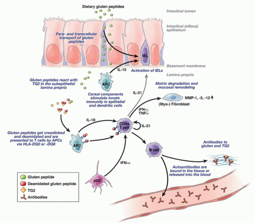
Figure 17-5. Pathogenesis of celiac disease. Gluten is transported across the epithelium by yet unclear mechanisms where crosslinking and particularly deamidation peptides in the tissue by transglutaminaase2 (TG2) creates potent immunostimulatory epitopes that are presented via HLA-DQ2 or HLA-DQ8 on antigen-presenting cells. Subsequently, CD4+ T cells are activated, secreting mainly Th1 cytokines such as IFN-γ, which induces the release and activation of metalloproteinases (MMPs) by myofibroblasts, finally resulting in mucosal remodeling and villus atrophy. Additionally, Th2 cytokines are also produced driving the production of (auto-)antibodies to gluten and TG2. Other cytokines such as IL-18, IFN-α, or IL-21 seem to play a role in polarizing and maintaining the Th1 response. The scheme is simplified. It does not show that T cells circulate to mesenteric lymph nodes where they encounter and are primed by antigen-presenting cells (mainly dendritic cells) and from where they home back to the lamina propria, a process that is driven by the lymphocyte homing receptors CCR9 and integrin α4β7. (From Schuppan D, Junker Y, Barisani D. Celiac disease: from pathogenesis to novel therapies. Gastroenterology. 2009;137(6):1912-1933.)

There are two criteria for the diagnosis of celiac sprue. One is the demonstration of the characteristic severe flat lesion. Although the lesion is in the nonspecific category, it is virtually always indicative of celiac sprue in North America and Europe. Nevertheless, celiac disease is increasingly being recognized in countries that were thought not to have much celiac disease (India, other parts of Asia), which may be occurring as the diet changes from rice to include gluten containing foods including “fast” foods.43, 44 Paralleling this, it is increasingly common to see biopsies in immigrants to North America with typical features of celiac disease. The possibility of other causes of an intraepithelial lymphocytosis always has to be borne in mind, but we suspect this trend will continue to increase.

The second criterion for diagnosis is the demonstration that malabsorption disappears when gluten is removed from the diet. In most instances, especially in adults, the clinical response alone (e.g., an almost immediate feeling of well-being, cessation of diarrhea, and weight gain) to a gluten-free diet is sufficient to clinch the diagnosis. Before obvious weight gain occurs, there is often an increased sense of wellbeing; this is especially true in infants, who within
1to 2 days may become much less irritable. In addition to these, the serologic tests have become equally important in establishing the diagnosis of celiac disease. TTG and antiendomysial antibodies appear to be the highly sensitive and specific serologic markers for celiac disease.34, 35 Of these, the antiendomysial antibody is considered more specific; however, it is also more expensive. Hence, in practice most often only TTG is obtained as a part of serologic testing. Some authors have advocated that following TTG titers may be a good way of monitoring a response to a glutenfree diet and thus confirming the diagnosis.45

The mucosal lesion of celiac sprue is most severe in the duodenum and the proximal jejunum.46 The ileum is usually spared or less severely affected with a mild lesion characterized primarily by an increased number of intraepithelial lymphocytes. In general, the degree of malabsorption is proportional to the length of the involved small intestine. Thus, folate and iron deficiency anemias are more common because these nutrients are absorbed in the proximal small intestine. Vitamin B12 deficiency is much less common because vitamin B12 is absorbed primarily in the ileum. In patients with severely symptomatic celiac sprue, the ileal mucosa may also be markedly abnormal with moderate to severe lesions.47

Associations of celiac sprue. Apart from the complications of celiac sprue such as lymphoma, ulcerative jejunoileitis, and collagenous sprue, which are discussed subsequently, celiac sprue has been found in association with a number of disorders. The long list of associated disorders includes some that may be considered dubious, without clear evidence that they occur more than by chance alone.48 Associated disorders include selective IgA deficiency that may be associated with giardiasis, abnormalities of thyroid function, and diabetes mellitus,49 association with cow’s milk hypersensitivity,46, 50 bronchopulmonary disorders,51 cavitation of mesenteric lymph nodes,52 intestinal pseudo-obstruction,53 pneumatosis intestinalis,54 and both lymphocytic and collagenous colitis.55 Lymphocytic gastritis (see Chapter 13), generally unaccompanied by Helicobacter pylori, was reported in approximately 50% of patients with small bowel lesions compatible with celiac sprue.52, 56 In our experience, lymphocytic gastritis is more often present in children with celiac disease than in adults. Lymphocytic colitis is also associated with celiac disease as 15% of patients with lymphocytic colitis have been found to have celiac disease and up to 31% of celiac patients have been reported to have lymphocytic colitis.57, 58 It is important to know of these associations as failure of patients with celiac disease to respond to a gluten-free diet should prompt consideration of an associated colitis, and vice versa.

There is clearly an association between celiac disease and hepatitis. Many studies have found that celiac diseases patients have transaminase elevations that go away once they respond to a gluten-free diet.59 The histologic changes described in these patients vary from fairly nonspecific to autoimmune hepatitislike, and rarely cirrhosis or fulminant liver failure.

Endoscopic appearance. Three endoscopic appearances of untreated celiac sprue have been described (Fig. 17-6). There may be scalloping of the folds59 or a paucity of valvulae conniventes.60 In severe villous atrophy, there may be a mosaic pattern to the mucosa. When this occurs, typically the superficial layer is missing and the submucosal vasculature becomes quite prominent. These patterns should sometimes help the endoscopist detect unsuspected celiac sprue, and can direct biopsies to more severely affected areas. However, their absence should not deter the endoscopist from obtaining distal duodenal biopsy specimens when the diagnosis is suspected. In fact, these endoscopic findings are neither sensitive nor specific for celiac disease; hence the pathologist should not be influenced by the presence or absence of these changes when rendering a histologic diagnosis.

Where to Biopsy Biopsy specimens in untreated patients are classically taken from the second part of the duodenum. The reason for this is that historically villi from the duodenal bulb are often shorter than more distal villi because of the presence of underlying Brunner’s glands, and also likely to be involved when gastric Helicobacter are present. However, as Helicobacter becomes less common, the latter reason is not only far less of an issue, but can be easily circumvented by taking biopsies of the gastric antrum and body. The “ideal” set of biopsies are therefore distal duodenum (4 biopsies from 2nd or 3rd part),4 duodenal bulb (2 biopsies),2 and gastric antral and body (two each) to provide not only the greatest chance of making the diagnosis, but of anticipating any issues that may arise. Indeed, the maxim of “if biopsying the duodenum, always take gastric biopsies for Helicobacter” should be standard of practice. About 10% of children will have flat duodenal bulb biopsies in the presence of normal biopsies in the more distal duodenum, while in adults the figure is about 2%.60, 61, 62

Pathology. The typical biopsy in symptomatic untreated celiac disease is a diffuse, flat lesion (Fig. 17-7). Specimens from individuals with subclinical sprue, such as those with dermatitis herpetiformis or family members of patients with celiac sprue, may rather have patchily distributed lesions of mild or moderate severity. There was a time when the gold
standard for diagnosis was only histology. Nowadays, the serological testing is so robust with very positive and negative predictive values that the real clinching of a celiac disease diagnosis is made with an indicative biopsy together with positive serology. If a patient has a biopsy suggestive of celiac disease, but negative serology testing then an alternative diagnosis should be entertained. Alternatively, if a patient has positive TTG/endomysial antibody test (the most specific of the serological markers) but a completely normal duodenal biopsy then it is likely the patient has preclinical celiac disease and repeat biopsies at some time in the future may show histological changes typical of celiac disease.


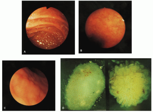
Figure 17-6. Endoscopic and dissecting microscopic appearances of the proximal small bowel in celiac sprue. A: Normal mucosal appearance with valvulae conniventes. B: Appearance in celiac sprue; valvulae are absent. C: Scalloped, ridged appearance of the mucosa on a close-up view. D: Dissecting microscopic appearance of a normal biopsy specimen (right) and a flat biopsy specimen (left). (Courtesy of Cyrus E. Rubin.)

In a biopsy consistent with celiac disease, surface epithelial cells are often but not invariably cuboidal, demonstrate enhanced basophilia, and characteristically contain numerous intraepithelial lymphocytes (Fig. 17-7C,D). The intraepithelial lymphocytes appear to be the hallmark of the mucosal immune response to gluten.63 There may be vacuoles within the surface epithelial cells. These represent trapped fat (also known as lipid hang-up), which cannot be “exported” to the lacteals due to impaired lipoprotein synthesis in the diseased enterocytes. This results in failure to produce chylomicrons, which are a prerequisite for fat transport into the lymphatic system (Fig. 17-7E). Crypts are elongated and contain increased numbers of mitoses, some of which may almost reach the surface epithelium (Fig. 17-7G). In the uncommon circumstance when frank megaloblastic anemia is present, epithelial cell macrocytosis may also be seen. There is a marked increase in the number of lamina propria round cells, predominantly plasma cells. In addition, scattered increased numbers of lamina propria eosinophils, free neutrophils, and mast cells64, 65) may be observed. Much more rarely seen are crypt abscesses and sparsely distributed neutrophils in the surface epithelium, but they disappear quickly following gluten withdrawal (Fig. 17-7H). The presence of more neutrophils than lymphocytes in the epithelium should make one
reconsider the diagnosis of celiac disease and worry more about either Crohn’s disease, peptic duodenitis, autoimmune enteropathy (AIE), or celiac disease, and a second disease that may be associated (e.g. Crohn’s disease) or unrelated (e.g. NSAID ingestion). Paneth cell numbers may appear reduced. It was once claimed that this might portend a suboptimal response to therapy, but this has not been confirmed. In our experience, a marked reduction in Paneth cells is rare, they may be reduced in severe lesions,66 but this does not indicate that the mucosal response will be refractory to gluten withdrawal. A complete lack of Paneth or goblet cells or both should raise the possibility of AIE. There are also reports that the numbers of certain GI endocrine cells are increased, although the clinical implications of these findings are unclear.67, 68, 69, 70


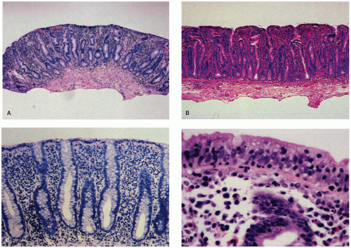
Figure 17-7. Untreated celiac sprue. Small bowel biopsy specimens showing a nonspecific severe, “flat” mucosal lesion. A,B: Lowpower views. B has an undulating surface but, at low power, is still appreciated as being flat, albeit not as flat as A. C: Medium-power magnification of superficial mucosa showing replacement of normal columnar lining epithelium by cuboidal epithelial cells. Note also the dense lymphoplasmacytic infiltrate of the lamina propria. D: High-power magnification of surface epithelium shows nuclear stratification and many small, dark-staining intraepithelial lymphocytes.

Striking abnormalities have also been demonstrated in the absorptive cells of patients with celiac sprue by histochemistry and electron microscopy. For example, histochemically there is a marked reduction in dipeptidase, disaccharidase, enterokinase, and oxidoreductase activity. Additionally, enzymes related to microsomes, mitochondria, and lysosomes are also greatly reduced.71, 72, 73 Ultrastructurally, the microvilli of the columnar cells are reduced in number, short and stubby, and often fused. The cytoplasm often shows degenerative changes such as cytoplasmic and mitochondrial vacuolization and many large lysosomes. In addition, there are structural abnormalities of tight junctions between absorptive cells. This condition probably produces disruption of the barrier separating the luminal contents from the paracellular spaces, resulting in greater permeability.74

Only gold members can continue reading. Log In or Register to continue

Stay updated, free articles. Join our Telegram channel

Jun 8, 2016 | Posted by in PATHOLOGY & LABORATORY MEDICINE | Comments Off on Small Bowel Mucosal Disease

Full access? Get Clinical Tree

Get Clinical Tree app for offline access