Skeletal Muscle
Staci Bryson, MD
Larissa V. Furtado, MD
Key Facts
Embryology
Skeletal muscle derives from mesoderm
Pharyngeal arches give rise to muscles of head
Paraxial mesoderm gives rise to muscles of trunk
Lateral plate mesoderm gives rise to visceral and limb muscles
Paraxial and lateral plate mesoderm are formed by cells migrating laterally from the primitive streak in week 3 of gestation
Paraxial mesoderm develops into somitomeres and then somites during week 3
Somites form from cranial to caudal at rate of 3-4 per day up until day 30 of gestation
Somites differentiate into myotomes (and sclerotomes and dermatomes) during week 4 of gestation
Myotomes further subdivide into dorsal and ventral parts, which are separately innervated by spinal nerves (dorsal and ventral primary rami)
Neck and trunk muscles contract spontaneously by week 7
Embryo can respond to skin stimulation, and some postural reflexes are present by week 12
Muscle development progresses to the point where fetal movements are felt by mother by weeks 16-20
Macroscopic Anatomy
Muscle comprises 25% of body weight at term
Individual muscles vary greatly in size and shape
Individual muscles are composed of varying numbers of muscle fibers, up to 1 million in gastrocnemius
Muscles are connected at both ends by tendons or epimysium
Blood supply to individual skeletal muscles is not well delineated, and arterial supply can vary from person to person
Most skeletal muscles receive blood from multiple arteries, making skeletal muscle relatively resistant to ischemia
Gross color of muscle generally progresses from pink to red over course of fetal life due to accumulation of myoglobin and increasing vascular supply
Microscopic Anatomy
Earliest cells with distinct muscle differentiation are myoblasts, containing desmin
Myoblasts are spindle-shaped cells with finely granular cytoplasm, oval nuclei, and prominent nucleoli
Myoblasts begin to form primary myotubes, precursors to muscle fibers, by week 7
Primary myotubes are characterized by longitudinally arranged nuclei
Cytoplasm is eosinophilic due to presence of mitochondria, glycogen, and myofibrils; cross-striations are not yet present
Myofibrils accumulate in myoblasts even as myotube formation continues
Myofibrils are composed of thin actin and thick myosin filaments
Secondary myotubes form during early fetal period by fusion of myoblasts, progress toward multinucleated myocytes
Secondary myotubes mature into myofibers, recognizable at midpoint of 2nd trimester
Myofibers are marked by development of cross-striations and progression to peripheral nuclei
Early muscle fibers are undifferentiated as far as type 1 vs. type 2
Differentiation to type 1 fibers occurs from 16-20 weeks gestation
Differentiation of type 2 fibers occurs later with type 2b fibers appearing at ˜ 25 weeks and type 2a at ˜ 35 weeks
Most muscle fibers have differentiated by term
Age Variation
Myoblasts continue to divide and myotube formation can continue up until ˜ 30 weeks gestation
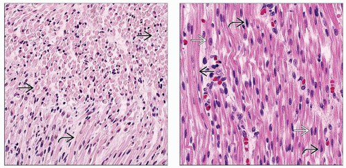 (Left) At 16 weeks gestation, the muscle is a mix of myotubes
 , identifiable in cross section as cells with a central clear zone, and early muscle fibers , identifiable in cross section as cells with a central clear zone, and early muscle fibers  . (Right) At 19 weeks, secondary myotubes have begun to mature into myofibers. Note the elongated shape of the fibers . (Right) At 19 weeks, secondary myotubes have begun to mature into myofibers. Note the elongated shape of the fibers  , with some having central nuclei , with some having central nuclei  and others peripheral nuclei and others peripheral nuclei  . .Stay updated, free articles. Join our Telegram channel
Full access? Get Clinical Tree
 Get Clinical Tree app for offline access

|