Sinonasal Hamartoma
Bruce M. Wenig, MD
Key Facts
Terminology
READ hamartoma: Benign acquired nonneoplastic overgrowth of indigenous glands of sinonasal tract and nasopharynx
CORE hamartoma: Related to READ hamartoma but has additional feature of chondroid tissue
NCH: Tumefactive process of sinonasal tract comprised of admixture of chondroid and stromal elements with cystic features analogous to chest wall hamartoma
Clinical Issues
Conservative, but complete surgical excision treatment of choice
Cured following surgical resection
Microscopic Pathology
READ hamartoma
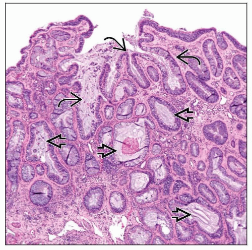
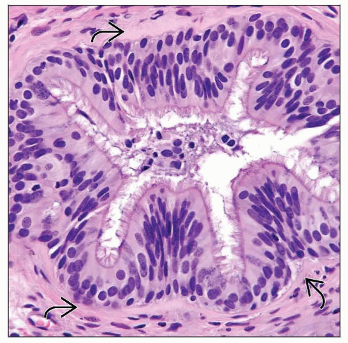
Changes dominated by presence of glandular proliferation enveloped by stromal hyalinization
CORE hamartoma
Similar features to READ hamartomas but with intimate admixture of cartilaginous &/or osseous trabeculae
NCH
Characterized by presence of nodules of cartilage varying in size, shape, and contour with varying differentiation
Chondromesenchymal elements are relatively cellular and “immature”
TERMINOLOGY
Abbreviations
Respiratory epithelial adenomatoid (READ) hamartoma
Chondro-osseous and respiratory epithelial (CORE) hamartomas
Nasal chondromesenchymal hamartoma (NCH)
Synonyms
Glandular hamartoma
Seromucinous hamartoma
Nasal hamartoma
Definitions
READ hamartoma: Benign acquired nonneoplastic overgrowth of indigenous glands of sinonasal tract and nasopharynx
Arising from surface epithelium
Devoid of ectodermal, neuroectodermal, &/or mesodermal elements
CORE hamartoma
Related to READ hamartoma but has additional feature of chondroid tissue
NCH: Tumefactive process of sinonasal tract
Composed of admixture of chondroid and stromal elements with cystic features analogous to chest wall hamartoma
Histologic similarities to READ and CORE hamartomas; may be within spectrum of same lesion type
ETIOLOGY/PATHOGENESIS
Idiopathic
No association with any specific etiologic agent
Developmental
READ hamartomas arise in setting of inflammatory polyps
Raises possible developmental induction secondary to inflammatory process
CLINICAL ISSUES
Epidemiology
Incidence
Rare lesions
Age
READ hamartoma
Occurs in adult patients
Range from 3rd to 9th decades
Reported median in 6th decade
CORE hamartoma
Ranges from 11-73 years
NCH
Most of these lesions occur in newborns within 1st 3 months of life but may occur in 2nd decade of life
Occasionally in adults
Gender
READ hamartoma
Equal gender distribution
CORE hamartoma
Equal gender distribution
NCH
Male > Female
Site
Majority occur in nasal cavity, particularly posterior nasal septum
Involvement of other intranasal sites occurs less often and may be identified
Along lateral nasal wall, middle meatus, and inferior turbinate
Other sites of involvement include nasopharynx, ethmoid sinus, and frontal sinus
Majority of lesions are unilateral, but occasionally bilateral lesions may occur
Presentation
READ hamartoma
Clinical presentation may include one or more of the following symptoms
Nasal obstruction, nasal stuffiness, epistaxis, and chronic (recurrent) rhinosinusitis; associated complaints include allergies
Symptoms may occur over months to years
Nondestructive lesion
CORE hamartoma
Polypoid mass lesion
NCH
Respiratory difficulty and intranasal mass or facial swelling may be present
May erode into cranial cavity (through cribriform plate area), clinically simulating appearance of meningoencephalocele
Treatment
Surgical approaches
Stay updated, free articles. Join our Telegram channel

Full access? Get Clinical Tree


