Sentinel Lymph Node Biopsy for Breast Cancer
Anees B. Chagpar
DEFINITION
Sentinel node biopsy is a minimally invasive means to accurately stage the axilla in breast cancer patients.
PATIENT HISTORY AND PHYSICAL FINDINGS
As always, a complete history and physical exam is warranted. If the patient has obvious clinically enlarged axillary lymph nodes on physical exam, ultrasound, and/or fine needle aspiration (FNA) or core needle biopsy may provide diagnostic information. If the biopsied node is positive, one could proceed to neoadjuvant chemotherapy and/or axillary dissection if primary surgery is planned. Alternatively, if the biopsied node is negative, sentinel node biopsy is still indicated for definitive evaluation.
IMAGING AND OTHER DIAGNOSTIC STUDIES
Lymphoscintigraphy is commonly performed in conjunction with sentinel lymph node (SLN) biopsy for breast cancer, although it is not absolutely necessary.1 Depending on how the radionucleotide tracer is injected, lymphoscintigraphy may show drainage to the internal mammary nodes (see Part 5, Chapter 9). If patients have had previous sentinel node biopsy and/or axillary node dissection, repeat sentinel node biopsy may be considered for staging ipsilateral recurrent or new primary disease. In this circumstance, alternative drainage patterns are possible, and therefore, preoperative lymphoscintigraphy may be useful.2
SURGICAL MANAGEMENT
Sentinel node biopsy is indicated for staging of patients with invasive breast cancer or those with ductal carcinoma in situ undergoing mastectomy.
Preoperative Planning
Timing of sentinel node biopsy vis-à-vis neoadjuvant chemotherapy is controversial; some opt to perform this procedure prior to initiation of neoadjuvant chemotherapy so as to most accurately stage the axilla, whereas others will do a sentinel node biopsy after completion of neoadjuvant chemotherapy so as to potentially spare patients who have had a pathologic complete response to the morbidity of a complete axillary dissection.
Positioning
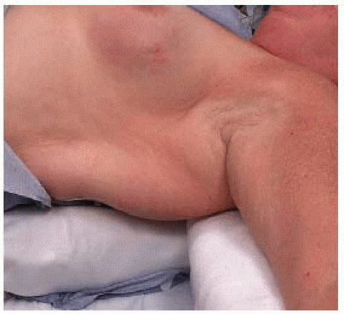
Patients are positioned supine. A roll may be placed under the ipsilateral shoulder so as to elevate the latissimus dorsi muscle. Care should be exercised to ensure that the arm is supported on folded sheets so as to avoid a brachial plexus stretch injury (FIG 1).
Intravenous lines, pulse oximeter devices, and blood pressure cuffs should be placed on other extremities if possible.
TECHNIQUES
INJECTION OF RADIOACTIVE TRACER AND/OR BLUE DYE
Use of dual tracer has been shown to be associated with higher identification and lower false-negative rates3; however, particularly in patients undergoing skin-sparing mastectomy, surgeons may wish to forego blue dye, as the dye may discolor the skin and make it more difficult to evaluate for skin ischemia or necrosis.
In general, the radioactive tracer used is technetium Tc 99m sulfur colloid. For patients not undergoing lymphoscintigraphy, the tracer can be injected in the preoperative holding area. However, injection of this material is painful, and adequate counts can often be obtained with intradermal injection after induction of anesthesia. The radiotracer can be injected peritumoral, intradermal (in the skin over the location of the cancer), or periareolar (four intradermal injections around the areola). If injection is done on the same day as the operative procedure, a dose of 0.5 mCi is adequate. If injection is done the day prior to surgery, 2.5 mCi should be used.
Both isosulfan and methylene blue dye have been used for SLN biopsy; they vary in their color, complication profile, and cost. Isosulfan blue is a more azure blue (which is easier to distinguish from venous structures) but is associated with less than 1% risk of anaphylaxis4 and is significantly more costly than methylene blue. Methylene blue is darker, associated with a higher rate of skin necrosis, but is far cheaper.5
In general, 5 mL of these tracers are used; if methylene blue is chosen, it should be diluted 1:2 with saline.
Stay updated, free articles. Join our Telegram channel

Full access? Get Clinical Tree


