Secondary Syphilis
Gonzalo De Toro, MD
Key Facts
Terminology
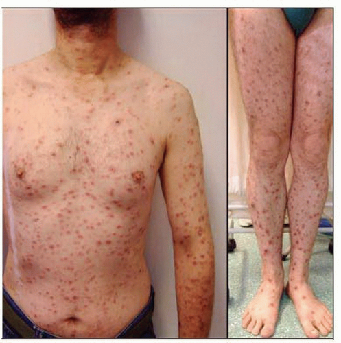
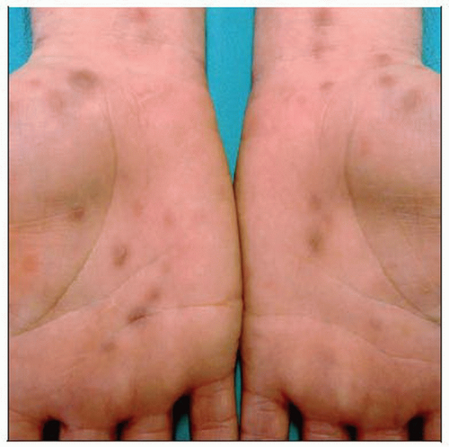
SS occurs 4-8 weeks after primary chancre, usually as maculopapular, scaly patches or plaques characteristically involving palms and soles, extremities, trunk, and elsewhere
Microscopic Pathology
Lesions of secondary syphilis have great histologic variability (considered the “great simulator”)
Perivascular lymphocytes are often mixed with plasma cells and sometimes neutrophils
Top Differential Diagnoses
Lichen planus
Psoriasis
Pityriasis lichenoides
Psoriasiform drug reactions
Lichenoid hypersensitivity reaction
 This HIV-positive patient shows a widespread maculopapular eruption indicative of secondary syphilis. (Courtesy G. Strauch, MD.) |
TERMINOLOGY
Abbreviations
Secondary syphilis (SS)
Synonyms
Lues
Definitions
Occurs 4-8 weeks after primary chancre, usually as maculopapular, scaly patches or plaques characteristically involving palms and soles, extremities, trunk, and elsewhere
ETIOLOGY/PATHOGENESIS
Infectious Agents
Results from hematogenous and lymphatic dissemination of Treponema pallidum and multiplication of microorganisms in different tissues
Occurs after untreated primary syphilis
Circulating immune complexes (which contain treponemal outer membrane proteins), human fibronectin, antibodies, and complement play a role in pathogenesis of lesions
CLINICAL ISSUES
Presentation
Broad spectrum of clinical manifestations involving the skin as well as other tissues
Usually presents as maculopapular, scaly patches or plaques involving palms, soles, extremities, trunk
Early (10%)
Generalized eruption, nonpruritic, roseola-like, discrete macules, initially distributed on flanks and shoulders
Stay updated, free articles. Join our Telegram channel

Full access? Get Clinical Tree


