Sebaceoma (Sebaceous Epithelioma)
David Cassarino, MD, PhD
Key Facts
Terminology
Sebaceous epithelioma is older term (discouraged)
Benign proliferation of mature sebaceous cells with predominant basaloid cell population
Etiology/Pathogenesis
Some cases are part of Muir-Torre syndrome
Autosomal dominant disease due to mutations in mismatch repair genes MLH1, MSH2, MSH6
Multiple sebaceous tumors including sebaceous adenomas > carcinomas and sebaceomas
Clinical Issues
Rare tumors; typically occur in adults
Most common on the face
Usually single but may be multiple, especially in MTS patients
Microscopic Pathology
Nodular, dermal-based adnexal tumor
Well-circumscribed, noninfiltrative appearing
Composed mostly of basaloid cells
Minor population of clear cells with multivacuolated cytoplasm, consistent with mature sebaceous cells
Show nuclear hyperchromasia with indentations
Basaloid cells may show mild cytologic atypia and increased mitotic figures
Sebaceous cells do not show significant atypia or mitotic activity
Top Differential Diagnoses
Basal cell carcinoma with sebaceous differentiation, sebaceous carcinoma, sebaceous adenoma, trichoblastoma with sebaceous differentiation
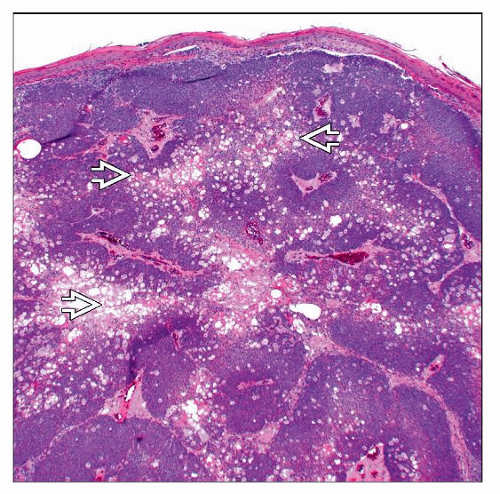 Low magnification of a sebaceoma shows a dermal-based basaloid to clear cell neoplasm with peripheral basaloid cells surrounding central collections of clear cells  and sebaceous secretions. and sebaceous secretions. |
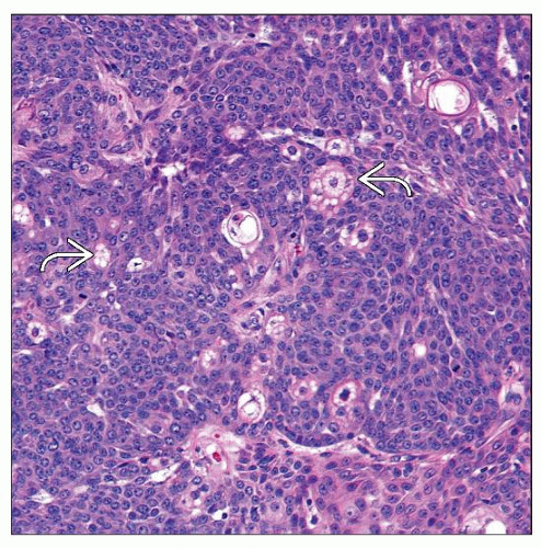 High magnification of a sebaceoma shows a predominantly basaloid population of cells surrounding several large clear cells  with abundant, multivacuolated cytoplasm. with abundant, multivacuolated cytoplasm. |
TERMINOLOGY
Synonyms
Sebaceous epithelioma (older term, should be discouraged)
Definitions
Benign proliferation of mature sebaceous cells associated with a predominant basaloid cell population
ETIOLOGY/PATHOGENESIS
Unknown in Most Cases
Some cases are part of Muir-Torre syndrome (MTS)
Autosomal dominant disease due to mutations in mismatch repair genes MLH1, MSH2, MSH6
Patients present with multiple sebaceous tumors including sebaceous adenomas, sebaceomas, and sebaceous carcinomas
Also associated with internal malignancies including gastrointestinal carcinomas (most common), genitourinary, breast, and ovarian tumors
Rare cases arise in nevus sebaceous of Jadassohn
CLINICAL ISSUES
Epidemiology
Incidence
Rare tumors
Age
Typically occur in adults
Site
Often occur on face, but may also present on trunk
Presentation
Slow-growing papular to nodular lesion
Usually single, but may be multiple, especially in MTS
Flesh-colored to yellowish
Treatment
Surgical approaches
Complete conservative excision is curative
Prognosis
Excellent; very low malignant potential
Possible transformation to sebaceous carcinoma in longstanding lesions
MACROSCOPIC FEATURES
Size
Usually measure between 0.5-3 cm in diameter
MICROSCOPIC PATHOLOGY
Histologic Features
Nodular, dermal-based adnexal tumor
Usually located in mid to upper dermis
Well-circumscribed, noninfiltrative appearing
May show association with hair follicle in some cases
Epidermal attachments in rare cases
Cysts and (sebaceous) ductal structures often present
Rare apocrine differentiation has been reported
Composed mostly of immature-appearing basaloid cells
Basaloid cells may show mild cytologic atypia and increased mitotic figures
Atypical mitoses should be absent
Minority of tumor consists of clear cells with multivacuolated cytoplasm, consistent with mature sebaceous cells
Show nuclear hyperchromasia with indentations, typical of sebocytes
Sebaceous cells do not show significant atypia or mitotic activity
Rarely, overlying changes similar to verruca vulgaris or seborrheic keratosis may be seen
Stay updated, free articles. Join our Telegram channel

Full access? Get Clinical Tree


