Sarcomas
Primary sarcomas of the lung are very rare. Most sarcomas in the lung are metastases from other primary sites. The histopathologic features of sarcomas in the lung are generally the same whether they are rare primary tumors or metastases from other sites.
Part 1 Sarcoma of the Pulmonary Artery and Vein
Mojghan Amrikachi
Timothy C. Allen
Sarcomas of the pulmonary artery and vein are rare primary pulmonary sarcomas, generally occurring in adults in the fourth to sixth decades of life. Grossly, sarcomas of the pulmonary artery and vein form fleshy, occasionally polypoid, masses, distending the pulmonary trunk or pulmonary artery by gray tumor mixed with thrombus. The tumor may extend to the pulmonary valve and right ventricle. Extravascular extension of the tumor from the vessel lumen or wall may occur. Metastases, especially to the distal lung, may occur.
Histologic Features
Hemorrhagic lesion with a uniform spindle-cell proliferation within the vessel lumen and tumor cells infiltrating vascular intima and media.
Some tumors may demonstrate marked pleomorphism with occasional giant cells (undifferentiated intimal sarcoma).
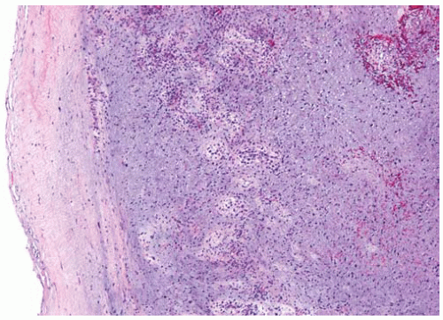 Figure 11.2 Artery with a spindle-cell proliferation filling the lumen and infiltrating the vessel wall. |
Part 2 Angiosarcoma
Mojghan Amrikachi
Timothy C. Allen
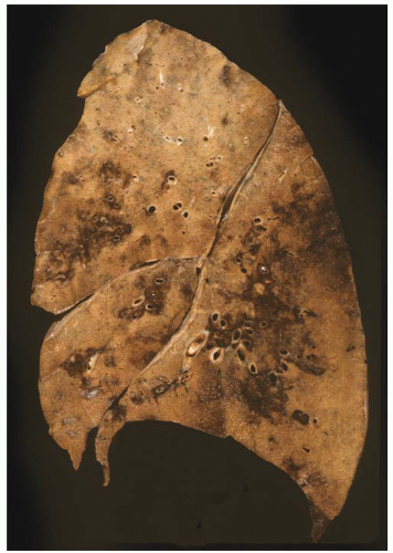
Angiosarcomas generally occur in older adults. Angiosarcomas in the lung are generally metastatic tumors; however, primary pulmonary angiosarcomas may very rarely occur. Angiosarcomas in the lung often present with hemoptysis and commonly appear grossly as ill-defined hemorrhagic areas. The histologic features of primary pulmonary angiosarcomas are similar to angiosarcomas arising from other sites.
Histologic Features
Malignant tumors with endothelial differentiation.
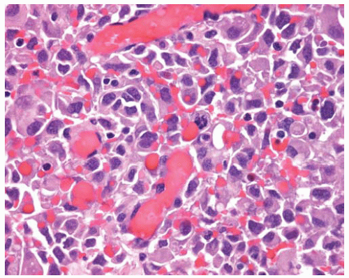
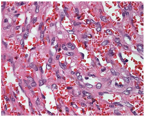
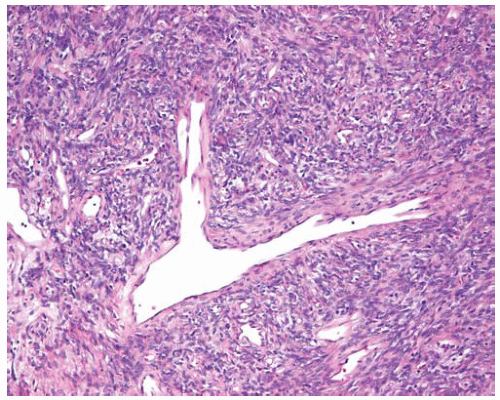
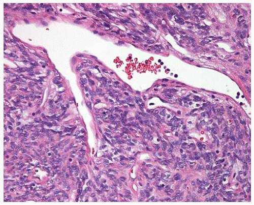
Well to moderately differentiated angiosarcomas show irregular anastomosing vascular spaces lined by large endothelial cells, hyperchromatic nuclei, and papillary tufting along the lumina.
Poorly differentiated angiosarcomas show high-grade pleomorphic spindle cells with rudimentary lumen formation in some areas and prominent mitotic activity.
Ultrastructurally show tight junction between cells, pinocytic vesicles, and Weibel-Palade bodies (in small number of angiosarcomas).
Generally immunopositive for CD34, CD31, and von Willebrand factor (less sensitive in high-grade tumors).
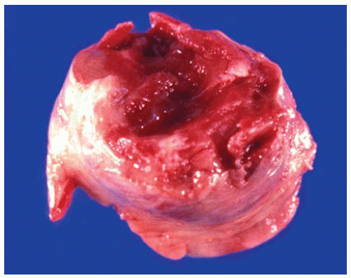
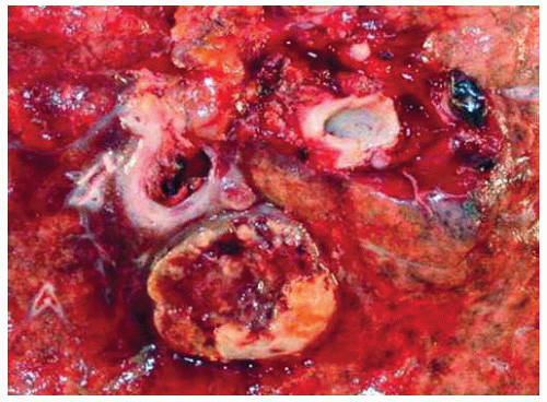 Figure 11.4 Gross figure of angiosarcoma, fleshy tumor containing necrosis and hemorrhage, lying near a large bronchus. |
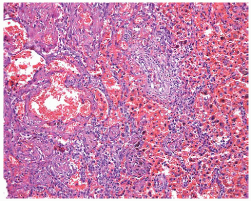 Figure 11.6 Angiosarcoma with irregular anastomosing vascular spaces and blood in airspaces peripheral to the tumor. |
Part 3 Epithelioid Hemangioendothelioma
Mojghan Amrikachi
Mary L. Ostrowski
Alvaro C. Laga
Epithelioid hemangioendothelioma is a vascular tumor typically occurring around medium-sized and large veins in the soft tissue of adults. Although epithelioid hemangioendotheliomas occurring in the lung are most often metastases, rare primary pulmonary epithelioid hemangioendotheliomas occur. Differential diagnosis includes epithelioid angiosarcoma.
Cytologic Features
Large, polygonal eosinophilic cells, often vacuolated, with periodic acid-Schiff (PAS)-positive stroma.
Histologic Features
Usually a low-grade sarcoma with little mitotic activity.
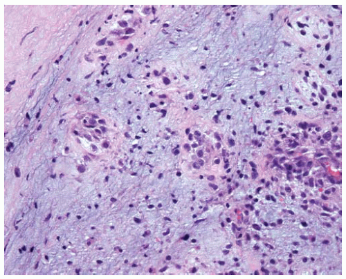
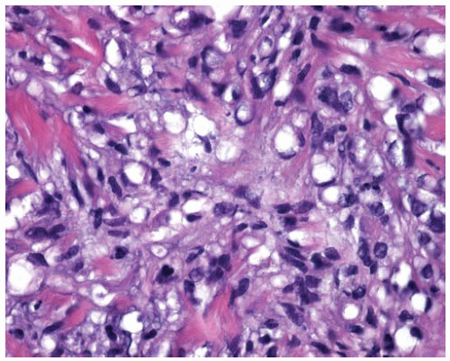
Cords, strands, and nests of cells infiltrating a myxoid or hyalinized stroma.
Round to spindled cells with pale eosinophilic cytoplasm, vesicular nuclei, and inconspicuous nucleoli.
Intracytoplasmic vacuoles with occasional intraluminal erythrocytes occur.
High-grade tumors contain solid sheets of cells with marked nuclear atypia and increased mitotic activity.
Generally immunopositive for CD31 and von Willebrand factor; approximately one-third can be focally immunopositive for cytokeratin.
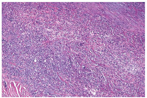 Figure 11.9 Nests, cords, and clusters of round to spindled cells within a myxoid and hyalinized stroma. |
Part 4 Kaposi Sarcoma
Mojghan Amrikachi
Carlos Bedrossian
Abida K. Haque
Timothy C. Allen
Kaposi sarcoma is commonly associated with acquired immunodeficiency syndrome (AIDS) and may arise as a primary pulmonary neoplasm. Grossly, lung lesions generally occur as multiple hemorrhagic lesions. Differential diagnosis includes angiosarcoma, fibrosarcoma, arteriovenous malformation, spindle-cell hemangioendothelioma, inflammatory granulation tissue, and bacillary angiomatosis.
Cytologic Features
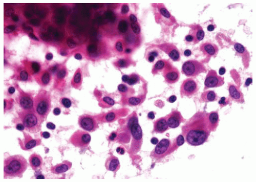
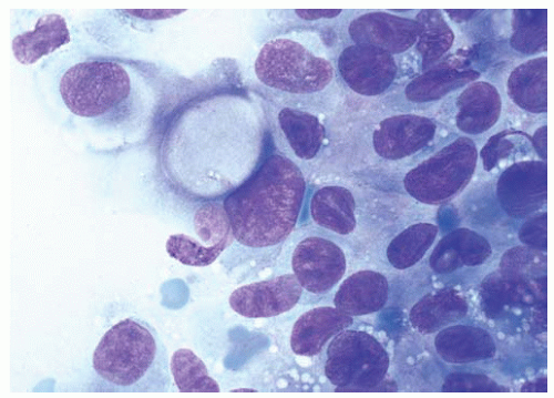
Bland to mildly atypical oval to spindled cells with a background of red blood cells.
Histologic Features
Diffuse infiltrative lesion along lymphatic pathways, interlobular septa, and pleura.
Bland monomorphic spindle cells separated by slit-like vascular spaces.
Spindle cells resemble fibroblasts and form a network of irregular vascular channels, with erythrocytes scattered between spindle cells.
Sparse to abundant plasma cells and lymphocytes may occur.
PAS-positive, diastase-resistant intracellular and extracellular hyaline globules may occur.
Generally immunopositive for CD34, CD31, and HHV-8.
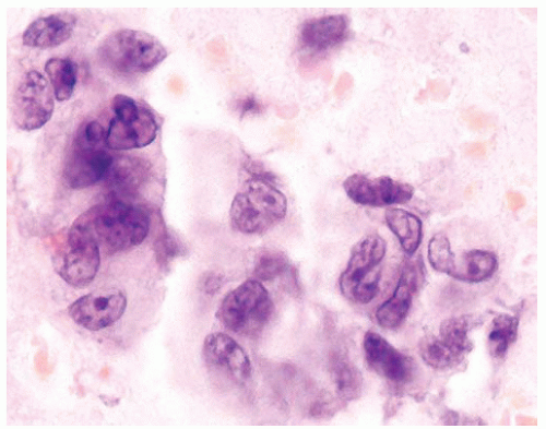 Figure 11.12 Cytology figure showing mildly atypical oval to spindled cells with a background of red blood cells. |
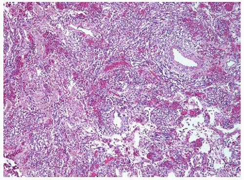 Figure 11.13 Kaposi sarcoma showing nests of spindled cells and surrounding lung parenchyma containing hemorrhage and hemosiderin-laden macrophages. |
Part 5 Hemangiopericytoma
Mojghan Amrikachi
Alvaro C. Laga
Timothy C. Allen
Philip T. Cagle
Most, if not all, neoplasms previously designated as hemangiopericytoma are now considered solitary fibrous tumors. Tumors designated hemangiopericytomas have been described in the lungs as rare primary and metastatic tumors. Tumors diagnosed as hemangiopericytomas in the lung typically appear grossly as well-circumscribed masses with gray-white cut surfaces and areas of cystic degeneration.
Histologic Features
Dilated branching blood vessels with staghorn configuration surrounded by tightly packed round to fusiform small cells.
Faint cytoplasm with indistinct cytoplasmic borders, round to oval nuclei, and rare to absent mitotic figures.
Foci of spindle-cell changes and myxoid changes can occur.
Generally immunopositive for CD34, and immunonegative for CD31, actin, desmin, and CD99.
Part 6 Undifferentiated Sarcomas
Mojghan Amrikachi
Carlos Bedrossian
Alvaro C. Laga
Philip T. Cagle
Sarcomas without differentiating features, including those formerly referred to as “malignant fibrous histiocytomas,” are generally metastatic in the lung and very rarely occur as primary lung tumors. Generally sarcomas in the lung form gray-white masses, commonly with areas of necrosis.
Cytologic Features
Mixture of malignant spindle cells, and occasional giant cells, with areas of myxoid stromal tissue.
Histologic Features
Spindle cells in short fascicles or storiform pattern, with bizarre pleomorphic spindled cells including multinucleated cells.
Some may have myxoid stroma.
Abnormal mitotic activity is common.
Stay updated, free articles. Join our Telegram channel

Full access? Get Clinical Tree


