Rosai-Dorfman Disease
Rose Anton, MD
John M. Stewart, MD, PhD
Key Facts
Clinical Issues
Usually asymptomatic adenopathy in young patients
Spontaneous regression occurs in most patients
No specific therapy is required
Cytopathology
RDD histiocytes
Large size with abundant eosinophilic cytoplasm
Defined cell border
Emperipolesis is usual
Round vesicular nucleus
Distinct central nucleolus
Ancillary Tests
Immunohistochemistry
S100(+), CD1a(-)
Top Differential Diagnoses
Langerhans cell histiocytosis
Chronic granulomatous inflammation
Kikuchi lymphadenitis
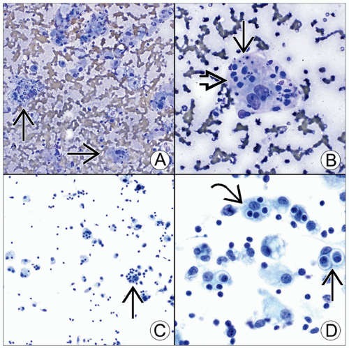 (A) Emperipolesis
 is subtle in this Diff-Quik-stained smear and can easily be dismissed as just tingible body macrophages due to inflammation/repair. (B) Diff-Quik-stained smear shows an RDD histiocyte containing lymphocytes is subtle in this Diff-Quik-stained smear and can easily be dismissed as just tingible body macrophages due to inflammation/repair. (B) Diff-Quik-stained smear shows an RDD histiocyte containing lymphocytes  and sparse tingible debris and sparse tingible debris  . (C) Pap-stained smear shows RDD histiocytes with variable numbers of lymphocytes. The eye is quickly drawn to cells with significant emperipolesis . (C) Pap-stained smear shows RDD histiocytes with variable numbers of lymphocytes. The eye is quickly drawn to cells with significant emperipolesis  . (D) Pap-stained smear shows a high-power view of emperipolesis. Note that the lymphocytes . (D) Pap-stained smear shows a high-power view of emperipolesis. Note that the lymphocytes  and plasma cells and plasma cells  appear viable and enclosed in a defined space within the cytoplasm of the RDD histiocyte. appear viable and enclosed in a defined space within the cytoplasm of the RDD histiocyte.Stay updated, free articles. Join our Telegram channel
Full access? Get Clinical Tree
 Get Clinical Tree app for offline access

|