Right and Left Pneumonectomy
M. Victoria Gerken
Phillip C. Camp
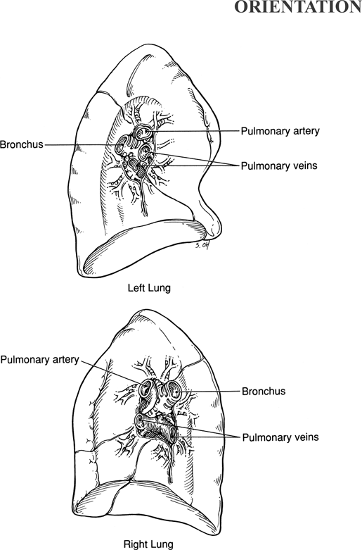
Pneumonectomy is most commonly performed for carcinoma of the lung or for removal of trapped and necrotic lung after cavitary diseases. In this chapter, the operations of right and left pneumonectomy are described and the hilar anatomy of the right and left lung is illustrated.
Steps in Procedure
Posterolateral thoracotomy incision, 4 or 5 intercostal space
Explore and determine extent of disease
Retract lung inferiorly and dissect pleura inferior to azygos vein (right pneumonectomy) or along superior hilum (left pneumonectomy)
Identify and mobilize main pulmonary artery; secure and divide it (suture ligature or vascular stapler)
Divide pleura as it reflects on the lung at the anterior surface of the hilum
Dissect and divide superior pulmonary vein (suture ligature or vascular stapler)
Retract lung anteriorly and superiorly
Identify and divide inferior pulmonary ligament to level of inferior pulmonary vein
Secure and divide the inferior pulmonary vein
Incise pleural reflection interiorly and posteriorly to reveal bronchus
Divide bronchus with stapler
Cover bronchial stump with pleura
Close chest without chest tubes
Hallmark Anatomic Complications
Bronchial stump leak (devascularization)
Injury to phrenic nerve
List of Structures
Mediastinum
Azygos vein
Hemiazygos vein
Accessory hemiazygos vein
Superior vena cava
Phrenic nerve
Pericardiophrenic artery
Vagus nerve
Recurrent laryngeal nerve
Esophagus
Aorta
Pericardium
Right Lung
Right pulmonary artery
Right main-stem bronchus
Right superior pulmonary vein
Right inferior pulmonary vein
Bronchial arteries
Right bronchial vein
Left Lung
Inferior pulmonary ligament
Left pulmonary artery
Left superior pulmonary vein
Left inferior pulmonary vein
Left main-stem bronchus
Right Pneumonectomy
Exposure of the Hilum and Division of the Pulmonary Artery (Fig. 26.1)
Technical Points
Enter the chest in the fourth or fifth intercostal space using a standard posterolateral thoracotomy incision. Examine the mediastinum and hilum to confirm that the diseased area does not extend into the mediastinum, chest wall, or apex and is thus resectable. Retract the lung inferiorly to reveal the superior hilum. Inferior to the azygos vein, dissect the pleura carefully at the apex with Metzenbaum scissors or electrocautery.
Identify and mobilize the main pulmonary artery by careful blunt dissection with a “peanut” dissector. Pass a large
right-angled clamp carefully around the artery in preparation for double ligation. For security, first tie the proximal pulmonary artery with heavy silk (usually number 1). Place a transfixion suture ligature (usually one size smaller than the freehand tie) just distal to the freehand tie. Control the distal end of the artery (specimen side) with a freehand tie and divide the pulmonary artery. Alternatively, a linear stapler with vascular staples is an expedient way to secure the proximal side of this large, fragile vessel.
right-angled clamp carefully around the artery in preparation for double ligation. For security, first tie the proximal pulmonary artery with heavy silk (usually number 1). Place a transfixion suture ligature (usually one size smaller than the freehand tie) just distal to the freehand tie. Control the distal end of the artery (specimen side) with a freehand tie and divide the pulmonary artery. Alternatively, a linear stapler with vascular staples is an expedient way to secure the proximal side of this large, fragile vessel.
 |
Anatomic Points
Review the location of mediastinal structures and the relationships of major structures in the root of the lung before surgery. Mediastinal structures of concern include the azygos vein, superior vena cava, phrenic and vagus nerves, and esophagus. The unpaired azygos vein provides a reliable landmark, for the superior aspect of the right hilum. This vein, lying on the side of the thoracic vertebral bodies, drains the right intercostal spaces and receives the termination of the hemiazygos vein on the left, then arches anteriorly to enter the superior vena cava immediately superior to the hilum of the lung. The right bronchial vein, which drains the lung parenchyma, also empties into the azygos vein. Division of the azygos vein, if necessary, is permissible owing to the abundant collateral venous return of the chest wall.
The right pulmonary artery lies immediately anterior to the right main-stem bronchus and is the first hilar structure to be encountered as dissection proceeds from above downward. The superior vena cava, just inferior to the termination of the azygos vein, is still extrapericardial. It is immediately anterior to the right pulmonary artery.
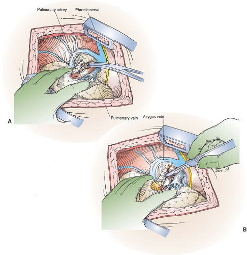 Figure 26-1 Exposure of the Hilum and Division of the Pulmonary Artery
Stay updated, free articles. Join our Telegram channel
Full access? Get Clinical Tree
 Get Clinical Tree app for offline access

|