Rare Benign and Low Malignant Potential Tumors
Michael J. Thrall, MD
Key Facts
Clinical Issues
Hamartoma results from overgrowth of mature mesenchymal elements
Sclerosing hemangioma arises from pneumocytes though lesions resemble vascular tumors
Solitary fibrous tumor usually arises from pleura but may arise elsewhere and is not always single
Clear cell (sugar) tumor arises from perivascular epithelioid cells (PEComa)
Cytopathology
Hamartoma consists of mixed benign stromal and glandular elements
Sclerosing hemangioma neoplastic pneumocytes form loose clusters surrounding stromal cores
Solitary fibrous tumor features bland spindle cells with collagen in background
Clear cell (sugar) tumor consists of bland cells with abundant finely vacuolated glycogen-rich cytoplasm
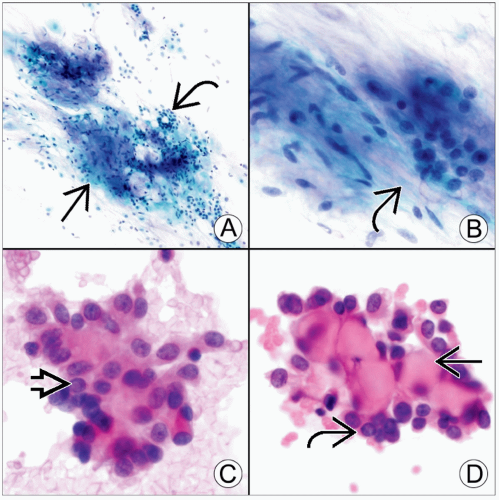 (A) Aspiration of a pulmonary hamartoma will yield mixed benign mesenchymal
 and epithelial and epithelial  elements, as seen in this Pap stain. (B) A closer view from the same case highlights the fibrillary appearance of the cartilaginous matrix elements, as seen in this Pap stain. (B) A closer view from the same case highlights the fibrillary appearance of the cartilaginous matrix  . (C) The surface cells of sclerosing hemangioma form loose clusters with fine chromatin, seen here in an H&E touch preparation. Nuclear contour irregularities, including pseudoinclusions . (C) The surface cells of sclerosing hemangioma form loose clusters with fine chromatin, seen here in an H&E touch preparation. Nuclear contour irregularities, including pseudoinclusions  , may raise concern for well-differentiated adenocarcinoma. (D) Another fragment from same case shows central collagenous stromal cores , may raise concern for well-differentiated adenocarcinoma. (D) Another fragment from same case shows central collagenous stromal cores  & smaller, rounded stromal cells & smaller, rounded stromal cells  . .Stay updated, free articles. Join our Telegram channel
Full access? Get Clinical Tree
 Get Clinical Tree app for offline access

|