Rare Adenomas
Pulmonary adenomas include alveolar adenomas, papillary adenomas, and mucinous cystadenomas. All are extremely rare neoplasms.
Part 1 Alveolar Adenoma
Alvaro C. Laga
Timothy C. Allen
Philip T. Cagle
Alveolar adenomas are extremely rare benign multicystic neoplasms that histologically resemble alveolar type 2 pneumocytes and mesenchyme. Alveolar adenomas typically present in older women as a solitary peripheral nodule.
Histologic Features
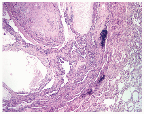
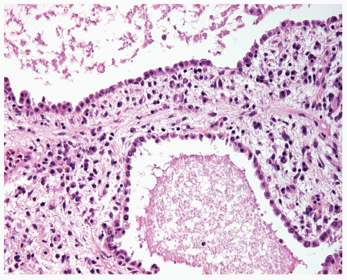
Variably sized cystic spaces lined by hobnail or cuboidal to flattened cells (type 2 pneumocytes) that are generally immunopositive for keratin and TTF-1 and focally immunopositive for CEA.
Cystic spaces tend to be larger in the center of the lesion, and squamous metaplasia may be seen.
Spaces contain eosinophilic material and occasionally scattered macrophages.
The interstitium underlying the lining of type 2 pneumocytes contains a myxoid spindle-cell matrix; the spindle cells are typically immunopositive for smooth-muscle actin and muscle-specific actin.
 Figure 22.1 Alveolar adenoma with cystic spaces surrounded by intervening septa of variable thickness; cystic spaces are larger centrally. |
 Figure 22.2 Higher power showing cuboidal to hobnail cells lining the cystic spaces.
Stay updated, free articles. Join our Telegram channel
Full access? Get Clinical Tree
 Get Clinical Tree app for offline access

|