Radial Artery Cannulation
The radial artery is cannulated for monitoring purposes. A catheter in the radial artery can be used for direct measurement of arterial pressure and for sampling arterial blood for blood gas determinations. It is almost always possible to cannulate the radial artery percutaneously, particularly if Doppler ultrasound guidance is used in difficult cases. Under rare circumstances, a patient with significant vascular disease or shock may require direct cutdown on the artery, with subsequent introduction of the catheter under direct vision. Both procedures are described in this chapter.
Steps in Procedure
Confirm Patent Palmar Arch by Allen Test
Ask patient to clench fist
Occlude both radial and ulnar arteries by direct pressure
Have patient open hand, which should be blanched
Release ulnar artery—hand should become pink within 3 seconds
Alternatively, use Doppler ultrasound
Secure hand on armboard with wrist slightly cocked
Palpate radial artery
Inject lidocaine around artery
Introduce catheter at approximately 45-degree angle
If Cutdown is Necessary:
Transverse incision over radial artery
Isolate and elevate radial artery
Cannulate under direct vision
Hallmark Anatomic Complication
Ischemia of digits or hand due to lack of adequate collateral circulation
List of Structures
Radial Artery
Superficial palmar branch of radial artery
Principal artery of the thumb
Radial artery of the index finger
Deep Palmar Arch
Palmar metacarpal arteries
Ulnar Artery
Deep palmar artery
Superficial palmar arch
Common palmar digital arteries
Radius
Radial styloid process
Ulna
Palmaris longus tendon
Brachioradialis tendon
Tendon of the flexor carpi radialis
Tendons of the flexor digitorum superficialis
Tendon of the flexor carpi ulnaris
Median nerve
Ulnar nerve
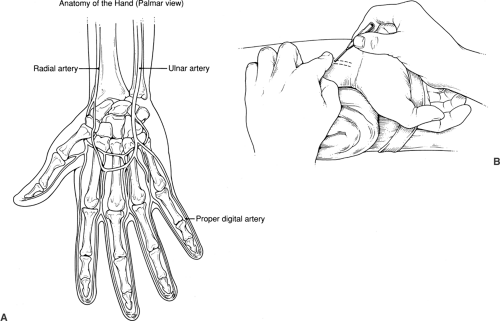
Position of the Extremity and Identification of Landmarks (Fig. 35.1)
Technical Points
Before inserting an indwelling radial artery catheter, perform an Allen test to assess the adequacy of collateral circulation of the ulnar artery across the palmar arch. Because the arch is variable, the adequacy of circulation must be checked in each individual and in each extremity. Instruct the patient to clench the fist tightly. Use both of your hands to occlude both the radial and ulnar arteries. Then have the patient open the fist, which should be blanched. Release pressure on the ulnar artery and note the time required for the hand to become pink. The hand should become pink within 3 seconds after release of
occlusion. Alternatively, a Doppler ultrasound stethoscope may be used as a more objective means of determining the adequacy of circulation. Place the Doppler stethoscope over the palmar arch and do the test as previously described. In this case, use the appearance of Doppler flow in the palmar arch as evidence of collateral flow by the ulnar artery.
occlusion. Alternatively, a Doppler ultrasound stethoscope may be used as a more objective means of determining the adequacy of circulation. Place the Doppler stethoscope over the palmar arch and do the test as previously described. In this case, use the appearance of Doppler flow in the palmar arch as evidence of collateral flow by the ulnar artery.
 Figure 35.1 Position of the Extremity and Identification of Landmarks
Stay updated, free articles. Join our Telegram channel
Full access? Get Clinical Tree
 Get Clinical Tree app for offline access

|