Proliferating Pilar (Tricholemmal) Cyst/Tumor
David Cassarino, MD, PhD
Key Facts
Terminology
Proliferating pilar tumor (PPT), proliferating pilar cyst (PPC), proliferating trichilemmal cyst/tumor
Multicystic squamous neoplasm composed of mature keratinocytes lining keratin-filled spaces
Clinical Issues
Typically occur on scalp (90% of cases)
Most cases of PPT behave in benign fashion, but malignant PPTs are aggressive tumors that have a high rate of metastasis
Microscopic Pathology
Cystic spaces are irregularly formed and anastomosing
Cysts show keratinization without granular layer (unlike PEC)
Large, multicystic, dermal-based tumor with squamous lining and spaces containing dense keratin
Peripheral palisading of basilar layer is typically present, and there may be thickened basement membrane
Occasional mitotic figures are present, but no high-grade atypia or increased mitotic activity should be present
Top Differential Diagnoses
Pilar/tricholemmal cyst
Proliferating epidermoid cyst (PEC)
Malignant PPT (squamous cell carcinoma arising in PPT)
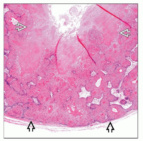 Scanning magnification of PPT shows a smooth-bordered  , circumscribed tumor composed of thickened cords of squamous cells surrounding a large cystic cavity , circumscribed tumor composed of thickened cords of squamous cells surrounding a large cystic cavity  containing abundant keratin material. containing abundant keratin material. |
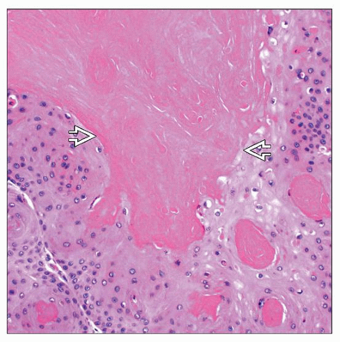 High-magnification examination of a benign PPT shows bland squamous keratinocytes with abundant eosinophilic cytoplasm surrounding keratin-filled spaces. Note the lack of an intervening granular layer  . . |
TERMINOLOGY
Abbreviations
Proliferating pilar tumor (PPT)
Proliferating pilar cyst (PPC)
Synonyms
Proliferating trichilemmal cyst/tumor
Definitions
Multicystic squamous neoplasm composed of mature keratinocytes lining keratin-filled spaces
ETIOLOGY/PATHOGENESIS
Unknown
Postulated that most cases arise in preexisting pilar (tricholemmal) cyst; may be related to chronic inflammation or trauma
CLINICAL ISSUES
Epidemiology
Incidence
Uncommon tumors
Age
Typically occur in older adults
Gender
Much more common in females than males
Site
Typically occur on scalp (90%); also may occur on face, trunk, and extremities
Treatment
Surgical approaches
Complete surgical excision is recommended in order to prevent recurrence and malignant transformation
Prognosis
PPTs behave in benign fashion, but malignant PPTs are aggressive tumors that have a high rate of metastasis
MACROSCOPIC FEATURES
General Features
Often multicystic dermal-based tumors that may involve subcutis
Size
Large tumors, 6 cm or greater in diameter
MICROSCOPIC PATHOLOGY
Histologic Features
Large, multicystic, dermal-based tumor with squamous lining and spaces containing dense keratin
Typically symmetric and well circumscribed at lowpower examination
Cystic spaces are irregularly formed and anastomosing
Stay updated, free articles. Join our Telegram channel

Full access? Get Clinical Tree


