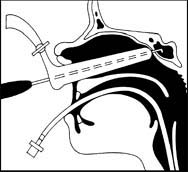
Pituitary tumors
Constituting 10% of intracranial neoplasms, pituitary tumors typically originate in the anterior pituitary (adenohypophysis). They occur in adults of both sexes, usually during the third and fourth decades of life. The three tissue types of pituitary tumors are chromophobe adenoma (90%), basophil adenoma, and eosinophil adenoma.
The prognosis is fair to good, depending on the extent to which the tumor spreads beyond the sella turcica.
Causes
Although the exact cause is unknown, a predisposition to pituitary tumors may be inherited through an autosomal dominant trait. Some are part of a hereditary disorder called multiple endocrine neoplasia 1. Pituitary tumors aren’t malignant in the strict sense; however, because their growth is invasive, they’re considered a neoplastic disease.
Chromophobe adenoma may be associated with the production of corticotropin, melanocyte-stimulating hormone, growth hormone, and prolactin; basophil adenoma, with evidence of excess corticotropin production and, consequently, with signs of Cushing’s syndrome; and eosinophil adenoma, with excessive growth hormone.
Signs and symptoms
As pituitary adenomas grow, they replace normal glandular tissue and enlarge the sella turcica, which houses the pituitary gland. The resulting pressure on adjacent intracranial structures produces typical clinical features.
Neurologic features
Frontal headache
Visual symptoms, beginning with blurring and progressing to field cuts (hemianopias) and then unilateral blindness
Cranial nerve involvement (III, IV, VI) from lateral extension of the tumor, resulting in strabismus; double vision, with compensating head tilting and dizziness; conjugate deviation of gaze; nystagmus; lid ptosis; and limited eye movements
Increased intracranial pressure (secondary hydrocephalus)
Personality changes or dementia, if the tumor breaks through to the frontal lobes
Seizures
Rhinorrhea, if the tumor erodes the base of the skull


