Pilonidal Cystectomy
Whether pilonidal cysts are congenital or acquired lesions remains a topic of debate. The frequent appearance of an epithelialized tract in the midline, leading to a cyst that may or may not contain hair, suggests a congenital origin. A pilonidal cyst (or sinus) characteristically occurs in the posterior midline skin overlying the sacrum. Secondary infection within the cyst or sinus causes pain, drawing attention to the cyst. Incision and drainage may be needed, but are not definitive treatment, and recurrent bouts of infection are typical. Definitive treatment involves either marsupialization or excision of the cyst.
The variety of techniques that are available for dealing with pilonidal cysts indicates that complications have occurred with all approaches. Marsupialization, the simplest procedure, is described in this chapter. A more complex procedure for primary closure, Z-plasty, may be used in some circumstances. Consult the references at the end for alternative approaches.
Steps in Procedure
Prone jackknife position
Tape buttocks open to enhance exposure
Cannulate tract
Inject mixture of 50% Methylene blue and 50% hydrogen peroxide
Marsupialization
Completely open all tracts
Excise overlying skin to saucerize the wound
Curette the base of the cyst to healthy tissue
Suture skin edges to edges of cyst
Pack wound open
Z-plasty
Excise median tract with overlying skin
Include any side tracts—if extensive side tracts are encountered, consider marsupialization
Carry excision down to fascia
Outline top and bottom bars of Z
Elevate flaps at fascial level
Transpose flaps and close skin over a closed-suction drain
Hallmark Anatomic Complications
Recurrence
Delayed wound healing
List of Structures
Sacrum
Lateral sacral crest
Intermediate sacral crest
Median sacral crest
Posterior sacral foramina
Sacral promontory
Ilium
Sacroiliac joint
Coccyx
Lumbodorsal fascia
Gluteus maximus muscle
Gluteus medius muscle
Gluteus minimus muscle
Intergluteal cleft
Gluteal aponeurosis
Sacral nerves
Posterior femoral cutaneous nerve
Gluteal branches
Anus
Rectum
Anococcygeal ligament
Levator ani muscles
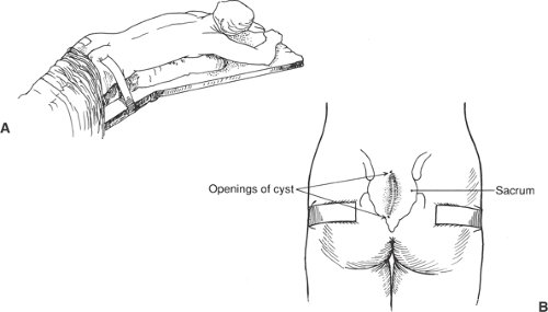 Figure 98-1 Positioning the Patient |
Positioning the Patient (Fig. 98.1)
Technical Points
Place the patient in a prone jackknife position. Use tincture of benzoin on the lateral buttocks to prepare the skin. Place tape on the lateral buttocks and use this tape to pull laterally, spreading the intergluteal cleft. Shave the region of the cyst and the gluteal region.
Anatomic Points
The prominent and important structures in this area are all musculoskeletal. The bony sacrum forms the posterior part of the bony pelvic ring and is the distal continuation of the vertebral column. Formed by the fusion of the five sacral vertebrae (the number of vertebrae that fuse to form the sacrum varies from four to six, but is commonly five), the sacrum is a complexly curved and heavy bone that is shield shaped when viewed from behind. The posterior surface is roughened and has two paramedian crests—the lateral sacral and intermediate crest—which, with the prominent midline median sacral crest, form points of attachment for fascial and aponeurotic structures. Four broad posterior sacral foramina between the five fused vertebrae are points of ingress and egress for the dorsal rami of the sacral spinal nerves. Viewed from the side, a prominent anterior concavity, commonly termed the hollow of the sacrum, is obvious. This forms a space in which lie the rectum, muscles of the pelvic diaphragm, neurovascular structures, and a variable amount of fat. At the top of this concavity, the sacral promontory (located at the point of articulation of the body of the lowest lumbar vertebra with the sacrum) forms an easily palpable bony landmark for the surgeon operating within the pelvis. The sacrum is shorter and wider in the female pelvis than in the male pelvis, contributing to the wider, rounder gynecoid shape that is designed to accommodate the head of a full-term infant at the time of delivery.
The coccyx is composed of three to five remaining vertebrae (commonly, four). These small, nubbin-like, rudimentary vertebrae articulate with the sacrum. Only the first coccygeal vertebra possesses identifiable transverse processes and homologues of pedicles (coccygeal cornua). No vertebral canal is present. The mobility of the coccygeal vertebrae varies considerably from individual to individual, and the terminal three coccygeal vertebrae are commonly fused.
Stay updated, free articles. Join our Telegram channel

Full access? Get Clinical Tree


