Peripheral Nerve Sheath Tumor
Key Facts
Terminology
Peripheral nerve sheath tumor (PNST)
Synonyms: Malignant schwannoma, neurofibrosarcoma, neurogenic sarcoma
Clinical Issues
These tumors are more common in posterior mediastinum but can also occur in anterior mediastinum
Symptoms
Chest pain
Cough
Dyspnea
Neurofibromatosis
Prognosis
These tumors follow aggressive behavior, and prognosis is poor
Worse prognosis in patients with neurofibromatosis
Incidence
Unusual in mediastinal location
May account for approximately 5% of all mediastinal tumors
Top Differential Diagnoses
Neurofibroma
Leiomyosarcoma
Solitary fibrous tumor
Diagnostic Checklist
Solid spindle cellular proliferation
Perivascular hyalinization
Hemorrhage &/or necrosis
Nuclear atypia and mitotic activity
 Mediastinal peripheral nerve sheath tumor shows a solid spindle cell proliferation with a vague herringbone-like pattern of growth. |
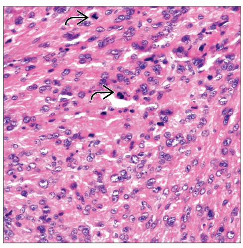 High-power view of a mediastinal peripheral nerve sheath tumor shows more epithelioid areas with cellular pleomorphism, nuclear atypia, and mitotic activity  . . |
TERMINOLOGY
Abbreviations
Peripheral nerve sheath tumor (PNST)
Synonyms
Malignant schwannoma, neurofibrosarcoma, neurogenic sarcoma
Definitions
Malignant tumor of neural sheath origin
ETIOLOGY/PATHOGENESIS
Etiology
These tumors may occur as manifestation of neurofibromatosis (von Recklinghausen disease)
PNST may also occur post radiation
CLINICAL ISSUES
Epidemiology
Incidence
Unusual in mediastinal location
May account for approximately 5% of all mediastinal tumors
Age
More common in adults in 3rd, 4th, and 5th decades of life
Gender
No apparent gender predilection
Patients with neurofibromatosis are commonly male and young adults
Site
More common in posterior mediastinum but can also occur in anterior mediastinum
Presentation
Chest pain
Cough
Dyspnea
Neurofibromatosis
Treatment
Surgical approaches
Complete surgical resection
Adjuvant therapy
Radiation &/or chemotherapy will be determined on individual basis and depending on extent of tumor at time of diagnosis
Prognosis
Tumors follow aggressive behavior, and prognosis is poor
Worse prognosis in patients with neurofibromatosis
MACROSCOPIC FEATURES
General Features
Lobulated large but not encapsulated tumors
Light tan in color
Areas of necrosis and hemorrhage may be seen
Size
Usually > 5 cm in diameter
MICROSCOPIC PATHOLOGY
Histologic Features
DIFFERENTIAL DIAGNOSIS
Neurofibroma
Does not show increase in mitotic activity or cellular pleomorphism
Stay updated, free articles. Join our Telegram channel

Full access? Get Clinical Tree


