Pepsin
History of pepsinogen
Pepsinogen is a potent digestive enzyme secreted by the chief cell of the gastric gland into the lumen of the stomach. This agent is likely to play a significant role in the pathogenesis of peptic ulcer disease. Although abnormalities in this protein have been epidemiologically linked to gastric carcinoma and its precursors, no specific pathogenic role exists for this potent enzyme.
The story of this poorly understood enzyme began in the Berlin laboratory of J. Müller, where, in 1836, Theodor Schwann described a water-soluble factor in gastric juice that digested egg white. He called it pepsin, after the Greek word for digestion.
Schwann had just obtained his MD degree and was continuing as an assistant in Müller’s laboratory. His investigation of gastric glands was initiated by the latter, who had asked him to attempt to subject the physiologic properties of either an organ or a tissue to physical measurement. Schwann initially developed a muscle balance and became the first to establish the basics of the tension-length diagram. Thereafter, while successfully measuring secretion from the gastric gland, he stumbled on a proteolytic enzyme whose properties he characterized and soon thereafter published.
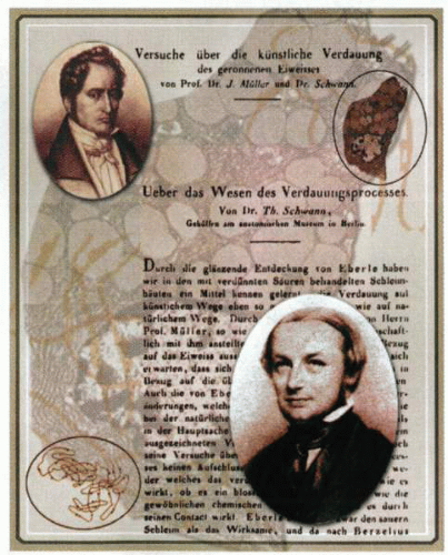 In 1835, Theodor Schwann (bottom right), while working in the laboratory of Johannes Müller (top left), stumbled on a proteolytic enzyme whose properties he characterized and soon thereafter, in 1836, published (right). At the time, they were involved in measuring secretion from the gastric glands (top). Both articles were published sequentially in the same journal; Müller’s work was given precedence. It is of interest that the second reference in Müller’s work on the artificial digestion of proteins is to William Beaumont’s experiments and observations on the gastric juice and physiology of digestion (of 1830). In his studies, Schwann was able to extract a crude preparation of a digestive enzyme from gastric juice, which he demonstrated to have the ability to convert egg-white albumin to peptones in vitro. He named this water-soluble factor pepsin, after the Greek word for digestion. In the course of his studies, both Müller and Schwann noted that no gas evolved during pepsin digestion of food. These findings were thus able to dispel a notion that had been held for 3 centuries: that digestion was a fermentation-like process. The identification of the cell involved in pepsin secretion (top right) would require another half century, whereas the identification of the prolate ellipsoid structure of pepsinogen (the protein precursor of this enzyme), required an additional 100 years. Despite the identification of pepsin and its properties in 1836, neither the role of this factor in gastric pathology (peptic ulceration) nor its extragastric function has been entirely elucidated. |
Unfortunately for the science of the stomach, this observation became lost in the subsequent spate of diverse investigative work that emanated from the young man. He moved on to fermentation and was the first to demonstrate the importance of oxygen both for alcoholic fermentation in yeast and for putrefaction. His work with yeast led to probably his greatest discovery, which was the assertion of a common cellular basis for all living matter. However, his failure to win a chair at the University of Bonn appeared to have damaged his impetus for scientific investigation—his last useful digestive observation, which appeared 40 years before his death, was the necessity of bile for digestion (and for survival). He had modified a biliary fistula model for these experiments, which also happened to be the last physiologic experiments he would conduct.
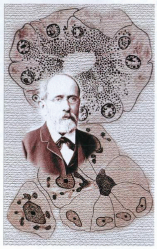 Rudolf Peter Heidenhain (1834-1897) (left) was the eldest of the 22 children of the physician Heinrich Jacob Heidenhain (1808-1868). Born January 29, 1834, in Marienwerder, East Prussia, he died October 13, 1897, in Breslau, Germany, having revolutionized many spheres of physiology. After completion of his secondary education in his native town at the age of 16 years, he began the study of nature on an estate near his home but soon turned to medicine at the University of Königsberg. He subsequently studied at a number of institutions before undertaking in 1867 a systematic investigation of the physiology of glands and of the secretory and absorption process, which remained his chief field of interest for the rest of his life. Heidenhain noted in the stomach two types of cells in the gastric glands and demonstrated that one produced pepsin and the other hydrochloric acid. By the late nineteenth century, investigators were able to differentiate between these chief cells (Hauptzellen, top) and granules from Belegzellen (parietal cells), which possessed canaliculi (bottom). |
Three years after the initial identification of pepsin by Schwann, Wasmann was able to isolate the protein and thereby establish the premise for protein digestion. In 1846, Claude Bernard wrote extensively on the digestive ferments of the pancreas, whereas the possibility of a proenzyme, pepsinogen, was formally postulated by Epstein and Grutzner in 1854. The first evaluation of the protein products of gastric digestion was described by Meisner in 1859. Heidenhain, while studying the pancreas, was soon thereafter able to describe the secretory mechanisms of proteolytic zymogens. Eight years into his tenure as Professor of Physiology at Breslau, Heidenhain began a systematic study of glands that would occupy him for almost the next 30 years. During this time, his experimental work suggested to him that all secretory phenomena were intracellular rather than mechanical processes. From these, he was able to draw some important conclusions about gastric physiology. First, he concluded that the secretion of saliva was an indicator of blood flow, and second, that there were two kinds of cells in gastric glands: those that secrete hydrochloric acid, which he named Belegzellen, and those that secrete pepsin, Hauptzellen. His histologic notes also describe a yellow-stained small cell situated adjacent to these cells that 100 years later would be identified as the endocrine ECL cell. His use of a surgical pouch for investigating acid physiology later became the standard model for such investigations.
Heidenhain’s observations were followed by those of Kühne, who theorized that because the stomach itself was not digested by pepsin, the gastric ferments must have inactive protein precursors (e.g., pepsinogen). It was Kühne who
developed the term zymogen for these precursors, as well as the term enzyme. He also identified the proteolytic pancreatic enzyme, trypsin, in 1868 and influenced the work of his later English admirers.
developed the term zymogen for these precursors, as well as the term enzyme. He also identified the proteolytic pancreatic enzyme, trypsin, in 1868 and influenced the work of his later English admirers.
 The obituary notice of John Newport Langley (1852-1925): “The main achievements of Langley’s research work are familiar to the readers of the Journal of Physiology and need little here in the way of description or comment. They stand permanently in their place, not merely as additions here and there to knowledge, but as indispensable stepping stones along which at this point, or that, the progress of knowledge has actually made its way. Each gain he made was a step placed securely and finally, and few indeed of them as the road has become more firmly and widely trodden by others following, have been found wrongly placed. All his chief works keep, and must always keep, their place in the significant history of Animal Physiology. The bare titles of his papers and books deployed there along the years give the plainest testimony to me to his unvarying, unhalting service to science. No single year in all that series, extending well nigh for half a century, from youth to age, appears without its contribution of effective work.” W. M. Fletcher. J Physiol 1926. |
However, it remained for Langley at Cambridge in the 1880s to formalize the study of pepsinogen and the mechanisms of its secretion. Langley’s introduction to the gastric gland was driven by chance. His mentor, the great physiologist Foster, suggested he study the effects of the drug jaborandi (pilocarpine) on the heart. These studies in 1874 led him toward studying its effects on secretion. After an initial prelude in the submaxillary gland, Langley dove into the regulation of secretion in the stomach, which he would pursue for the better part of the next 20 years.
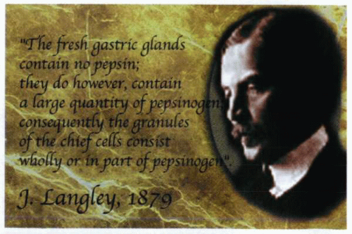
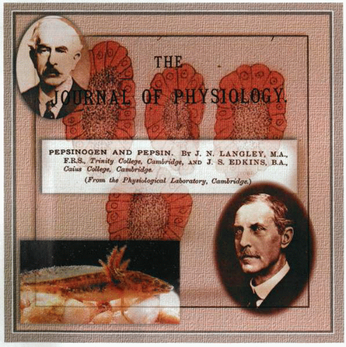 The study of pepsinogen and the mechanisms of its secretion was formalized by John Langley (bottom right) of Cambridge. Langley succeeded Sir Michael Foster (1836-1907) as the editor of the Journal of Physiology and trained J. S. Edkins (1863-1940) (top left). His introduction to the gland was by chance, because his initial assignment had been to elucidate the effects of the drug jaborandi (pilocarpine) on the heart. In 1874, this work led him toward the investigation of its effects on secretion. After an initial prelude in the submaxillary gland, he began to address the regulation of secretion in the stomach, a field of endeavor that would take him the better part of 20 years. Using the salamander as a model (bottom left), he undertook histologic studies of the gland structure in activity and rest and checked the interpretation of the appearance of killed and stained cells with that of direct observation of living gland cells. He correlated these findings with the effect of nervous influence on the glands and linked these observations to chemical estimations of the changes in the quality of pepsinogen secretion under different circumstances. In the background are esophageal glands of Rana temporana. This drawing by Langley (September 1879) shows the end tubes of the esophageal gland of a frog fed with a sponge for 35 hours before it was expelled by vomiting. The animal was killed 10 hours thereafter and the tissue placed in absolute alcohol for 24 hours and then dilute carmine for a further 24 hours. These cells show stained nongranular zones and unstained granular zones. This is consistent with contemporary biologic understanding of apical exocytosis of pepsinogen granules. Indeed, these drawings and sketches, although now more than 100 years old, attest to his clear understanding of the nature of zymogen secretion and the general mechanisms of its stimulation. In addition, Langley was so impressed with Heidenhain’s contribution that he was to borrow and translate the former’s terms for the cells in the stomach into English as border (Haupt) and chief (Beleg) cells, respectively, and also coined the term oxyntic to identify the role of the acid-secreting cells. In a series of publications between 1879 and 1882, Langley established the basic morphology and secretory characteristics of the pepsin-forming glands of the stomach and esophagus (center) and was, in addition, able to correct Heidenhain by demonstrating that, contrary to previous reports, gland cells became less granular as secretion took place. Langley demonstrated that granules were stored up during rest and discharged during secretion not only in the pancreas, but also in the stomach and salivary glands and that during this event, a chemical change in the zymogen occurred. To quote: “The fresh gastric glands contain no pepsin; they do however contain a large quantity of pepsinogen; consequently the granules of the chief cells consist wholly or in part of pepsinogen.” Langley’s pupil, Edkins, entered Cambridge University as a scholar of Caius College in 1881. Such was his ability that he was awarded two scholarships, one in mathematics and the other in natural sciences. After Cambridge, he worked with C. S. Sherrington (1857-1952) in Liverpool (who was later to attain a Nobel Prize in Neurophysiology). Edkins taught with great distinction at St. Bartholomew’s in London and subsequently became Chairman of Physiology at Bedford College for Women in 1914. In this capacity, he was responsible for training the majority of women physiologists in England between 1914 and 1930. Apart from his fundamental observations in regard to gastrin (he documented the existence of a novel antral stimulant of acid secretion-gastrin in 1905), he investigated the presence of spiral bacteria (Spirella regaudi) (Helicobacter felis) in the gastric mucosa of cats and their relevance to digestion (1923). |
Stay updated, free articles. Join our Telegram channel

Full access? Get Clinical Tree


