Penis/Scrotum


Penis/Scrotum


5.1 EXTRAMAMMARY PAGET DISEASE VS. HIGH-GRADE PENILE INTRAEPITHELIAL NEOPLASIA


Extramammary Paget Disease


High-Grade Penile Intraepithelial Neoplasia (PeIN)


Age


Adults. Sixth to eighth decades


Younger adults


Location


Scrotum and scrotal penile junction most common locations


Shaft, glans, and perimeatal region


Symptoms


Pruritus, pain


None


Signs


Erythematous, eczematous, or ulcerated plaque-like lesion with well-defined borders


Flat to slightly raised plaque


Etiology


Unknown in majority of cases. Occasionally arise in association with underlying colorectal, urogenital, or skin adnexal carcinoma


High-risk HPV related to undifferentiated (basaloid) PeIN (most cases) and differentiated PeIN (˜50% of cases)


Histology


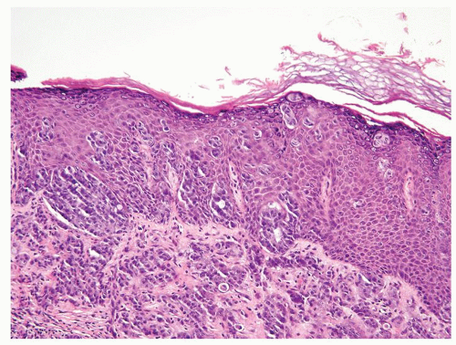
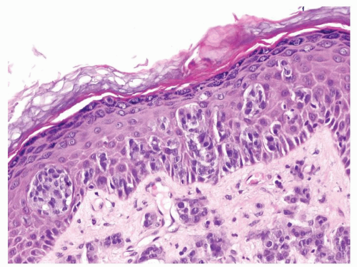
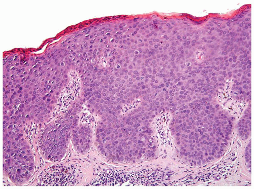
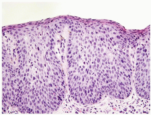
  1. Scattered more loosely cohesive atypical cells and cell clusters with abundant pale vacuolated cytoplasm in a background of normal epithelial cells (Figs. 5.1.1 and 5.1.2)


  2. Nuclei large vesicular with prominent nucleoli


  3. Vacuolated pale cytoplasm with mucinous material occasionally discernible


  4. Many suprabasally located and separated from basement membranes by normal basal cells. Tends to cluster at tips of the rete ridges


  5. Lacks basaloid appearance


  6. Dyskeratotic cells not seen. May see mitoses and apoptosis


  7. Invasion into dermis or lamina propria occasionally encountered


  8. Associated hyperkeratosis, parakeratosis, and papillomatosis commonly present


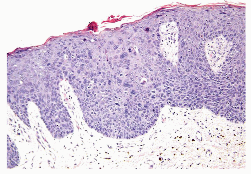
  1. Full-thickness cohesive atypical neoplastic cells


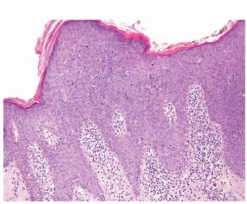
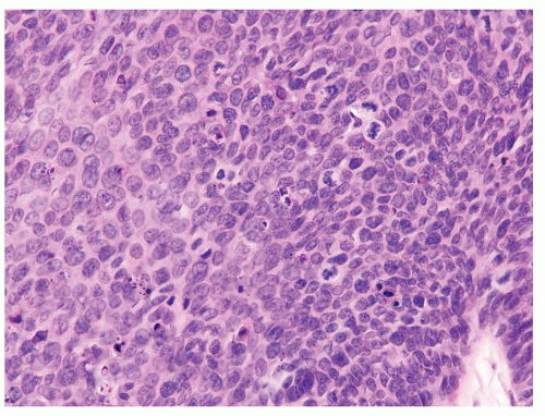
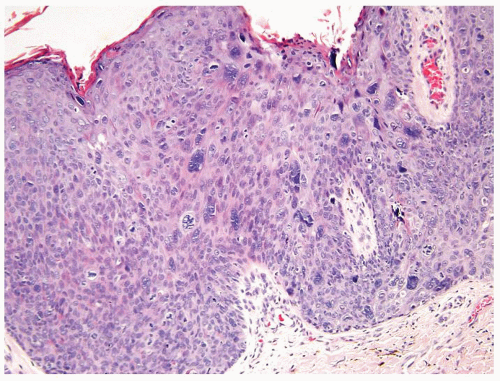
  2. Nuclei typically not as pleomorphic in undifferentiated PeIN (Figs.5.1.3, 5.1.4, 5.1.5, 5.1.6) compared to differentiated PeIn (Figs. 5.1.7 and 5.1.8)


  3. Cytoplasm eosinophilic identical to normal squamous cells


  4. No sparing of the basal cell layer


  5. Basaloid PeIN with smaller cells with high N/C ratio


  6. Mitoses, individually keratinized dyskeratotic cells and apoptotic bodies seen


  7. Invasion into dermis absent by definition


  8. Associated surface hyperkeratosis and parakeratosis commonly present


Special studies


  • Intracytoplasmic mucin on histochemical stains


  • CEA+, CK7+, HER2+, CAM5.2+


  • Negative for high-risk HPV using in situ hybridization (ISH)


  • Intracytoplasmic mucin not demonstrable on histochemical stains


  • Typically CEA-, CK7-, HER2-, CAM5.2-


  • High-risk HPV can be detected using ISH


Treatment


Wide local excision of the skin and subcutaneous tissues. Sentinel lymph node biopsy and/or regional lymphadenectomy warranted for invasive Paget disease


Complete conservative excision


Prognosis


If noninvasive, favorable prognosis. Invasion with 2-y overall survival rate approaching 50%. Depth of invasion and lymphovascular involvement prognostic


Excellent


Figure 5.1.1 Extramammary Paget disease. Intraepidermal proliferation of atypical cells with abundant pale vacuolated cytoplasm. Invasion into underlying dermis is seen.


Figure 5.1.2 Same case as Figure with nest of cells with pale cytoplasm spread within otherwise normal epidermis. Invasive Paget disease is seen in the dermis (bottom).


Figure 5.1.3 Undifferentiated high-grade PeIN with full-thickness proliferation of small basaloid cells.


Figure 5.1.4 Same case as Figure 5.1.3 with numerous mitotic figures and dyskeratotic cells. The nuclei are smaller with high N/C ratio.


Figure 5.1.5 Undifferentiated high-grade PeIN.


Figure 5.1.6 Undifferentiated grade PeIN.


Figure 5.1.7 Differentiated high-grade PeIN with cells having significant pleomorphism and abundant cytoplasm.


Figure 5.1.8 Differentiated high-grade PeIN.


5.2 PSEUDOEPITHELIOMATOUS SQUAMOUS CELL CARCINOMA VS. PSEUDOEPITHELIOMATOUS HYPERPLASIA


Pseudoepitheliomatous Squamous Cell Carcinoma


Pseudoepitheliomatous Hyperplasia


Age


Adults. Median 69 y (53-76 y)


Adults


Location


Frequently multifocal. Preferentially affects inner foreskin mucosa but also glans surface, coronal sulcus, and frenulum


Gland, coronal sulcus, and foreskin


Symptoms


None


None


Signs


Flat to slightly raised mass on inner preputial mucosa


Flat to slightly raised mass


Etiology


Majority of cases arise in association with lichen sclerosus et atrophicus (LS&A) not associated with HPV infection


Associated inflammatory dermal conditions including LS&A


Gross and Histology


  1. Flat to only slightly raised, pearly whitish gray surface with irregular borders, measuring 0.4-3.5 cm (mean 2 cm)


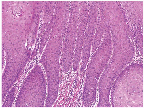
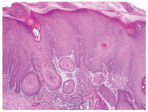
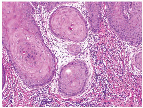
  2. Downward proliferation of very well-differentiated squamous epithelial columns and nests (Figs.5.2.1, 5.2.2, 5.2.3)


  3. Nests and columns more haphazard, deeply invasive, and irregular frequently angulated border. Abundant eosinophilic cytoplasm at the invading edge is seen with a lack of a palisaded basal layer


  4. Neoplastic cells extremely well differentiated almost indistinguishable from normal epithelial cells, with only minimal atypia of basal layer


  5. Adjacent flanking mucosa frequently reveals associated differentiated PeIN and LS&A


  1. Flat to only slightly raised, pearly whitish gray surface with irregular borders


  2. Downward proliferation of benign mature squamous epithelial columns that may give the impression of nests on cut surface


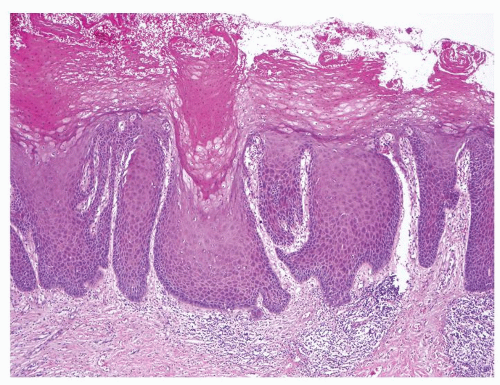
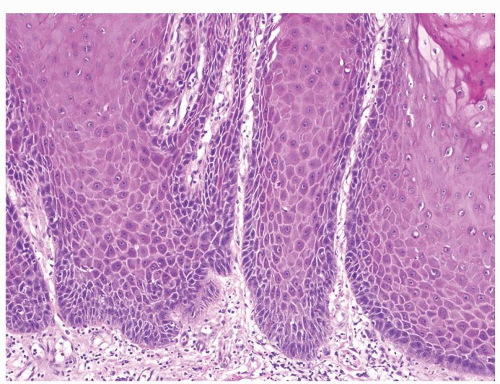
  3. Columns and nests reach only a superficial level of depth ending at a linear limiting front paralleling the surface mucosa/skin. A palisaded basal layer at the interface of columns/nests with stroma is present (Figs.5.2.4, 5.2.5, 5.2.6)


  4. Hyperplastic epithelial cells indistinguishable from normal epithelial cells. Keratin pearls and individual cell keratinization not seen


  5. Adjacent flanking mucosa or skin reveals changes of underlying inflammatory etiology but no PeIN


Special studies


Not helpful in differential. Both are negative for HPV on IHC and molecular studies and share similar cytokeratin profile


Not helpful in differential


Treatment


Very conservative surgical excision. At time, circumcision sufficient. Prophylactic inguinal lymphadenectomy not indicated


Directed toward underlying etiology


Prognosis


Only rare recurrence. No lymph node metastases have ever been reported


Benign


Figure 5.2.1 Pseudoepitheliomatous squamous cell carcinoma.


Figure 5.2.2 Same case as Figure 5.2.1 with downward proliferation of very well-differentiated squamous epithelial columns and nests.


Figure 5.2.3 Nests and columns more haphazard and lack palisaded basal layer compared to hyperplasia. Also some islands have abundant eosinophilic cytoplasm at the invasive edge. Cytologic atypia is also noted (upper right).


Figure 5.2.4 Pseudoepitheliomatous hyperplasia.


Figure 5.2.5 Same case as Figure 5.2.4 with columns and nests reaching only a superficial level of depth ending at a linear limiting front paralleling the surface mucosa/skin.


Figure 5.2.6 Same case as Figures 5.2.4 and 5.2.5 with a palisaded basal layer at the interface of columns/nests with stroma.


5.3 VERRUCOUS SQUAMOUS CELL CARCINOMA VS. CONDYLOMA ACUMINATUM


Verrucous Squamous Cell Carcinoma (Verrucous SCC)


Condyloma Acuminatum


Age


Older adults. Mean age at diagnosis 70-80 y


Older adults. Mean age at diagnosis 60 y


Location


Usually unifocal. Glans or foreskin


Usually unifocal but multifocality occasionally seen. Glans, prepuce, or shaft


Symptoms


None


None


Signs


Cauliflower-like mass, slow growing


Cauliflower-like mass, soft to moderately firm slow growing


Etiology


Unknown. Not associated with HPV infection


Strongly associated with low-risk HPV infection (HPV6 and HPV11). Higher incidence in sexual partners of HPV-related cervical or anal lesions


Gross and Histology


  1. Cauliflower-like large soft masses, white-gray cobblestone surface


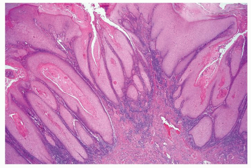
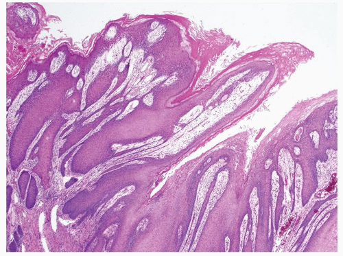
  2. Broad base bulbous papillae characterized by marked acanthosis. Fibrovascular cores very inconspicuous or absent (Fig. 5.3.1)


  3. Intraepithelial keratin plugs frequently found


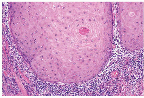
  4. Base of papillae broad based and rounded (Fig. 5.3.2)


  5. Prominent acanthosis


  6. Koilocytes absent and parakeratosis ranges from mild to prominent


  7. Chronic inflammatory stromal reaction, ranging from moderate to severe frequently observed. Usually only invades lamina propria and occasionally up to corpus spongiosum with infrequent extension


  8. Extremely well-differentiated identical to normal squamous epithelium (Fig. 5.3.2)


  1. A very small subset can reach large size (>8 cm). Grossly, cauliflower-like soft masses, white-gray cobblestone surface


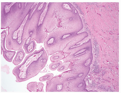
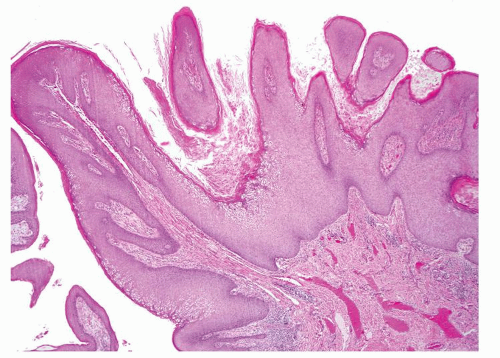
  2. Complex papillae with irregularly shaped prominent fibrovascular cores (Figs. 5.3.3 and 5.3.4)


  3. Intraepithelial keratin plugs present


  4. Base of papillae broad based and rounded


  5. Prominent acanthosis


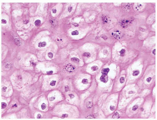
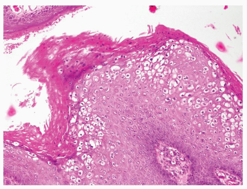
  6. Extensive and prominent nuclear koilocytosis (perinuclear halo) with nuclear irregular membrane and frequent bi/multinucleation (Figs. 5.3.5 and 5.3.6). Mild dysplasia almost always present with a small subset showing focal moderate to severe dysplasia


  7. Tumor base can have invaginations with inverted pushing base. Does not invade corpus spongiosum or beyond


  8. Extremely well-differentiated identical to normal squamous epithelium


Special studies


IHC negative for HPV


Overall majority positive for low-risk HPV (6, 11) with only rare lesions showing other types of HPV (16, 18, 31, 32)


Treatment


Conservative surgical excision. No lymph node metastases ever been reported in pure verrucous SCC. Prophylactic inguinal lymphadenectomy not indicated


Complete conservative resection


Prognosis


Higher metastatic and recurrence rates encountered in cases with verrucous carcinoma associated with usual SCC. Generous sampling advised in verrucous SCC to rule out presence of usual SCC foci


Benign tumors even when locally destructive. A very small minority give rise to SCC


Figure 5.3.1 Verrucous squamous cell carcinoma with broad base bulbous papillae characterized by marked acanthosis. Fibrovascular cores are very inconspicuous or absent.


Figure 5.3.2 Same case as Figure 5.3.1 with broad base bulbous papillae and not nuclear atypia.


Figure 5.3.3 Condyloma acuminatum with complex papillae with irregularly shaped prominent fibrovascular cores.


Figure 5.3.4 Condyloma acuminatum with well-formed papillae with prominent fibrovascular cores.


Figure 5.3.5 Condyloma acuminatum with extensive koilocytosis.


Figure 5.3.6 Same case as Figure 5.3.5 with koilocytosis, binucleated cells, and irregular nuclei.


5.4 VERRUCOUS SQUAMOUS CELL CARCINOMA VS. PAPILLARY SQUAMOUS CELL CARCINOMA


Verrucous Squamous Cell Carcinoma (Verrucous SCC)


Papillary Squamous Cell Carcinoma (Papillary SCC)


Age


Older adults. Mean age at diagnosis 70-80 y


Older adults. Mean age at diagnosis 40-50 y


Location


Usually unifocal. Glans or foreskin


Usually unifocal. Glans or foreskin


Symptoms


None


None


Signs


Cauliflower-like mass, slow growing


Cauliflower-like mass, slow growing


Etiology


Unknown. Not associated with HPV infection


Unknown. Not associated with HPV infection


Gross and Histology


  1. Grossly, cauliflower-like large soft masses, white-gray cobblestone surface


  2. Broad base bulbous papillae characterized by marked acanthosis. Fibrovascular cores very inconspicuous or absent (Figs. 5.4.1 and 5.4.2)


  3. Intraepithelial keratin plugs frequently found


  4. Prominent acanthosis


  5. Koilocytes absent and parakeratosis ranges from mild to prominent


  6. Tumor base is broad and pushing (Fig. 5.4.3). Usually only invades lamina propria and occasionally up to corpus spongiosum


  7. Extremely well-differentiated identical to normal squamous epithelium (Fig. 5.4.4)


  1. Cauliflower-like large soft masses, white-gray cobblestone surface


  2. Complex papillae with irregularly shaped fibrovascular cores (Figs. 5.4.5 and 5.4.6). Tend to have some areas exhibiting condylomatous well-formed papillae and others with a more verrucous-like aspect lacking prominent fibrovascular cores


  3. Intraepithelial keratin plugs not seen


  4. Prominent acanthosis


  5. Koilocytes absent and parakeratosis not prominent


  6. Jagged tumor-stroma interface. Tends to infiltrate deeper into penile tissues compared to verrucous SCC


  7. Neoplastic cells range in grade from well to moderately differentiated, with a minority of the cases poorly differentiated (Figs.5.4.7, 5.4.8, 5.4.9, 5.4.10)


Special studies


IHC negative for HPV


HPV detection rate is very low or even absent


Treatment


Conservative surgical excision. No lymph node metastases have ever been reported in pure verrucous SCC. Prophylactic inguinal lymphadenectomy not indicated


Managed according to penile risk-group stratification systems, taking into account histologic grade, anatomical level of maximum tumor infiltration, and the presence of vascular and perineural invasion


Prognosis


Higher metastatic and recurrence rates are encountered in cases with verrucous carcinoma associated with usual SCC. Generous sampling is advised in verrucous SCC in order to rule out the presence of usual SCC foci


Inguinal metastatic rate very low. Less than one-fifth of all patients present with inguinal involvement, and even in these cases, the mortality rate is low. Even when tumors invade penile erectile tissues, prognosis is good as long as no high-grade areas are identified

Only gold members can continue reading. Log In or Register to continue

Stay updated, free articles. Join our Telegram channel

Jul 9, 2016 | Posted by in PATHOLOGY & LABORATORY MEDICINE | Comments Off on Penis/Scrotum

Full access? Get Clinical Tree

Get Clinical Tree app for offline access