Pediatric Exploratory Laparotomy for Trauma, Malrotation, or Intussusception
Raphael C. Sun
Graeme J. Pitcher
While there are many different incisions for laparotomy in the adult population, the two most common approaches for exploratory laparotomy in a pediatric patient are the vertical midline and the transverse abdominal incision. In most situations, the pediatric surgeon prefers a transverse abdominal incision over the traditional vertical midline incision. However, it is important to consider the indication for surgery and take into account the area of concern when choosing an incision.
In general, a child younger than 5 years of age has a round or elliptical abdomen with the width being relatively longer than the length. The costal margin in children is proportionately higher from the iliac crest than the adult, making the abdominal cavity proportionately larger compared to the adult. Given these differences in body habitus and anatomy, it is understandable that the pediatric surgeon prefers the transverse abdominal incision.
Most of the time, a transverse abdominal incision provides adequate exposure for all four quadrants. However, in certain penetrating trauma situations, the traditional vertical incision is preferred. This incision can also be more easily extended into a sternotomy should that become necessary. It is important to understand the technical and anatomic points for each approach.
This chapter discusses the general conduct of exploratory laparotomy in infants and children, and then describes specific management of two common conditions: Malrotation and intussusception.
SCORE™, the Surgical Council on Resident Education, classified emergency operation for malrotation and emergency operation for intussusception as “ESSENTIAL UNCOMMON” procedures.
STEPS IN PROCEDURE
Make a vertical midline or transverse abdominal incision
Gain entry into the abdomen
Pack all four quadrants if there is severe bleeding
Inspect the entire abdomen by quadrants or organ systems
Perform the necessary procedure
Achieve hemostasis
Close fascia and skin or leave abdomen open if indicated
HALLMARK ANATOMIC COMPLICATIONS
Making a paramedian or off midline incision unintentionally
Injury to bowel or other organs upon entry
Missed injury or pathology
LIST OF STRUCTURES
External oblique muscle and aponeurosis
Internal oblique muscle and aponeurosis
Transversus abdominis muscle and transversalis fascia
Preperitoneal fat
Peritoneum
Linea alba
Remnant of the umbilical vein (falciform ligament)
The Vertical Midline Incision (Fig. 91.1)
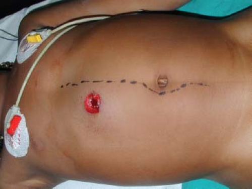
The traditional vertical midline incision is most often used in trauma situations. The reason in choosing this incision is because it allows access to all four quadrants of the abdomen and provides better exposure to areas such as the aortic hiatus and the pelvis and can be performed quickly. There is usually minimal blood loss if the surgeon stays in the midline. The incision may be extended in either direction and can even be extended into the chest if a sternotomy is necessary.
The Transverse Abdominal Incision
There have been many clinical studies to observe the differences and outcomes between surgical incisions in the pediatric patient population. Studies have concluded that the transverse abdominal incision has decreased the occurrence of hernia defects, fascial or wound dehiscence compared to the vertical midline incision. The cosmetic appearance of the transverse incision is superior if placed accurately in the skin creases or Langer’s lines. This incision allows all four quadrants to be exposed adequately and may easily be extended laterally if needed. Therefore, transverse abdominal incision is the preferred approach for exploratory laparotomy for nontraumatic indications in a child younger than 5 years of age.
Technical Points and Anatomic Points
Make the intended incision by using a knife to cut through the skin and dermis. The child’s skin is much thinner and easier to cut compared to the adult. The subcutaneous layer and fat layer may be separated using electrocautery. If electrocautery is used, a low setting should be used in the “blend” mode. In general, the younger the patient is, the lower the electrocautery settings should be.
For a midline vertical incision, use the anatomic landmarks of the xiphoid and the pubic symphysis to guide the incision. The incision should sweep gently around the umbilicus. If you anticipate the creation of an ostomy, the curvilinear incision should be placed on the opposite side of planned ostomy site. The linea alba is the reference landmark to confirm that the midline is reached and is seen more easily above the umbilicus than below. The midline incision may be superior or inferior to the umbilicus. When extending below the umbilicus in an infant, remember that the bladder occupies an intra-abdominal position and needs to be swept aside to avoid injury to it on entry.
 Figure 91.1 Midline incision preferred in small infant with penetrating abdominal trauma for ease of access to pelvis and aortic hiatus. |
Keep in mind that the umbilicus is lower in the abdomen compared to an adult, so a supraumbilical transverse incision will give excellent access to most structures in the upper and central abdomen. As you gain entry into the abdomen, the falciform ligament (remnant of the umbilical vein) is usually encountered, and will need to be ligated and divided to expose the liver, stomach, esophagus, and duodenum.
Close the fascia of a midline incision with absorbable sutures such as Vicryl or PDS. The fascia is normally closed in a standard running fashion from one end to the other. Make sure that each bite of fascia closure includes the anterior rectus sheath below the umbilicus and are adequately “deep” and closely spaced. The thin skin and subcutaneous tissues dictate that knots should be buried, even with absorbable material to prevent unsightly and uncomfortably prominent “bumps” during healing.
In situations where tension is encountered such as when closing the abdomen for abdominal wall defects, interrupted suturing techniques can be used. These sutures are usually simple or mattress sutures but some prefer the Smead-Jones technique. This technique is a double stitch on each side of the fascia in a far-near-near-far fashion as shown (for the adult case) in Chapter 44, Figure 44.6. Be careful not to injure bowel contents underneath the fascia. Retraction and exposure is important to allow the surgeon to visualize each bite of suture going through the fascia. This will help avoid injuries while closing the abdomen. A narrow malleable retractor placed on the bowel surface and projecting out of the abdomen (to ensure its removal) facilitates safe closure.
When closing a transverse incision, two options are available. Most surgeons prefer layered closure where the posterior rectus sheath, internal oblique and transversus abdominis muscles are approximated in the deep layer, and the external oblique muscles and the anterior rectus sheath are sutured in the second layer. A mass closure with a single running suture line encompassing all tissue layers is also acceptable and may in fact be preferable in very premature babies.
Skin closure is achieved by the use of absorbable sutures wherever possible in children to avoid the need for painful and fear-inducing suture removal postoperatively. Skin clips are generally avoided for this reason except for situations where hemostasis in skin edges is required. Running subcuticular sutures of Vicryl or Monocryl are commonly used. The choice of dressings is personal.
Drains or catheters need to be securely fixed to avoid premature removal by an often uncooperative child postoperatively. Ensure that all devices are covered by secure dressings before the patient emerges from anesthesia.
Management of Malrotation (Figs. 91.2–91.4)
The intestines undergo their first stages of development in an extracelomic position by lengthening around the superior mesenteric artery (SMA) pedicle at around the sixth to eighth week of gestation. Malrotation refers to a variety of anatomic abnormalities which are the result of failure of the intestinal tract to complete its normal rotation and fixation, following its return to the abdominal cavity between the eight and twelfth week of development. There are two distinct components:
Stay updated, free articles. Join our Telegram channel

Full access? Get Clinical Tree


