Papillary Carcinoma


Papillary Carcinoma


Yuri E. Nikiforov

N. Paul Ohori


INCIDENCE AND EPIDEMIOLOGY

Papillary carcinoma is the most common type of thyroid cancer. Its incidence in absolute numbers and in proportion to other types of thyroid cancer has been steadily increasing in the United States and many other countries around the world.1,2,3,4 In the United States, based on the Surveillance, Epidemiology and End Results (SEER) data, the annual incidence of papillary carcinoma has more than tripled during the last 35 years, as it was 2.7 per 100,000 persons in 1973 and 11.2 per 100,000 in 2008.5 Whereas in 1973 papillary carcinoma accounted for 74% of all thyroid cancer cases in the United States, its proportion increased to 86% in 2004 to 2008.5 Papillary cancer more frequently affects women, with the female:male ratio of about 3:1 in the United States and many other countries.5,6 The increase in incidence affects both genders but is more pronounced in females (Fig. 11.1).

Several factors may be responsible for the increase. First would be a widespread use of ultrasonography, which can detect thyroid nodules down to 0.2 cm in size and guide a fine needle aspiration (FNA) procedure to sample these small lesions. This leads to the detection of occult papillary carcinomas of <1 cm in size, which are common in the general population based on autopsy series.7,8 Indeed, papillary carcinomas of 1 cm or smaller in size are responsible for over 50% of the observed increase in incidence (Fig. 11.2, left).2,5,9,10 However, another half of the increase is still attributed to the tumors >1 cm in size. Overall, currently about 40% of newly diagnosed cases are 1 cm or less in size. Second, the increase may be due to the better recognition of the follicular variant of papillary carcinoma and changes in the histopathologic criteria for its diagnosis. As a result, tumors with partially developed nuclear features of papillary carcinoma are more likely to be interpreted as malignant today than 15 to 20 years ago.11,12,13 This may not, however, provide a full explanation for the increase because the increase in incidence has affected both classic papillary carcinoma and the follicular variant (Fig. 11.2, right).


FIGURE 11.1. Incidence of papillary carcinoma in the United States in 1973-2008. Based on the SEER data.5

Overall, it is unlikely that the detection of small, clinically irrelevant cancers due to better imaging and change in the diagnostic criteria are solely responsible for the entire increase in thyroid cancer incidence. In fact, recent studies, although limited and restricted to specific types of papillary carcinoma, suggest that the proportion of tumors carrying a BRAF mutation, a marker of more aggressive cancer, has been constant or even increasing during the last decades.14,15 Therefore, other factors may contribute to the rapidly growing incidence of papillary carcinoma, such as more common exposure to radiation and/or a more iodine-rich diet, as discussed later in this section.

Worldwide, the incidence of papillary carcinoma has significant geographic variability, which largely follows the variation in the cumulative rates of thyroid cancer observed in different countries (see Chapter 7, Fig. 7.2). The incidence also varies among different ethnic groups. In the United States, the average incidence in whites between 1973 to 2003 was 11.5 per 100,000 per year in females and 4.8 in males, which was higher than in blacks, where the incidence was 8.1 in females and 3.7 in males.9 The incidence was observed to be higher among Israeli Jews than among Israeli Arabs.16

Although uncommon in early childhood, papillary carcinoma accounts for the most thyroid cancers in this age group.17 The incidence increases sharply during adolescence and early adulthood. Surprisingly, the age of patients who develop papillary carcinoma has been continuously increasing over the last three decades (Fig. 11.3). Based on the SEER data, the mean age of patients was 37.8 years in 1975, 41.6 years in 1985, 43.7 years in 1995, and 46.8 years in 2005.5


ETIOLOGIC FACTORS

Although familial occurrence of follicular carcinomas has gained more recognition in the recent years, most tumors are sporadic, nonfamilial cases.


Ionizing Radiation

Exposure to ionizing radiation is a major known risk factor for papillary carcinoma (Table 11.1). Both external X-ray and λ-radiation as well as internal exposure to radioiodine during childhood lead to an increased cancer risk. This includes external beam radiation therapy for malignant and benign conditions as well as accidental exposure to radioiodine or λ-radiation as a result of nuclear weapon explosion or nuclear reactor accidents. Widespread use
of external beam radiation therapy for benign conditions of the head and neck, common in the United States and several other countries in the 1930s to 1940s, resulted in a significant increase of thyroid cancer incidence in individuals exposed during childhood.18,19 However, the use of radiation for treatment of benign conditions was abandoned in the 1950s. As a result, the impact of radiation as an etiologic agent has been gradually decreasing. For example, a history of radiation exposure was documented in 12% of patients with thyroid cancer diagnosed in 1978 to 1980 as compared with 5% of patients diagnosed in 1996.20,21 However, radiation therapy for malignant tumors, such as Hodgkin disease, remains a source of radiation-induced papillary carcinoma. The Chernobyl nuclear power accident in 1986 has lead to the development of papillary carcinomas in >5,000 individuals exposed to radioiodines during childhood and adolescence.22,23,24 The increase in cancer risk due to diagnostic X-ray procedures, occupational radiation exposure, and high dose 131I therapy for hyperthyroidism has not been found.21,25


FIGURE 11.2. (Left) Incidence of papillary carcinomas 1 cm or less and >1 cm in 1988-2008 based on the SEER data.5 (Right) Incidence of papillary carcinoma, not otherwise specified (PC, NOS) and the follicular variant (PC, FV) in 1973-2008 based on the SEER data.5

The risk of radiation-induced cancer has a linear correlation with dose received by the thyroid in the dose range of 0.2 to 2 Gy, and it reaches a plateau at higher doses.18,26,27 The risk has strong inverse correlation with age at exposure and is highest in children exposed during infancy. The excess relative risk of cancer after childhood exposure ranges widely in different studies and depends on the source of radiation, age at exposure, and length of follow-up, but most frequently is in the range of 5 to 9 per 1 Gy of radiation.18,26,27,28,29,30,31 The risk of exposure during the adulthood is significantly lower and may not be detectable at all.31


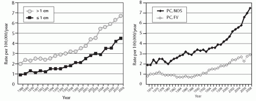
FIGURE 11.3. Mean age of patients with papillary carcinomas in the United States in 1974-2008. Based on the SEER data.5

The shortest time interval between exposure and cancer development is 4 years, and the risk remains elevated for 50 years and longer.32,33,34 Papillary carcinomas is by far the most common type of radiation-induced thyroid cancer, and many of them reveal a solid or mixed solid-follicular growth pattern.24,35,36 Molecular mechanisms of radiation-associated papillary carcinogenesis involve primarily chromosomal rearrangements, such as RET/PTC, whereas point mutations in BRAF, RAS, and other genes are rare in these tumors.37,38


Iodine-rich Diet

High iodine consumption represents another potential risk factor. The incidence of papillary carcinoma appears to be higher in regions with high dietary iodine intake, such as Iceland, Japan, and the Pacific islands.39,40 In addition, the ratio of papillary carcinoma to follicular carcinoma is significantly higher in the areas of high iodine intake as compared with the areas of moderate and low intake.41 In areas of severe iodine deficiency, widespread iodine supplementation coincided with the reduction in the prevalence of follicular and anaplastic carcinoma and a proportional increase in papillary carcinoma.42,43,44 It remains unclear, however, whether the increase was caused by an iodine-rich diet or it reflected the reduction in other thyroid tumor types and a general increase in the incidence of papillary cancer observed in many countries.

A recent study of thyroid cancer in several regions of China with very high iodine content in drinking water and regions with normal iodine content has shown a significantly higher ratio of papillary cancer to non-papillary cancer in the former regions (7 to 8:1 vs. 3 to 4:1).45 Moreover, the study has also identified a significantly higher prevalence of BRAF V600E mutation in papillary carcinoma from regions with high iodine content (69% vs. 53%), suggesting that correlation between this mutation and iodine-rich diet may serve as a biologic mechanism behind the increasing incidence of papillary carcinoma.


Table 11.1 Radiation Exposure and Thyroid Papillary Carcinoma


Radiation type: External (X-ray, γ-) radiation and internal exposure to 131I impose increased cancer risk


Dose dependence: Risk is dose dependent and close to linear in 0.2-2 Gy dose range


Age dependence: Risk has strong inverse correlation with age at exposure; highest risk is in youngest children


Shortest time interval between exposure and cancer development: 4 years


Persistence of elevated risk: 50 years and longer after exposure


Histologic features: Many radiation-induced papillary cancers have solid or mixed solid-follicular architecture


Typical genetic alterations: Chromosomal rearrangements, particularly RET/PTC; point mutations (BRAF, RAS) are rare


Other Environmental Factors

A recent study of thyroid cancer in Sicily has identified a significantly higher incidence of papillary cancer, but not follicular or medullary cancer, in the volcanic region with high concentration of boron, iron, vanadium, manganese, and other chemicals in drinking water as compared with the neighboring regions with normal concentration of these elements.46 Moreover, this region had a significantly higher incidence of BRAF mutations in papillary carcinomas (52% vs. 33% in other regions of Sicily).46 This finding raises the possibility that mutagenesis leading to BRAF point mutation may be related to the excessive exposure to specific chemical compounds found in the volcanic areas. In support of this possibility, Hawaiian islands and Iceland belong to the regions of the world with the highest incidence of thyroid cancer, and they host multiple volcanoes, many of which are active.6 However, no data linking papillary cancer and BRAF mutation to exposure to specific chemical compounds are available yet.


Preexisting Benign Thyroid Disease and Thyroid-Stimulating Hormone levels

A history of benign thyroid disease is a well-documented risk factor for papillary carcinoma. Preexisting solitary thyroid nodules and/or adenomas increase the carcinoma risk by 27 to 29 folds and multiple hyperplastic nodules (multinodular goiter) by 6 to 9 folds.47,48 The mechanisms underlying the association between benign thyroid disease and papillary cancer are not fully understood. Mutations in the RAS genes are common in follicular adenomas and may predispose to malignant transformation.49 Another possibility is an increased cell proliferation rate in adenomas and hyperplastic nodules, which may promote carcinogenesis by increasing the chances for mutation to occur in dividing cells50 or by cooperating with an oncogenic event in tumor progression, particularly in patients with higher levels of thyroid-stimulating hormone (TSH). Three epidemiologic studies have found the association between higher serum TSH levels and risk of thyroid cancer, particularly of papillary carcinoma, in patients with thyroid nodules.51,52,53 The risk of cancer was found to increase incrementally with higher levels of TSH, even within the normal range. Recently, strong experimental evidence has been offered indicating that expression of mutant BRAF at physiologic levels requires simultaneous TSH stimulation in order to transform thyroid cells and initiate papillary carcinoma development in transgenic mice.54

The association between papillary carcinoma and two other benign thyroid diseases, Graves disease and chronic lymphocytic (Hashimoto) thyroiditis, remains controversial. Several studies have reported a higher than expected frequency of papillary carcinoma in surgically removed thyroid glands of patients with Graves disease and Hashimoto thyroiditis.55,56 However, epidemiologic case-control and large retrospective cohort studies failed to establish the association between these conditions and increased cancer risk.57,58,59,60 Recent retrospective61 and prospective62 studies confirmed the lack of an increased risk of cancer in patients with Hashimoto thyroiditis. Moreover, although some reports have suggested that RET/PTC rearrangement, a molecular event characteristic for papillary carcinoma, can be found in most thyroid glands affected by Hashimoto thyroiditis,63,64 the presence of this molecular alteration has not been confirmed in the recent studies.65,66 As a result, to date there is no undisputable evidence for the causal association between Hashimoto thyroiditis and papillary carcinoma.67

In the United States, a personal history of Graves disease, thyroiditis, and goiter is reported in 2%, 8%, and 14%, respectively, of patients with papillary carcinoma.20


Hormonal and Reproductive Factors

The role of hormones and reproductive factors in papillary carcinoma development is suggested by the higher cancer incidence in women of reproductive age. However, no definitive association between estrogens or other specific hormonal factors or interventions and cancer development has been unraveled yet. A pooled analysis of 14 case-control studies demonstrated that the risk of papillary carcinoma is slightly increased in those with miscarriage of the first pregnancy (odds ratio of 1:7 as compared with nulligravidae) and in those with artificial menopause (odds ratio 1:7 as compared with premenopausal).68 Some studies have found a slight increase in thyroid cancer risk in association with the number of pregnancies, late age at first birth, and use of estrogencontaining preparations including oral contraceptives, although other studies have not confirmed those associations.39 Obesity has been linked to thyroid cancer in several, but not all, studies, with no specific reference to papillary carcinoma.21,69,70,71


Hereditary Factors

The risk of developing papillary carcinoma is 5- to 9-fold higher among first-degree relatives of patients with thyroid cancer than in the general population.21,72,73 About 5% of papillary carcinomas are familial; the familial cancers occur in two settings: (1) as a component of known hereditary multi-cancer syndromes and (2) as tumors occurring in families with isolated thyroid cancer and little or no risk of other tumors.


In the first group, the best recognized association is between papillary carcinoma and familial adenomatous polyposis (FAP). FAP is an autosomal dominant disease caused by a germline mutation of the APC gene located on 5q21 and characterized by numerous colonic adenomatous polyps, colon cancer, and various extracolonic tumors and nonneoplastic lesions. Approximately 1% to 2% of the affected patients develop thyroid tumors, chiefly papillary carcinomas, and the incidence may be higher when thyroid glands are examined by ultrasonography.74,75 Papillary carcinomas in this setting frequently manifest during the third decade of life and affect predominantly females (female:male ratio 8:1).73,76 In fact, women under the age of 35 with FAP have 100- to 160-fold higher risk of thyroid carcinoma than unaffected individuals.76,77 The FAP-associated papillary carcinomas are more frequently multifocal and exhibit distinct histologic features characteristic of the cribriform-morular variant, as described later in the chapter.

A higher incidence of papillary carcinoma is seen in patients with Carney complex78 and possibly in those with Werner syndrome,79 although both are rare diseases and the overall number of reported cases of papillary carcinoma in these patients is low.

Another subset of familial papillary carcinomas encompasses those cases which are not associated with known hereditary multi-cancer syndromes. The inherited nature of these papillary carcinomas can be established with certainty when three or more first-degree relatives develop thyroid cancer, whereas those with two affected family members have an equal chance of having either inherited or sporadic tumors.80,81 Familial papillary carcinomas are likely to show autosomal dominant inheritance with incomplete penetrance, the latter increasing with age.82,83 Familial cancers show more common multifocality and coexistence of multiple benign thyroid nodules, but typically do not differ from sporadic cancers with regard to female predominance and age at diagnosis.73,84 Several susceptibility loci have been mapped, although specific genes have not been identified yet. A locus on 19p13.2 has been linked to familial papillary carcinoma with and without oncocytic (oxyphilic) change,85,86 a locus on 1q21 to papillary carcinoma associated with papillary renal neoplasms,87 and a locus on 2q21 to familial papillary carcinoma associated with multinodular goiter.83,88


PATHOGENESIS AND MOLECULAR GENETICS


Clonality and Multifocality

Most papillary carcinomas have a monoclonal origin, which means that they originate from a single cell.89 Clonality assays, such as the human androgen receptor assay assay (see Chapter 20), have also revealed that multiple distinct foci of papillary carcinoma found within the thyroid gland are often (50%) of different clonal origin and therefore represent independent primary tumors rather than intraglandular spread from a single tumor.90 Additional confirmation of the independent clonal origin of several tumor nodules within one gland comes from the studies showing that different tumor nodules frequently have distinct genetic alterations, such as different types of RET/PTC rearrangement, or vary in the presence of BRAF mutation.91,92,93


DNA Ploidy

Aneuploidy, which reflects losses or gains of whole chromosomes or large chromosomal regions, is not a common feature of papillary carcinomas. These tumors typically show normal DNA content, and aneuploidy is found in <10% of papillary carcinomas, which is significantly lower than in follicular carcinomas and even follicular adenomas.94,95 The follicular variant of papillary carcinoma may have a slightly higher frequency of aneuploidy.96


Cytogenetic Abnormalities

Cytogenetic abnormalities are relatively infrequent in papillary carcinomas and are found by conventional karyotyping in 20% to 40% of all tumors.97,98 Approximately half of those are numerical changes, which typically involve one or several chromosomes, the most common of which are loss of chromosome Y or 22, and gain of chromosome 7. Trisomy 17 as a sole abnormality is more commonly seen in encapsulated follicular variant of papillary carcinoma.99 Some tumors reveal structural abnormalities, the most frequent of which is inversion inv(10)(q11.2;q21),100 which leads to RET/PTC1 rearrangement. Other translocations or inversions involving 10q11.2, the region where the RET gene resides, correspond to less frequent types of RET/PTC rearrangements that are discussed below. Single cases of translocation involving breakpoints at 1p32-36, 1q22, 3p25-26, and 7q32-36, among others, have been reported.98,101,102

Using comparative genomic hybridization (CGH), chromosomal imbalances are detected in about 40% of papillary carcinomas, and their frequency may be higher in aggressive tumors and in the tall cell variant.103,104,105 The most common losses detected by CGH are at 22q and 9q (particularly 9q21.3-32) and gains are at 17q, 1q, and 9q33-qter. Wreesmann and colleagues96 have found that the pattern of chromosomal imbalances detected by CGH in follicular variant of papillary carcinoma was closer to that of follicular carcinoma and adenoma than to the pattern seen in classic papillary carcinoma.


Loss of Heterozygosity

Loss of heterozygosity (LOH) results from a deletion of discrete chromosomal regions and frequently correlates with the loss of important tumor suppressor genes residing in these areas. Papillary carcinomas are characterized by a quite stable genotype and low overall rate of LOH. For example, a meta-analysis of the data reported in the literature demonstrates only a 2.5% average rate of LOH per chromosome arm in papillary carcinomas in contrast to a 20% rate in follicular carcinomas (for illustration see Chapter 10, Fig. 10.03).106 The foci on 3p, 4q, and 10q are among the more frequently deleted.106,107 The frequency of LOH may be higher in the follicular, oncocytic, and tall cell variants as compared with classic papillary carcinoma.108


Somatic Mutations

The pathogenesis of papillary carcinoma involves the perturbation of multiple signaling pathways, the most essential of which is the mitogen-activated protein kinase (MAPK) pathway that regulates cell growth, differentiation, and survival (Fig. 11.4).109 Activation of this pathway in thyroid cells results from point mutation of the BRAF and RAS genes or chromosomal rearrangement involving the RET and NTRK1 genes. These mutational events rarely overlap in the same tumor, and one of these alterations is found in >70% of papillary carcinomas.110,111,112 Despite the common ability to activate the MAPK pathway, each of these mutations is likely to have additional and unique effects on cell transformation, as they are associated with distinct phenotypical and biologic properties of papillary carcinoma (Table 11.2).113


BRAF

Mutations of the BRAF gene represent the most common genetic alteration in papillary carcinomas as they are found in 40% to 45% of these tumors. The spectrum of mutations affecting this gene includes point mutations, small in-frame deletions or insertions, and chromosomal rearrangement. The most common mutation in papillary carcinoma is a point mutation that involves a thymine to adenine substitution at nucleotide position 1799,

resulting in a valine-to-glutamate replacement at residue 600 (V600E).110,114 BRAF V600E comprises 98% to 99% of all BRAF mutations found in thyroid cancer. Other alterations involve K601E point mutation and small in-frame insertions or deletions surrounding codon 600,115,116,117,118,119 as well as AKAP9/BRAF rearrangement.37 The rearrangement is a paracentric inversion of chromosome 7q leading to the fusion between the portion of BRAF gene encoding the protein kinase domain to the AKAP9 gene.37 All point mutations and the rearrangement lead to the activation of BRAF kinase and chronic stimulation of the MAPK pathway.


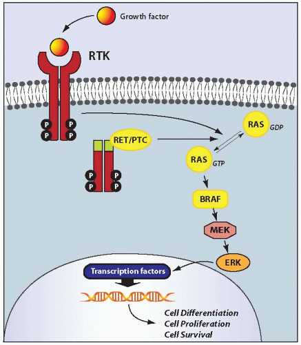
FIGURE 11.4. Schematic representation of the MAPK signaling pathway. Physiologically, binding of growth factors to a receptor tyrosine kinase (RTK), such RET and NTRK1, results in receptor dimerization and activation via autophosphorylation of tyrosine residues in the intracellular domain. The activated receptor through a series of adaptor proteins leads to activation of RAS, located at the inner surface of the plasma membrane, by substitution of GDP with GTP. The active, GTP-bound form of RAS binds to and recruits RAF proteins, mainly BRAF in thyroid follicular cells, to the plasma membrane. Activated BRAF phosphorylates and activates MEK and ERK. Once activated, ERK phosphorylates cytoplasmic proteins and translocates into the nucleus, where it regulates transcription of the genes involved in cell differentiation, proliferation, and survival. Activation of this pathway in papillary carcinoma occurs as a result of point mutation or chromosomal rearrangement affecting the RET, RAS, and BRAF genes.


Table 11.2 MAPK-activating Mutations in Papillary Carcinomas and Their Clinical and Phenotypical Associations


Mutation Type


Average Prevalence (%)


Common Associations


BRAF point mutation


40-45


Older age at presentation Classic papillary carcinoma, tall cell variant Extrathyroidal extension Higher tumor stage at presentation Higher rate of tumor recurrence Propensity for dedifferentiation


RET/PTC rearrangement


10-20


Younger age at presentation Classic papillary carcinoma, diffuse sclerosing variant History of radiation exposure Lymph node metastasis Lower tumor stage at presentation


RAS point mutation


10-20


Follicular variant of papillary carcinoma Tumor encapsulation More frequent vascular invasion Lack of lymph node metastasis Possibly more frequent distant metastasis


TRK rearrangement


<5


Not well defined


The causal role of BRAF mutation in tumor initiation has been confirmed in transgenic mice with thyroid-specific expression of V600E.54,120 These animals frequently developed papillary carcinomas with microscopic features similar to tumors found in humans, although when expressed at physiologic levels in thyroid cells, the mutant BRAF apparently requires TSH stimulation to initiate papillary carcinoma development.54

BRAF V600E mutation is typically found in classic papillary carcinomas and in the tall cell variant and is rare in the follicular variant.113,121,122 Tumors harboring BRAF K601E mutation usually have the follicular variant of papillary carcinoma histology.115,119

As discussed in more detail later in the chapter, the presence of BRAF V600E mutation correlates with aggressive tumor characteristics such as extrathyroidal extension, tumor recurrence, and distant metastases.123 The association between this mutation and older patient age has also been seen in many studies.123 BRAF association with more invasive tumor characteristics is likely due to the upregulation of expression of vascular endothelial growth factor (VEGF), matrix metalloproteinases, and other tumor cancer-promoting targets by mutant BRAF.124,125 The mutation may predispose to tumor dedifferentiation, as it is found in papillary carcinomas that undergo anaplastic transformation.121,126 These properties of mutant BRAF are confirmed in transgenic animals, which develop papillary carcinomas with frequent wide invasion into perithyroidal tissues and progression to poorly differentiated carcinoma.120 A process of dedifferentiation of BRAF-mutated cancers coincides with profound deregulation of the expression of genes involved in cell adhesion and intracellular junction, providing evidence for epithelialmesenchymal transition.127,128


Table 11.3 Types of RET/PTC Rearrangement in Papillary Carcinoma


N


RET/PTC Type


Partner Gene Fused with RET


Cytogenetic Alteration


Frequency among RET/PTC types


1


RET/PTC1129


H4 (CCDC6, D10S170)


inv(10)(q11.2;q21)


65%


2


RET/PTC2133


PRKAR1A


t(10;17)(q11.2;q23)


3%


3


RET/PTC3130,131 (RET/PTC4)132**


NCOA4 (RFG, ELE1)


inv(10)(q11.2;q10)


30%*


4


RET/PTC5134


GOLGA5 (RFG5)


t(10;14)(q11.2;q32)


Rare


5


RET/PTC6135


HTIF1 (TRIM24)


t(7;10)(q32;q11.2)


Rare


6


RET/PTC7135


TIF1G (RFG7, TRIM33)


t(1;10)(p13;q11.2)


Rare


7


ELKS/RET140


ELKS (RAB6IP2)


t(10;12)(q11.2;p13.3)


Rare


8


RET/PTC8137


KTN1


t(10;14)(q11.2;q22.1)


Rare


9


RET/PTC9136


RFG9


t(10;18)(q11.2;q21)


Rare


10


PCM1/RET138


PCM1


t(8;10)(p21;q11.2)


Rare


11


RFP/RET139


RFP (TRIM27)


t(6;10)(p21;q11.2)


Rare


12


HOOK3/RET141


HOOK3


t(8;10)(p11.21;q11.2)


Rare


** RET/PTC4 type has the same fusion partner genes as RET/PTC3, but a different location of a breakpoint in the RET gene.


RET/PTC

RET/PTC rearrangement is another genetic alteration found in a significant proportion of papillary carcinomas. It is formed by fusion among the 3’ portion of the RET gene, coding for the receptor tyrosine kinase (RTK), and the 5’ portion of various unrelated genes. The two most common rearrangement types, RET/PTC1 and RET/PTC3, are paracentric inversions because both RET and its respective fusion partner, H4 or NCOA4 (ELE1; RFG), reside in the long arm of chromosome 10.129,130,131,132 RET/PTC2 and nine more recently identified types of RET/PTC are all interchromosomal translocations (Table 11.3). Most of these rare RET/PTC types have been found in papillary carcinomas from patients with a history of exposure to ionizing radiation,133,134,135,136,137,138,139 with the exception of the ELKS/RET and HOOK3/RET fusions that have been identified in tumors from patients with no radiation exposure history.140,141

RET is not expressed in normal thyroid follicular cells in contrast to thyroid C cells. As a consequence of RET/PTC rearrangement, the portion of RET coding for the tyrosine kinase domain is fused in frame with an active promoter of the fusion partner gene. As a result, the truncated RET receptor becomes constitutively expressed and activated, stimulating the MAPK signaling. RET/PTC transforms thyroid cells in culture142 and leads to the development of papillary carcinomas in transgenic mice, which has been shown in animals with thyroid-specific expression of RET/PTC1 and RET/PTC3.143,144,145

The prevalence and specificity of RET/PTC rearrangement varies dramatically in the reported series.146,147,148 In part, this is due to true difference in the prevalence of this alteration in specific age groups and in individuals exposed to ionizing radiation. However, in many cases, this is due to heterogeneous distribution of this rearrangement within the tumor and variable sensitivity of the detection. This rearrangement can be present in a significant proportion of tumor cells and detected by multiple methods (clonal RET/PTC) or can occur in a small fraction or single cells within the lesion and can be detectable only using ultrasensitive detection techniques (nonclonal RET/PTC).149,150 Clonal RET/PTC occurs in 10% to 20% of papillary thyroid carcinomas and is specific for this tumor type,150,151 whereas nonclonal rearrangements have been reported with a significantly higher prevalence in papillary
carcinomas and also in various other thyroid tumors and benign lesions.146

Clonal RET/PTC is more common in papillary carcinomas from children and young adults and in patients with a history of radiation exposure. This includes individuals subjected to either accidental (mostly radioiodine) irradiation or therapeutic (mostly external beam) irradiation, as 50% to 80% of those papillary carcinomas harbor RET/PTC.152,153 RET/PTC can be induced by ionizing radiation in cultured human thyroid cells and in human thyroid tissue grafted into mice.154,155 The rearrangement may be a direct result of misrejoining of DNA breaks induced by radiation; this may be facilitated by close spatial positioning of chromosomal regions involved in RET/PTC generation, which can be seen by fluorescence in-situ hybridization (FISH) in normal human thyroid cells (Fig. 11.5).156,157

In most series of radiation-induced and sporadic tumors, RET/PTC1 is more common. The notable expression is a population of papillary carcinomas that developed in children shortly (4 to 10 years) after radiation exposure at Chernobyl. Among those, RET/PTC3 was the most prevalent rearrangement type.152,158

RET/PTC-positive papillary carcinomas typically present at younger age and have classic papillary histology, a high rate of lymph node metastases, but low stage at presentation. These findings are particularly characteristic of tumors harboring RET/PTC1.113 Among papillary carcinomas associated with radiation exposure, RET/PTC1 is associated with classic papillary carcinoma, whereas RET/PTC3 type is associated with the solid variant.38 RET/PTC-positive tumors lack the predisposition for progression to poorly differentiated and anaplastic carcinomas.159

The pathogenetic role of nonclonal RET/PTC rearrangement, i.e. RET/PTC detected at a very low level or in single cells within the thyroid nodule or in a gland affected by Hashimoto thyroiditis, is not clear.147,149 A claim that frequent detection of RET/PTC using highly sensitive techniques in glands affected by Hashimoto thyroiditis provides evidence for multiple occult papillary carcinomas that are not detectable histologically63,64 cannot be accepted at this time.67


FIGURE 11.5. Nuclear architecture predisposes to the generation of RET/PTC rearrangements in thyroid cells. Nucleus of normal human thyroid follicular cell hybridized with probes for the RET (green color), H4 (red), and NCOA4 (orange) genes showing close proximity of RET and NCOA4 (fusion partners in RET/PTC3) on one copy of chromosome 10 (upper left) and proximity of RET and H4 (fusion partners in RET/PTC1) on another copy of chromosome 10 (bottom).


RAS

Point mutations involving RAS genes are found in about 10% of papillary carcinomas and affect almost exclusively the follicular variant of this tumor.160,161 The mutations are located at several specific sites (codons 12, 13, and 61) of the NRAS, HRAS, and KRAS genes.162,163,164 The mutation stabilizes the protein in its active, GTP-bound conformation, resulting in chronic stimulation of several signaling pathways, most importantly the MAPK and phosphatidylinositol-3-kinase (PI3K/AKT) pathways. In addition to strong correlation with the follicular variant histology, this mutation is also associated with more frequent tumor encapsulation, less prominent nuclear features of papillary carcinoma, and a lower rate of lymph node metastases.113,160 Some studies have reported an association between RAS mutation and a higher frequency of distant metastases.165


TRK

Rearrangement of the NTRK1 gene, named TRK rearrangement, is the least frequent type of mutation capable of activating the MAPK pathway. NTRK1 is located on chromosome 1q22 and encodes a cell membrane receptor with tyrosine kinase activity.166,167 Similar to the RET gene, NTRK1 is not expressed in normal thyroid follicular cells. The rearrangement juxtaposes the portion of NTRK1 coding for the intracellular tyrosine kinase domain to the 5’ terminal sequence of one of three genes that are highly expressed in thyroid follicular cells.167 Two of them, the TPM3 gene168,169 and the TPR gene,170,171 are also located on chromosome 1q, and therefore, these fusions are intrachromosomal inversions. The third fusion partner, the TFG gene, resides on chromosome 3, and this fusion is a result of the t(1;3) translocation.172 All fusion types lead to the expression and activation of the tyrosine kinase domain of NTRK1. The fusion is tumorogenic for thyroid follicular cells, as TPR/NTRK1 drives the development of papillary carcinomas in transgenic mice.173

TRK rearrangements occur in <5% of papillary carcinomas,174,175 although in some regions of the world the reported frequency is in the range of 10% to 15%.167,176,177 All three fusion types are found with approximately similar incidence, and several tumors with NTRK1 fused to still unknown genes have been reported.176,177 Approximately 5% of radiation-induced papillary carcinomas carry this rearrangement, most commonly the NTRK1/TPM3 type.178


PAX8/PPARλ

This rearrangement is a prototypic alteration found in follicular thyroid carcinoma. However, the published data179 and our own experience indicate that this rearrangement can be found in a small proportion (1% to 5%) of the follicular variant papillary carcinoma. Single reports of a much higher prevalence of this rearrangement in the follicular variant of papillary carcinoma also exist,180 although the difference likely reflects the stringency of diagnostic criteria used to define the follicular variant of papillary carcinoma.


PI3K/PTEN/AKT Pathway Mutations

The PI3K/PTEN/AKT signaling pathway may be activated as a result of RAS mutation as well as mutation of the PIK3CA and PTEN genes (see Chapter 10, Fig. 10.5 for illustration). These mutations are common in anaplastic (undifferentiated) carcinoma and occur with lower prevalence in follicular carcinoma. However, PIK3CA and PTEN mutations are rare in papillary carcinomas, found in only 2% to 5% of tumors.181,182,183


Mutations in the Oncocytic Variant of Papillary Carcinoma

The oncocytic variant of papillary carcinoma reveals the characteristic nuclear features of papillary carcinoma along with cytoplasmic granularity due to accumulation of numerous and frequently abnormal mitochondria. The cause of the mitochondrial change and its relationship to the neoplastic process are not fully understood. It is likely that it may represent either a primary process induced by a distinct tumor initiation mutation or a secondary, probably compensatory change.184,185,186

Mutations of the GRIM-19 (NDUFA13) gene, which encodes a protein that regulates cell death as well as mitochondrial metabolism, occur in oncocytic thyroid tumors. In one study of 10 oncocytic papillary carcinomas, two tumors revealed somatic missense mutations and one a germline mutation of GRIM-19.187 The mutations may disrupt the function of this anti-apoptotic tumor suppressor gene and promote tumorigenesis. However, the prevalence of GRIM-19 mutations in oncocytic thyroid tumors and their role in carcinogenesis remains to be fully characterized.

In some studies, a high prevalence of RET/PTC rearrangements has been found in oncocytic variant papillary carcinomas using highly sensitive detection techniques.188,189,190 The significance of these findings is not clear because these ultrasensitive methods also led to the detection of nonclonal RET/PTC in many oncocytic follicular adenomas and carcinomas.


Alterations in Gene Expression Profiles

Using high-density cDNA microarrays, widespread alterations in gene expression can be detected in papillary carcinomas. Although the pathogenetic role of these changes awaits further analysis, several important observations have been made already. It has been found that gene expression profiles of papillary carcinoma (including follicular variant) are different from those of follicular carcinomas and other thyroid tumor types,191,192,193 supporting the current classification scheme that is based primarily on histopathologic criteria. Furthermore, distinct sets of differently expressed genes have been found in classic papillary carcinoma and in the follicular variant, and possibly in some other variants (such as tall cell variant), pointing toward the pathogenetic and biologic differences between these microscopic tumor variants (Fig. 11.6, left).192,194 It appears that variation in gene expression profiles between papillary carcinomas carrying BRAF, RAS, RET/PTC, and TRK mutations can be detected, providing molecular basis for distinct phenotypical and biologic features associated with each mutation type (Fig. 11.6, right).194,195


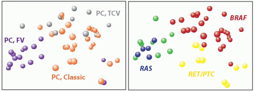
FIGURE 11.6. Correlation between gene expression profiles and histologic variants and mutations in papillary carcinoma. (Left) Principal component analysis based on the expression data for over 22,000 genes shows distinct clusters formed by cases of classic papillary carcinoma (orange spheres), follicular variant (purple), and tall cell variant (grey). (Right) Similar analysis with reference to mutational status demonstrates clusters of tumors carrying BRAF mutation (red), RET/PTC rearrangement (yellow), and RAS mutation (blue). Green spheres represent papillary carcinomas with no mutation. (Based on the data reported by Giordano TJ, Kuick R, Thomas DG, et al. Molecular classification of papillary thyroid carcinoma: distinct BRAF, RAS, and RET/PTC mutation-specific gene expression profiles discovered by DNA microarray analysis. Oncogene. 2005;24:6646-6656.)

Gene expression array studies have also confirmed the overexpression of several genes previously known to be upregulated in papillary carcinoma, such as MET, LGALS3 (galectin-3), and KRT19 (cytokeratin 19). Other characteristic findings included general downregulation of genes responsible for specialized thyroid function such as thyroid hormone synthesis, upregulation of many genes involved in cell adhesion, motility, and cell-cell interaction, and disregulation of the expression of genes coding for cytokines and other proteins involved in inflammation and immune response.191,193,194 Dysregulation of expression of genes involved in cell adhesion and intracellular junction, consistent with epithelial-mesenchymal transition, is particularly prominent during invasion and dedifferentiation of BRAF-mutated cancers.127,128


Alterations in miRNA Expression

MicroRNAs (miRNAs) are small endogenous RNA molecules that do not code for proteins but act as negative regulators of the expression of protein-coding genes.196,197 The miRNAs regulate the expression of well-known oncogenes and tumor suppressor genes. Upregulation and downregulation of specific miRNAs are common in cancer cells and may be involved in carcinogenesis. miRNA expression profiles of papillary carcinoma are different from those of follicular carcinoma and other thyroid tumors.198 Within the papillary carcinoma group, the patterns of miRNA expression correlate with specific somatic mutations found in these tumors.198 Several specific miRNAs, such as miR-221, miR-222, miR-187, miR-181b, and miR-146b, are consistently found to be strongly upregulated in papillary carcinomas, suggesting that they may play a pathogenetic role in the development of these tumors (Fig. 11.7).198,199,200 Possible target genes affected by these miRNAs are the regulators of the cell cycle p27(Kip1) gene and the thyroid hormone receptor gene.201,202 Other miRNAs upregulated in papillary carcinoma at lower levels are miR-155, miR-181b, miR-187, miR- 21, miR-31.199,203,204


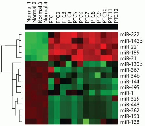
FIGURE 11.7. miRNAs dysregulation in papillary thyroid carcinoma. Cluster dendrogram demonstrates several upregulated (red) and downregulated (green) miR-NAs in papillary carcinomas (PTC) as compared with normal thyroid cells. (Based on data reported by Nikiforova MN, Tseng GC, Steward D, et al. MicroRNA expression profiling of thyroid tumors: biological significance and diagnostic utility. J Clin Endocrinol Metab. 2008;93:1600-1608 and Yip L, Kelly L, Shuai Y, et al. MicroRNA signature distinguishes the degree of aggressiveness of papillary thyroid carcinoma. Ann Surg Oncol. 2011;18:2035-2041.)

Differences in miRNA expression were also observed between papillary carcinomas with aggressive features as compared with nonaggressive tumors. Several miRNAs have been found to be significantly upregulated (miR-146b, miR-221, and miR-222) or downregulated (miR-34b and miR-130b) in aggressive tumors.205,206 The MET gene is a potential target for two downregulated miRNAs (miR-34b and miR-1), and significantly higher level of MET expression was observed in aggressive papillary carcinomas,205 suggesting that this may represent a mechanism of tumor progression.


CLINICAL FEATURES AND IMAGING

Patients typically present with a painless thyroid nodule. Other local symptoms, such as dysphagia, hoarseness, and stridor, are found in about 20% of patients and are indicative of vocal cord paralysis or tracheal compression.20 Enlarged cervical lymph nodes may sometimes provide the first evidence of disease. Overall, neck lymphadenopathy is found at presentation in 27% of patients.20 In some cases, patients are totally asymptomatic, and the nodule in the thyroid is identified incidentally using ultrasound or other imaging studies performed for other reasons. Thyroid function tests are typically normal.

Thyroid radioisotope scan typically reveals a “cold” nodule, although this diagnostic modality has fallen out of favor. Hyperfunctioning papillary carcinoma that appears as a “hot” nodule on thyroid scan have been described, but they are exceedingly rare.207 On ultrasound, papillary carcinoma typically appears as a hypoechoic or isoechoic solid nodule with ill-defined margins (Fig. 11.8). Cystic change may be present, although rarely extensive. Finding punctate microcalcifications (that correspond to psammoma bodies) and high central blood flow within the nodule on color Doppler are more commonly seen in papillary carcinoma although none of the ultrasonographic features are entirely diagnostic of malignancy.208,209 Tumor harboring BRAF mutations appear to have a more elongated shape and less frequently show microcalcifications.210 Cervical lymph nodes may reveal internal nodularity, cystic change, and microcalcification on ultrasound, all of which are suspicious for papillary carcinoma.

Other imaging modalities, such as CT, MRI, and [18F]fluorodeoxyglucose (FDG)-positron emission tomography/computed tomography (PET/CT), can be used to evaluate substernal masses and to assess the extent of extrathyroidal disease. Distant metastases at diagnosis are rare and occur in 2% to 5% of patients.211,212

FNA is performed on almost all solitary thyroid nodules. Cytologic evaluation of the FNA material typically establishes or at least raises the possibility of the diagnosis of papillary carcinoma, resulting in a referral for surgery.


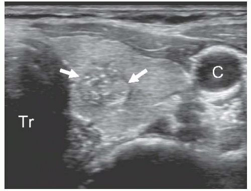
FIGURE 11.8. Ultrasonographic features of papillary carcinoma. Transverse grey scale image of the left thyroid lobe shows an irregularly-shaped isoechoic nodule (arrows) with ill-defined borders and several punctuate hyperechoic foci of microcalcifications, which correspond to psammoma bodies found on microscopic examination. Tr, trachea; C, carotid artery.


GROSS FEATURES

On gross examination, papillary carcinoma typically appears as a discrete but ill-defined nodule with irregular borders (Fig. 11.9 A,B). A capsule is typically absent. Some tumors, particularly the follicular variant of papillary carcinoma, may be well demarcated or encapsulated (Fig. 11.9 C). Tumor size varies widely but most frequently is in the range of 1 to 3 cm.20 The cut surface is tan-brown or grey-white, irregular, firm and solid or more friable with small or larger cystic spaces. Papillary structures may be evident or suspected based on a granular or shaggy texture of the cut surface. Irregularly-shaped whitish areas of fibrosis are frequently seen. Foci of hemorrhage and necrosis can be found in tumors following a recent FNA procedure, whereas spontaneous necrosis and hemorrhage are rare. Calcifications may be occasionally seen. Focal cystic change is identifiable in many tumors, and some tumors may show extensive cystic change, presenting as an ill-defined multicystic lesion. Tumor multifocality is fairly common. In rare instances, diffuse changes are noticed in the thyroid with no discrete tumor mass; this presentation is common for the diffuse sclerosing variant of papillary carcinoma.

Because papillary carcinoma is frequently associated with areas of fibrosis, all whitish, fibrotic-appearing foci found during gross examination should to be sampled for microscopic evaluation.


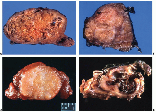
FIGURE 11.9. Gross appearance of papillary carcinoma. A, B: Two examples of classic papillary carcinoma. C: Follicular variant of papillary carcinoma. The tumor is well demarcated and has smooth borders but shows no capsule. D: Metastastatic papillary carcinoma to a lymph node with marked cystic change.

Perithyroidal lymph nodes may be received with a thyroidectomy specimen. Metastatic disease frequently manifests as an enlarged firm lymph node that may also contain cystic areas filled with brownish, hemorrhagic fluid (Fig. 11.9 D).


MICROSCOPIC FEATURES

On microscopic examination, papillary carcinoma typically shows infiltrative growth with an irregular, invasive border (Fig. 11.10). In some cases, the tumor may show a pushing border, although true encapsulation is rare with the exception of the follicular variant of papillary carcinoma. Characteristic microscopic features of papillary carcinoma include growth pattern, nuclear features, psammoma bodies, and tumor fibrosis, but only the nuclear features are required for the diagnosis (Table 11.4).


Architectural Patterns

The most characteristic growth pattern is papillary, although it is rarely pure and typically admixed with a variable proportion of neoplastic follicles. In approximately two-thirds of tumors, a papillary growth predominates, whereas another one-third have a predominantly follicular (including macrofollicular) architecture.213 Other growth patterns include solid and trabecular, which are seen in about 20% of cases but rarely predominate.213


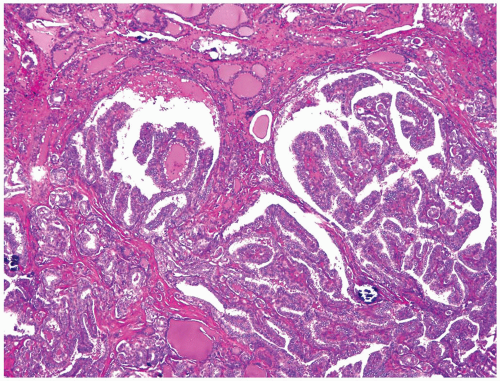
FIGURE 11.10. Low-power view of papillary carcinoma. Note an infiltrative border and a predominantly papillary architecture with occasional neoplastic follicles and scattered calcifications.

Papillae are finger-like projections composed of delicate strands of fibrovascular stroma that are covered by epithelial cells. The papillae are usually irregular, complex, arborizing, ramifying, and branching, with well-developed cores formed by cellular fibrous tissue and capillary blood vessels (Fig. 11.11). In some tumors, the stalks contain loose myxoid edematous stroma or virtually acellular hyalinized material. Occasionally, dense lymphoid infiltration can be seen in the tumor stroma extending into papillae stalks.

The papillae are typically covered by a single layer of epithelial cells. Although some polarity of nuclei with location closer to the basement membrane may be seen, many cells show haphazard (up and down) position of nuclei within the cell. Cellular overlapping and multilayering may be seen as a true phenomenon or more commonly due to tangential sectioning of papillae tips. Papillary growth is a valuable diagnostic feature. Nevertheless, it is important to remember that not all papillary carcinomas have a papillary growth pattern and not all papillary-patterned thyroid lesions are papillary carcinomas. An example of the former is the follicular variant of papillary carcinoma (discussed later in the chapter) and of the latter is papillary hyperplasia in goiter (see Chapter 5) and follicular adenoma with papillary hyperplasia (see Chapter 8). The hyperplastic papillae have a simple, typically nonbranching (nonforking) architecture, delicate hypocellular stalks, and uniform basally located nuclear of epithelial cells that lack the characteristic nuclear features of papillary carcinoma.


Tumor Cells

The cells typically have a cuboidal or low columnar shape and rest on the basal membrane that borders papillary stalks and neoplastic follicles. The cells are larger than adjacent nonneoplastic thyrocytes and usually have abundant, pale, eosinophilic cytoplasm.
The nuclei exhibit characteristic microscopic changes that serve as a core requirement for the diagnosis of papillary carcinoma. The following main diagnostic nuclear features are recognized:


Table 11.4 Main Microscopic Features of Papillary Carcinoma


I. Architecture


Papillary growth pattern


II. Nuclear features


1. Nuclear enlargement


2. Nuclear crowding and overlapping


3. Chromatin clearing


4. Irregularity of nuclear contours


5. Nuclear pseudoinclusions


6. Nuclear grooves


III. Psammoma bodies


IV. Tumor fibrosis


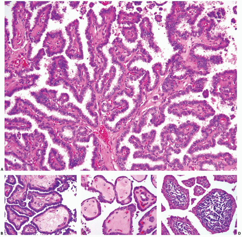
FIGURE 11.11. Papillae of papillary carcinoma. A: Typical tumor papillae with well-developed fibrovascular cores and complex branching. Some papillae may have stalks composed of loose myxoid stroma (B) or acellular hyalinized stroma (C) or contain dense lymphoid infiltration in the stalks (D).

1. Nuclear enlargement


The nuclei of papillary carcinoma cells are typically 2 to 3 times larger than those of nonneoplastic thyroid cells, as the nuclear area increases from 30 to 50 µm2 to 97 to 110 µm2.214,215 The difference in nuclear size is best appreciated when the tumor cells are compared side by side with the adjacent nonneoplastic cells (Fig. 11.12 A). A sharp distinction between the tumor cells containing large nuclei and the nonneoplastic cells with smaller nuclei is required to fulfill this criterion, as opposed to a gradual change in the nuclear size focally seen in benign reactive conditions such as Hashimoto thyroiditis.

2. Nuclear overlapping and crowding


Overlapping of nuclei in cells lining the neoplastic papillae or follicles is common. It reflects the larger size of the haphazardly arranged nuclei within the cell volume (Fig. 11.12 B). Tangentially sectioned tips of the papillae appear as “crowds” or “lakes” of overlapping nuclei, which is also described as the “egg-basket” appearance.216

3. Chromatin clearing


Dispersion of nuclear chromatin with its margination along the nuclear membrane appears microscopically as optically clear nuclei with thickened nuclear membrane (Fig. 11.12). This finding, also known as empty, pale, clear, water-clear, ground-glass or “Orphan Annie Eye” nuclei,217,218 had been considered for a long time as the most characteristic nuclear feature of papillary carcinoma. This nuclear
appearance requires a tissue fixation step, as it is not seen in frozen sections or FNA smears.217 Formalin fixation most consistently yields nuclear clearing, which is also seen after fixation in Bouin fluid, Zenker fluid, or B5.216 However, other fixatives such as SafeFix and HistoChoice tend not to show this nuclear appearance (Lester D. R. Thompson, unpublished data). Although changes in the distribution of chromatin and ribonucleoproteins have been seen in these tumors by electron microscopy,219,220 the exact mechanism of nuclear clearing is not well understood.


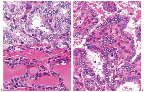
FIGURE 11.12. Nuclear features of papillary carcinoma. A: Enlarged size of tumor nuclei (top) is easier to appreciate when compared with adjacent normal thyroid tissue (bottom). B: Nuclear overlapping and crowding with “lakes” of overlapping nuclei. Many nuclei have finely dispersed chromatin and empty appearance with prominent nuclear membrane due to chromatin margination.

4. Irregularity of nuclear contours


In contrast to the round, smooth-contoured nuclei of normal thyroid cells or follicular lesions, the nuclei of papillary carcinomas cells have (1) elongated, oval shape, (2) asymmetric, irregular configuration with angulated, crescent-moon, and triangular shapes, and (3) highly irregular, jagged nuclear membrane with indentations, “rat bites,” and folds of various sizes and directions (Fig. 11.13 A). This light microscopic appearance reflects a marked irregularity of the nuclear contours that is seen ultramicroscopically (Fig. 11.13 B), as discussed later in the chapter. Importantly, the assessment of this feature should be performed in sections from tissue blocks that were not previously frozen during intraoperative consultation, as freezing introduces a prominent artifactual irregularity of the nuclear membrane.

5. Nuclear grooves


This is a direct consequence of marked irregularity of the nuclear contours, which manifests as a discrete, longitudinal fold typically situated along to the long axis of the nucleus.221 The fold may be linear and regular (a “coffee bean” appearance) or curved and irregular (a “piece of popcorn” appearance) (Fig. 11.14 A). By electron microscopy, it represents a deep invagination of the nuclear membrane (Fig. 11.14 B). This is a common finding and in most tumors, and one or more grooved nuclei are typically found in every high-power field.

6. Nuclear pseudoinclusions


This is another result of nuclear membrane irregularity, as an intranuclear cytoplasmic pseudoinclusion is formed by invagination of the nuclear membrane that drags a portion of the cytoplasm into the nuclear volume. The pseudoinclusion appears as a round or more irregular area within the nucleus that has staining qualities of the cytoplasm and is sharply demarcated by a thick nuclear membrane (Fig. 11.15).222,223 Nuclear pseudoinclusions are the least common feature seen in papillary carcinoma, identified in only about 50% of cases.113,221

The pseudoinclusions may be mimicked by punched out defects in the staining of nuclei, which is common in frozen sections (Fig. 11.16A). The pseudoinclusion-like nuclear bubbles or vacuoles may also be seen in routinely processed sections that are poorly fixed (Fig. 11.16 B).224,225 These artifactual formations typically have empty appearance or pale staining, are structureless, and lack a rim of the nuclear membrane. The bubbles may contain a nucleolus within its space, which rules out the possibility of a true cytoplasmic pseudoinclusion. In addition, these artifactual patches are frequently seen in numerous nuclei in a field, a finding that should immediately raise a question about their authenticity. This is because true intranuclear cytoplasmic pseudoinclusions are rarely seen in abundance and typically found in no more than 1 to 2 cells per high power field.226


FIGURE 11.13. Marked irregularity of the nuclear contours. This important feature of papillary carcinoma can be seen on light microscopy (A) and electron microscopy (B).


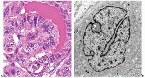
FIGURE 11.14. Nuclear grooves. These structures are often seen in papillary carcinoma cells on light microscopy (A). They are formed by linear invagination of the nuclear membrane that travels deep into the nuclear volume, which is best appreciated on electron microscopy (B).


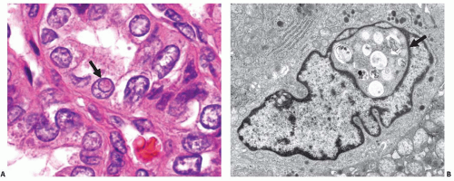
FIGURE 11.15. Nuclear pseudoinclusions. A: Light microscopy image showing a cell in the center of the field that contains an eosinophilic intranuclear body (arrow), which is demarcated by a rim of nuclear membrane and stained more like the cytoplasm than the remainder of the nucleus. B: Ultramicroscopic image of a cell containing a nuclear pseudoinclusion (arrow), which is surrounded by a nuclear membrane and contains phagolysosomes and other cytoplasmic organelles.


Frequency and Specificity of the Nuclear Features

In many papillary carcinomas, all or almost all of the nuclear features are readily identifiable, and this is diagnostic of papillary carcinoma irrespective of the growth pattern and other findings. However, some tumors exhibit most, but not all, of these features, or they are found focally within the nodule. There is no consensus on how many nuclear features are sufficient for the diagnosis and how widespread they should be.227 In our experience, most papillary carcinomas reveal at least four of these nuclear features, and they are identifiable in many areas of the tumor.113 We particularly rely on finding enlarged nuclei with nuclear contour irregularity. In our opinion, these two features are the most common and specific diagnostic features, particularly when they are present in a lesion sharply demarcated from the benign parenchyma. However, none of these features are pathognomonic for papillary carcinoma and a single feature may be seen, particularly focally, in many benign lesions.217,224,228,229 This is particularly true for the grooved nuclei, which can be found in scattered cells of various benign and malignant lesions of the thyroid. True nuclear pseudoinclusions have a relatively high specificity, but they are the least common feature and not found in a significant proportion of papillary carcinomas. Thyroid lesions composed of cells showing several, but not all, nuclear features are most challenging diagnostically, especially when they are encapsulated and lack papillary architecture, which is typically seen in the encapsulated follicular variant of papillary carcinoma.


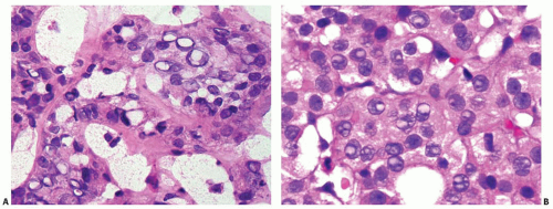
FIGURE 11.16. Structures mimicking nuclear pseudoinclusions. A: An intraoperative frozen section showing numerous nuclei with sharpededged, punched-out empty spaces. B: A routine formalin fixed section showing multiple nuclei with bubbles that are pale stained and lack the texture of the adjacent cytoplasm. Many of the bubbles contain nucleoli.


Psammoma Bodies

Psammoma (from Greek “salt-like”) bodies are distinctive calcifications that are found in 40% to 50% of papillary carcinomas.113,213 In order to qualify as a psammoma body, the calcification must have the following characteristics:


  • have a round or spherical shape,


  • show concentric layers of calcium deposition, and


  • be located in association with tumor cells, in the tumor stroma, or in a lymphatic channel, but not within the lumen of a follicle (Fig. 11.17).


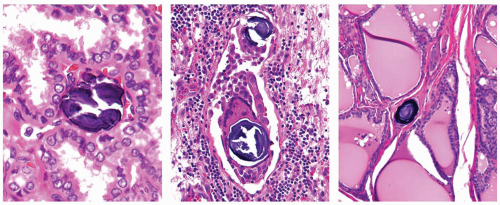
FIGURE 11.17. Psammoma bodies. True psammoma bodies are located either in a stalk of the papillae (left) or in association with tumor cells and multinucleated giant cells within a lymphatic channel (middle), or in isolation within thyroid stroma (right).

The intrafollicular calcifications can be seen in follicular adenomas and carcinomas, particularly of oncocytic type, and represent inspissated and calcified colloid. True psammoma bodies are believed to be formed by successive layering of calcium centered on a single or a small group of necrotic tumor cells, which serves as a nidus for calcium deposition.216,230 Cell necrosis may develop at the tip of the papillae as a result of vascular thrombosis of a vessel within the stalk or in the intralymphatic tumor aggregate.230 Psammoma bodies may be found in isolation, that is, with no tumor cells in the vicinity, and this is believed to represent a “tombstone,” marking the site of prior viable tumor cells (Fig. 11.17, right).231 However, the precise mechanism of psammoma body formation is not fully understood. The possible role of the immune response is suggested by the frequent presence of multinucleated giant cells intimately associated with psammoma bodies (Fig. 11.17, middle). Furthermore, macrophages in the vicinity of psammoma bodies overexpress a bone matrix protein osteopontin that participates in mineralization of bones and in ectopic calcification.232 Psammoma bodies are particularly abundant in the diffuse sclerosing variant of papillary carcinoma and in tumors that carry RET/PTC rearrangement.113 They are least common in the follicular variant and in tumors with RAS point mutations.

Psammoma bodies are not entirely specific for papillary carcinoma and have been reported in some benign lesions, such as nodular goiter with papillary hyperplasia.233 However, true psammoma bodies are exceedingly rare in lesions other than papillary carcinomas, and their finding in the lesion with at least moderately expressed nuclear features of papillary carcinoma is virtually diagnostic of this tumor. Finding isolated psammoma bodies in the lymphatic channels, lymph nodes, or in the stroma of otherwise normal thyroid tissue provides strong evidence for papillary carcinoma present either in the adjacent thyroid tissue or in the contralateral lobe of the thyroid. In one study of 29 cases of isolated psammoma bodies found either in thyroid parenchyma (27 cases) or in perithyroidal lymph nodes (2 cases), 27 (93%) cases revealed either papillary (25 cases) or oncocytic (2 cases) carcinoma.234 Two cases revealed no malignant tumor, but both of these patients underwent only lobectomy and the removed single lobe was not entirely submitted for microscopic examination. Of interest, most papillary carcinomas found in this study were incidental tumors <1 cm in size, and 44% of cases had psammoma bodies found in the opposite lobe from the carcinoma (intraglandular spread).


Tumor Fibrosis

In addition to the fibrous stroma of papillary stalks, discrete areas of fibrosis are frequently seen in papillary carcinomas. Discrete or interconnected fibrous bands traversing the tumor nodule in different directions or located at the periphery and forming a pseudocapsule may be found, as well as large, irregularly shaped, sometime stellate fibrous areas (Fig. 11.18). The fibrosis is typically paucicellular (sclerotic type) and less frequently cellular (desmoplastic type).229 Considerable fibrosis is found in 50% to 90% of all tumors.113,213,235 This type of densely eosinophilic fibrosis is not specific for papillary carcinoma, although it is far more characteristic of these tumors than of other thyroid follicular neoplasms. It serves as a particularly helpful diagnostic hint in two specific situations: (1) at the time of gross examination, when all small whitish, fibrotic-appearing areas should be sampled for microscopic evaluation as they often mark the areas of papillary carcinoma and (2) in the encapsulated follicular-pattern nodules, the presence of significant fibrosis not associated with degeneration or post-FNA injury provides support for the diagnosis of follicular variant of papillary carcinoma rather than follicular adenoma.


Other Microscopic Features

Colloid within neoplastic follicles frequently has a more intense, darkly eosinophilic tincture as compared with colloid in the adjacent nonneoplastic follicles (Fig. 11.19 A).236 Multinucleated giant cells and foamy macrophages can be seen in the lumen of neoplastic follicles and between papillae (Fig. 11.19 B).

Squamous metaplasia is seen in about 20% of papillary carcinomas213,237 and is more common in the diffuse sclerosing variant and tumors with classic papillary growth. In most cases, it appears as discrete concentric whorls of cells with keratinization and/or intercellular bridges (Fig. 11.19 C). Staining for thyroglobulin in these foci is often negative.224

Some degree of cystic change is common (Fig. 11.19 D), but it is usually not extensive. Approximately 10% of tumors reveal marked cystic transformation.213

Mitotic figures are uncommon. Typically, <1 to 2 mitoses per 10 high power fields (400× magnification: 40× objective combined with 10× eyepiece) are identified.238,239 Atypical mitoses are exceedingly rare. If high mitotic activity is found, it should raise the possibility of poorly differentiated thyroid carcinoma.


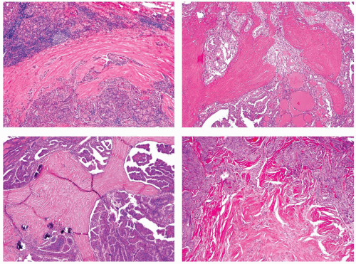
FIGURE 11.18. Various appearances of fibrosis in papillary carcinoma. Significant lymphocytic infiltration is found within the tumor stroma or in thyroid tissue surrounding the tumor in 25% to 40% of cases.113,213,237 Prominent stromal infiltration is a characteristic feature of the Warthin-like variant as described later in the chapter. Infiltration in the adjacent thyroid tissue may be either reactive (peritumor thyroiditis, see Chapter 4) or a part of chronic lymphocytic (Hashimoto) thyroiditis. As peritumoral lymphocytic infiltration is common in papillary carcinoma, this finding should not lead to the diagnosis of chronic lymphocytic (Hashimoto) thyroiditis, unless the same changes are found in thyroid tissue remote from the tumor, preferably in the opposite lobe.


SPREAD AND METASTASES


Multicentricity versus Intraglandular Spread

Papillary carcinoma presents as a multifocal disease in 22% to 35% of cases,213,240 which may represent either multiple independent primary tumors or intraglandular dissemination from a single primary tumor. As discussed earlier, clonality studies have shown that many discrete tumor foci in multifocal disease are of independent clonal origin.90,91,92,93 In addition to the true multicentricity, the intraglandular dissemination of the tumor via lymphatic channels is very common, and multiple small or larger satellite foci are found in the vicinity or even more remotely from the main tumor mass in 27% to 78% of cases.241,242 The distinction between independent primary tumors and intraglandular spread on one tumor is not always possible. In our experience, true independent primary tumors (judged based on the presence of different mutations) are more commonly: (1) located in different thyroid lobes, (2) have distinct histologic appearance and/or belong to different variants of papillary carcinoma, (3) show partial or complete encapsulation, and (4) show no smaller satellite tumor foci surrounding the main tumor nodule.


Lymphatic and Blood Vessel Invasion

Papillary carcinoma is frequently seen invading lymphatic vessels in the thyroid parenchyma. In contrast, invasion of blood vessel located within the thyroid or in adjacent soft tissues is seen in less than 10% of cases.213,243,244,245 Vascular invasion is more common in the follicular variant of papillary carcinoma than in other variants.160,245 In order to qualify for vascular invasion, the tumor aggregates should be seen attached to the vessel wall and covered by a layer of endothelial cells (see detail description and illustration of vascular invasion in Chapter 10). The presence of vascular invasion is an unfavorable prognostic factor, as it correlated with higher risk of distant metastases243,244,245 and tumor-related mortality.243


Extrathyroidal Extension

Papillary carcinomas typically have an infiltrative border and may extend beyond the confines of the gland by direct invasion. Extrathyroidal extension is defined as tumor penetration through the thyroid gland capsule into adjacent tissues. It is an important parameter used in the AJCC TNM-based staging system (Table 7.3.).246 For tumor staging, extrathyroidal extension is subdivided into minimal, which is tumor invasion into immediate perithyroidal soft tissues or sternothyroid muscle (corresponds to T3) (Fig. 11.20 A) and extensive, which is tumor invasion into
subcutaneous soft tissues, larynx, trachea, esophagus, or recurrent laryngeal nerve (corresponds to T4) (Fig. 11.20 B). Extrathyroidal extension is found in 20% to 25% of tumors213,240 and is more common in BRAF-positive papillary carcinomas and less common in the follicular variant.113 As discussed later in the chapter, extrathyroidal extension is an important prognostic factor, although some more recent studies suggest that only extensive extrathyroidal invasion correlates with prognosis.247,248


FIGURE 11.19. Additional changes seen in papillary carcinoma. A: Darker eosinophilic colloid in papillary carcinoma (right) as compared with adjacent thyroid (left). B: Multinucleated giant cell in a space between papillae. C: Two foci of squamous metaplasia. D: Cystic change in papillary carcinoma.


Local and Distant Metastases

Spread to the cervical lymph nodes is a common feature of thyroid papillary carcinoma, whereas hematogenous tumor spread to distant sites is rare. At the time of initial diagnosis, regional lymph node metastases are found in 30% to 50% and distant metastases in 2% to 5% of tumors.211,212,249 The most common pattern of tumor growth in the metastatic deposits in lymph nodes is papillary, even if the primary tumor has a predominantly follicular architecture (Fig. 11.21 A). Cystic changes in lymph node metastases are common, and they are prominent in about 25% of cases (Fig. 11.21 B).250 Tumor extension beyond the capsule of a lymph node can be seen in rare cases, and this is an unfavorable prognostic factor (Fig. 11.21 C).251


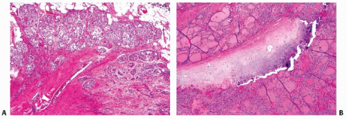
FIGURE 11.20. Extrathyroidal extension. A: Minimal extrathyroidal extension into adjacent soft tissue (TNM, T3). B: Extensive extrathyroidal extension involving tracheal cartilage (TNM, T4a).

Distant metastases most frequently involve lungs (70%) (Fig. 11.21 D), followed by bones (20%) and other rare sites such as soft tissues of the mediastinum and brain.252,253,254


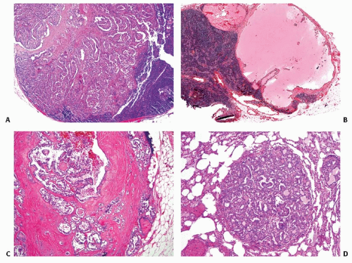
FIGURE 11.21. Papillary carcinoma metastases. A: Tumor metastasis to a lymph node. B: Lymph node metastasis with marked cystic change. C: Lymph node metastasis with tumor extension through the lymph node capsule. D: Tumor metastasis to the lung.


GRADING

Papillary carcinomas should not be graded because by definition they are well-differentiated tumors. Emergence of poorly differentiated thyroid carcinoma manifests by a solid, trabecular, or insular growth pattern and loss of characteristic nuclear features, frequently coupled with tumor necrosis and increased mitotic activity.255

Jul 9, 2016 | Posted by in PATHOLOGY & LABORATORY MEDICINE | Comments Off on Papillary Carcinoma

Full access? Get Clinical Tree

Get Clinical Tree app for offline access