Neuroendocrine Tumors of the Thyroid Gland


Neuroendocrine Tumors of the Thyroid Gland


Neuroendocrine tumors of the thyroid include medullary carcinoma, paraganglioma, and small cell carcinoma, majority being medullary carcinomas.

The neuroendocrine cells of the thyroid are referred to as C cells or calcitonin-producing cells, also known as “parafollicular cells” or “clear cells.” The C cells are located individually or in small groups within thyroid follicles. They are found at the periphery of the follicular wall within the basement membrane and without contact with the follicular lumen. They are in an intrafollicular rather than interfollicular position as confirmed by ultrastructural examination. The C cells are separated from the thyroid interstitium by the basal lamina. They are restricted to the midupper and upper thirds of the lateral lobes of the thyroid; hence, medullary carcinomas are found only in these locations (see Fig. 8.2C) and not in the isthmus or the extreme upper or lower third of the lateral lobes.

The C cells are derived embryologically from the remnants of the ultimobranchial body. By light microscopy, the C cells cannot be visualized in routine hematoxylin and eosin (H&E)-stained sections, but can be highlighted by special stains. Most C cells are round to polygonal. Special stains show that they are argyrophilic (Grimelius stain). They exhibit marked metachromasia with toluidine blue. Ultrastructurally, C cells contain the hallmark of polypeptide-producing cells, that is, membrane-bound neurosecretory granules. The latter are of two types: larger 250 to 280 nm and the smaller 130 nm. The C cells are rich with mitochondria and also contain abundant endoplasmic reticulin with lamellar stacking. Occasional C cells have prominent cytoplasmic processes that extend beyond the adjacent follicular cell. They stain intensely with calcitonin and calcitonin gene-related peptide as well as other neuroendocrine markers such as chromogranin, synaptophysin, neuron-specific enolase, and carcinoembryonic antigen (CEA).

C-cell hyperplasia is considered a precursor lesion for medullary carcinomas (Fig. 8.1A-C).


MEDULLARY CARCINOMA OF THE THYROID

Medullary carcinoma of the thyroid arises from the calcitonin-producing “C cells” comprising 3% to 10% of all thyroid malignancies. It occurs in two forms; roughly 20% are familial and the remaining as sporadic forms. The hereditary form of medullary carcinomas transmitted as an autosomal dominant trait includes three types: multiple endocrine neoplasia (MEN) syndromes—MEN 2A, MEN 2B, and non-MEN familial type (Table 8.1). The MEN 1 syndrome is associated with tumors of the other endocrine organs (see Table 1.1).


CLINICAL FEATURES

The sporadic form of medullary carcinoma occurs more commonly in women, primarily in the older individuals in fifth to sixth decades of life. They clinically present as a unilateral, palpable thyroid nodule and have a high incidence (up to 50%) of cervical lymph node metastases. Distant metastases via hematogenous spread to liver, lungs, bone, and occasionally to brain occur in roughly 15% of the cases. Occult medullary carcinomas with local metastatic disease involving the cervical lymph nodes have been described.

The familial forms are common in males and occur at a younger age. The non-MEN familial medullary carcinoma is slightly more common in women.

Majority of the patients present with a painless thyroid nodule that is cold on scan. Up to 50% of the patients will have nodal metastases and up to 15% may have distant metastases. Virtually all medullary carcinomas produce calcitonin. Elevated serum levels are diagnostic of the malignancy.

Medullary thyroid carcinomas in general project good clinical outcome, especially in sporadic forms. Aggressive clinical behavior with fatal outcome is described in some hereditary forms.


Medullary Carcinoma (Figs. 8.1 and 8.2)


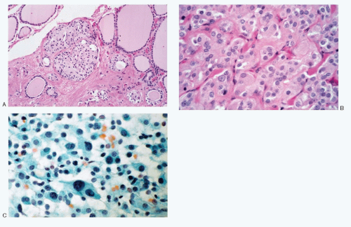
Fig. 8.1: A: Histologic section of a thyroid demonstrating C-cell hyperplasia. Note well-defined nests of cuboidal to polygonal cells in between the thyroid follicles. There was no grossly visible tumor. The patient had several members of the family with hereditary type medullary carcinoma. B: Higher magnification. These cells have abundant eosinophilic cytoplasm. C: Imprint of the surgically resected specimen showing discrete C cells with plasmacytoid appearance.


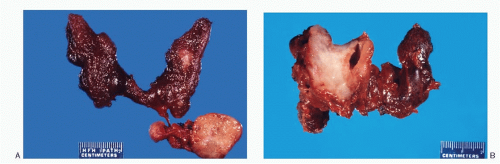
Fig. 8.2: A: A gross photograph of a nonfamilial (sporadic) form of medullary thyroid carcinoma located in the middle third of the left lobe and lymph node metastases (arrow). B: This nonfamilial medullary carcinoma is large replacing the entire right lobe. The tumor presents a flat cut surface.


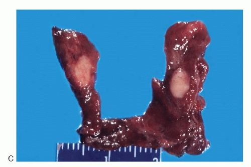
Fig. 8.2: (continued) C: An example of hereditary medullary carcinoma. Note the tumor is located in the middle third of the lobe and bilateral.


RADIOLOGIC FINDINGS

Medullary carcinomas present as hypofunctioning cold nodules. The plain x-rays of the neck may show dense calcification. Ultrasound reveals a hypoechoic mass. Imaging with ultrasonography using 131I metaiodobenzylguanidine demonstrates a positive uptake and is a useful diagnostic tool.


GROSS AND HISTOLOGIC FEATURES

The sporadic form of medullary carcinoma occurs as a unilateral, well-defined, nonencapsulated solitary nodule variable in size, ranging from <1 cm to several centimeters, and may replace the entire lobe (Fig. 8.2A,B). Its cut surface is tan-white to gray or pink, usually flat. The consistence is soft to firm, at times gritty, without areas of hemorrhage and necrosis. Calcification may be present. The hereditary forms are usually multifocal and bilateral (Fig. 8.2C). Their gross appearance is similar to the sporadic medullary carcinomas.


TABLE 8.1. HEREDITARY FORMS OF MEDULLARY THYROID CARCINOMA


MEN 2A


Medullary thyroid carcinoma


Adrenal pheochromocytoma


Parathyroid hyperplasia


Associated precursor lesions


C-cell hyperplasia


Adrenal medullary hyperplasia


MEN 2B


Medullary thyroid carcinoma


Adrenal pheochromocytoma


Mucosal neuromas


Gastrointestinal ganglioneuromas


Musculoskeletal abnormalities


Melanosis


Associated precursor lesions


C-cell hyperplasia


Adrenal medullary hyperplasia


Non-MEN familial type


MEN, multiple endocrine neoplasia.


Like many neuroendocrine tumors, the medullary thyroid carcinomas present an extremely wide spectrum of morphologic patterns that varies from tumor to tumor and within the same tumor. They also share morphologic similarities with other neuroendocrine tumors such as paragangliomas, carcinoid tumors, and small cell carcinomas. In addition, medullary carcinomas mimic not only several types of thyroid follicular cell derived tumors, such as follicular adenomas including hyalinizing trabecular adenoma, follicular carcinoma, papillary, insular, Hürthle cell, or anaplastic carcinomas, but also a wide array of nonthyroid neoplasms as well as nonneoplastic entities as described in differential diagnoses (see Table 8.4) The histologic patterns of medullary carcinomas (Fig. 8.3A-O) are similar in both the familial type and the sporadic type showing multiple growth patterns such as lobular, insular, trabecular, solid, rarely follicular or papillary, and fascicular, the latter formed by the spindle cells simulating a sarcomatous pattern. The nests of tumor cells vary in size, separated by variable stromal tissue, imparting an organoid pattern. The medullary carcinoma cell types vary in size and shapes, ranging from small, medium, to large with occasional giant forms. Their shapes vary from round to oval, carcinoid-like cells, cuboidal, plasmacytoid, polygonal or epithelioid, to spindle shape, triangular, and caudate to racket shaped. Their cell borders are well to poorly defined, and the cytoplasm varies from scant to abundant, clear, pale, to dense. Some may contain
mucin. Their nuclei can be monomorphic to pleomorphic, round, oval, oblong to spindle shape with stippled to coarsely granular chromatin popularly referred to as salt-pepper chromatin. Intranuclear cytoplasmic inclusions are almost always present. Hemorrhage, necrosis, and mitotic activity are infrequent findings in medullary carcinomas. A given tumor may be monomorphic showing a predominant cell type or pleomorphic with mixed patterns containing several cell types. The latter is more frequent.


Spectrum of Histopathologic Patterns of Medullary Carcinoma


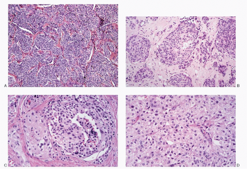
Fig. 8.3: A: Medullary carcinoma showing a lobular growth pattern and is composed of small round cells (H&E). B: Medullary thyroid carcinoma showing lobules of pleomorphic cells of varying size consisting of small to large polygonal and short spindle cells separated by amyloid containing stroma (H&E). C: Medullary thyroid carcinoma. The left half of the field shows tumor composed of large polygonal cells with abundant eosinophilic cytoplasm, while the rest consists of smaller round to cuboidal cells (H&E). D: Medullary carcinoma is composed predominantly of large polygonal cells with abundant eosinophilic cytoplasm bearing a morphologic resemblance to oncocytes (H&E).


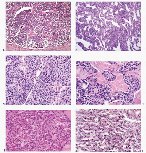
Fig. 8.3: (continued) E: Histologic section of a medullary carcinoma showing a lobular pattern. The lobules vary in size separated by bands of fibrous tissue septae. Also note clear change in the cytoplasm (H&E). F: Medullary carcinoma exhibiting a lobular and a trabecular growth pattern. The trabeculae vary in thickness from slender to broad and are interdigitating (medium power, H&E). G: Higher magnification of (F) showing broad trabeculae consisting of very pleomorphic, round to short spindle-shaped cells (H&E). H: Medullary carcinoma exhibiting nests of small cuboidal cells separated by amyloid (H&E). I: This medullary carcinoma is composed of uniform cuboidal cells forming small nests with a pseudofollicular growth pattern (H&E). J: A solid growth pattern of medullary carcinoma. The cells contain scant to indiscernible cytoplasm (H&E).


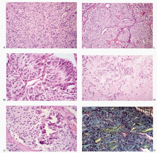
Fig. 8.3: (continued) K: Medullary carcinoma cells are short spindle shaped and separated by stroma (H&E). L: Medullary carcinoma with a spindle cell pattern. The spindle cells are forming large nests separated by fibrous tissue septae (H&E). M: Histologic pattern of this medullary carcinoma with nests of elongated spindle cells resembling a paraganglioma (H&E). N: Medullary carcinoma with abundant amyloid in the stroma. Fine needle aspirates from such areas may be poorly cellular (H&E). O: Medullary carcinoma with stromal fibrosis and calcification (H&E). P: Histologic section of medullary carcinoma showing apple green birefringence of amyloid stained with Congo red (H&E).


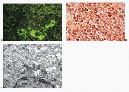
Fig. 8.3: (continued) Q: Bright green fluorescence of amyloid when stained with thioflavin T and viewed under ultraviolet light (H&E). R: Medullary carcinoma demonstrating a strong positive reactivity with calcitonin stain. S: Electron micrograph of medullary carcinoma showing membrane-bound, spherical neurosecretory granules with an electron-dense core (arrow). Uranyl acetate and lead citrate preparation (×42,000).

Medullary carcinoma is characterized by the presence of amyloid deposits in the stroma in up to 80% of the cases (Fig. 8.3H,N). The amyloid shows green birefringence under crossed polarized light when stained by Congo red (Fig. 8.3P) and green fluorescence under ultraviolet light when stained by thioflavin T (Fig. 8.3Q). Medullary carcinoma cells react strongly to calcitonin antibody (Fig. 8.3R).

The medullary carcinoma may also contain a significant amount of collagenous stroma, with (Fig. 8.3O) or without calcification. Several morphologic variants of medullary carcinoma have been described. These include follicular or tubular, papillary, small cell, giant cell, clear cell, melanotic (pigmented), oncocytic, squamous, amphicrine (composite calcitonin and mucin-producing), and paraganglioma-like.


HISTOCHEMISTRY

Argyrophilia as demonstrated by silver stain (Grimelius) is present in 90% of medullary carcinomas. The stroma shows periodic acid-Schiff stain and Alcian blue positivity. Both, intra and extracellular mucin are demonstrated by mucicarmine stain in a high proportion of cases.


IMMUNOPROFILE OF MEDULLARY


CARCINOMA

Positive reactivity to calcitonin is very specific for medullary carcinomas, although rare negative reactivity has been reported. The stain works well on alcohol-fixed cytologic preparations. The carcinoma cells react positively to a host of antibodies such as chromogranin and synaptophysin, aimed at neuroendocrine differentiation (Table 8.2). Medullary carcinoma cells show positive reactivity to CEA and low molecular weight cytokeratin but very rarely to high molecular weight keratin. Reactivity to vimentin is variable.


ULTRASTRUCTURE

Ultrastructurally, medullary carcinoma cells demonstrate characteristic membrane-bound secretory granules (Fig. 8.3S).


TABLE 8.2. IMMUNOPROFILE OF MEDULLARY CARCINOMA


Antibodies


Reactivity


Calcitonin


Positive


Low molecular weight cytokeratin


Positive


High molecular weight cytokeratin


Rarely expressed


Vimentin


Variable


Neuron-specific enolase


Positive


Synaptophysin


Positive


Chromogranin


Positive


CEA


Positive


Thyroglobulin


Negative


TTF-1


Positive


Somatostatin


Positive


CD56


Positive


TTF-1


Positive


CYTOPATHOLOGIC FEATURES

The specimens for cytopathologic diagnosis mostly represent fine needle aspiration (FNA) biopsies of thyroid nodules or neck masses such as enlarged lymph nodes involved by metastatic medullary carcinoma. Aspiration biopsies of organs such as lung, liver, bone, and subcutaneous masses are also examined to rule out a metastatic disease. Occasionally, specimens are obtained via bronchoscopy in cases of pulmonary involvement.

Cytopathologic features of medullary carcinoma encompass a broad spectrum (Table 8.3; Figs. 8.4,8.5,8.6,8.7,8.8,8.9,8.10,8.11,8.12,8.13,8.14,8.15,8.16,8.17,8.18,8.19 and 8.20), Depending on the histomorphology of a given medullary carcinoma, the cytologic presentation will be either monomorphic, that is, if only one pattern is evident, or pleomorphic with a combination of different morphologic patterns. The pleomorphic pattern is more frequently observed.

The aspirates of medullary carcinomas are usually adequate to hypercellular; scant cellularity is encountered with carcinomas containing extensive amyloid deposits and calcification.

The direct smears of the aspirate show malignant cells either isolated, presenting a dispersed cell pattern, or in loosely cohesive groups (Figs. 8.4A, 8.5, and 8.12B). Syncytial-type tissue fragments are also encountered although infrequently (Fig. 8.4B). A true papillary or follicular pattern is not identified. However, a pseudofollicular pattern may rarely be seen (see Fig. 8.29A). The spindle cells may be present in fascicles, dispersed, or aggregates with intertwining cytoplasmic processes (Figs. 8.9A,B; 8.13B; 8.14A; and 8.18A,B).

Medullary carcinoma cells are very pleomorphic; any size or shape may be present (Figs. 8.4C,D; 8.5; 8.12A; 8.13A; and 8.15A). The carcinoma cells can be small, round to cuboidal, reminiscent of carcinoid tumor cells (Fig. 8.6 and 8.7B), or oval to plasmacytoid (Fig. 8.12B). They may be triangular, polyhedral, racket shaped, caudate, or spindle-shaped (Fig. 8.18A-C). Their size also varies: the small, round cells are slightly larger than the thyroid follicular cells, and the larger cells are several microns in their largest dimension (Fig. 8.15A). A cellular aspirate with a dispersed cell pattern formed by pleomorphic cells is virtually pathognomonic of medullary carcinoma (Fig. 8.5), whereas a monomorphic pattern comprising only one type of cell is not frequently observed (Figs. 8.28A, 8.30A, 8.32B, and 8.33B). The latter does cause typing difficulties.

The nuclei of medullary carcinoma cells are always eccentric, regardless of the cell shape, cell size, or number of nuclei. Extreme marginal location of the nucleus is characteristic of the plasmacytoid cell type, and bi- and multinucleation occur very frequently (Fig. 8.4D). The nuclei are round, sometimes oval, and occasionally oblong or elongated as in spindle-shaped cells. Their chromatin is coarsely granular, stippled with salt-pepper pattern, characteristic of neuroendocrine tumors (Figs. 8.8 and 8.9). The presence of nucleoli is not a consistent finding. Bizarre nuclei, such as those seen in anaplastic carcinomas, are rare but do occur. A remarkable and consistent feature is the presence of intranuclear cytoplasmic inclusions (Figs. 8.4C and 8.14B).


TABLE 8.3. CYTOPATHOLOGIC FEATURES OF MEDULLARY CARCINOMA


Cellularity


Variable, depends on the stromal/amyloid content; usually very cellular


Presentation


Cells mostly isolated, in aggregates and infrequently in syncytial tissue fragments forming nests and trabeculae; follicular or papillary architecture not usually present; pseudofollicular pattern rarely present; dispersed cell pattern very characteristic; aggregates of spindle cells may present a “fish-school” pattern or occur in fascicles


Cells


Uniform to markedly pleomorphic; size ranging from small, medium-sized to large with frequent giant forms; varied shapes: round, cuboidal, plasmacytoid, polygonal, caudate, racket shaped, triangular, short to elongated spindle shaped; unipolar cells; admixture of various types is characteristic; cell borders well to poorly defined; variable N/C ratios


Nucleus


Location


Always eccentric with extreme marginal location


Numbers


Bi-multinucleation frequent


Shape


Round, oval, oblong to spindle-shaped; smooth nuclear membranes


Chromatin


Deep-staining, coarsely granular (salt-pepper) to chunky


Parachromatin clearing


Not common


Nucleoli


+/-


Nuclear grooves


Not observed


Intranuclear inclusions


Almost always present


Mitotic activity


Generally absent


Cytoplasm


Variable; scant, indiscernible to abundant; clear, pale, granular to dense; drawn into cytoplasmic processes; rudimentary tailing to delicate intertwining processes; azurophilic cytoplasmic granules in Romanowsky-stained preparations


Psammoma bodies


Extremely rare


Background


Usually clean; amorphous, fluffy to hyaline, acellular material staining positive for amyloid (Congo red or thioflavin T); strongly resembles colloid with Papanicolaou stain; necrosis generally absent


Histochemistry


Argyrophilic granules in the cytoplasm


Immunoprofile


Positive reactivity to calcitonin, low molecular weight keratin, CEA, neuroendocrine markers (chromogranin A, synaptophysin, somatostatin, neuron-specific enolase), and TTF-1; negative reactivity to thyroglobulin


Ultrastructure


Electron dense, membrane-bound neurosecretory granules


Spectrum of Cytopathologic Patterns of Medullary Thyroid Carcinoma (Figs. 8.4,8.5,8.6,8.7,8.8,8.9,8.10,8.11,8.12,8.13,8.14,8.15,8.16,8.17,8.18,8.19 and 8.20)


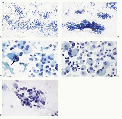
Fig. 8.4: FNA of a Medullary Thyroid Carcinoma. A: Low power showing the characteristic dispersed cell pattern. B: Medium power. Note the syncytial tissue fragments such as seen here are less frequently present. C: Higher magnification showing the discrete, medium-sized to large, pleomorphic, and plasmacytoid neoplastic cells. Binucleation is frequent. Note the intranuclear inclusions (arrow). D: The medullary carcinoma cells are large to giant form with multinucleation. The nuclei have coarse chromatin and exhibit extreme eccentric location. E: Same case, different field, showing a pleomorphic cell pattern with small round cells containing scant cytoplasm, while some cells have cytoplasmic processes.


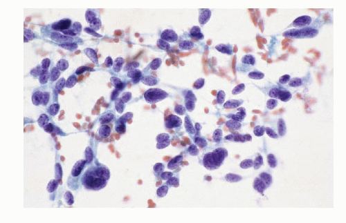
Fig. 8.5: FNA of a Medullary Carcinoma. A pleomorphic cell pattern such as seen here is the hallmark of medullary carcinoma.


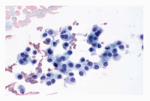
Fig. 8.6: FNA of a Medullary Carcinoma. The cells are very uniform, medium-sized, and plasmacytoid with moderate amount of cytoplasm. These cells strongly resemble either follicular cells or Hürthle cells and may be interpreted as follicular or Hürthle cell neoplasm.


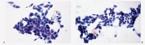
Fig. 8.7: FNA of a Medullary Carcinoma. A: Loosely cohesive medium-sized to large cells consisting of short spindle and cuboidal cells with high N/C ratios and dense nuclear chromatin. B: Another field from the same case showing predominantly small cells with poorly defined cell borders and high N/C ratios.


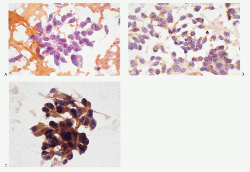
Fig. 8.8: FNA of a Medullary Carcinoma. A: The aspirate shows a pleomorphic cell pattern with coarsely granular nuclear chromatin pattern (salt-pepper) characteristic of a neuroendocrine tumor. Note nucleoli are not appreciated. The cells possess delicate cytoplasmic processes. B: Smear stained for calcitonin showing positive reactivity. C: Different example of medullary carcinoma. Reactivity to calcitonin is usually very strong as demonstrated by these medullary carcinoma cells.


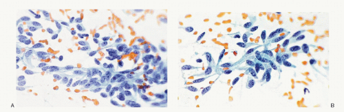
Fig. 8.9: A, B: FNA of a Medullary Carcinoma. A predominant spindle cell pattern with delicate, long, unipolar cytoplasmic processes and eccentric nuclei. Note the characteristic salt-pepper chromatin.


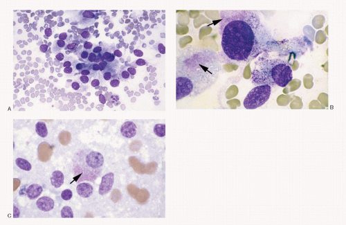
Fig. 8.10: FNA of a Medullary Carcinoma Stained with Romanowsky Stain. A: The cells are loosely cohesive. B, C: These medullary carcinoma cells contain the characteristic azurophilic granules in their cytoplasm (arrows).


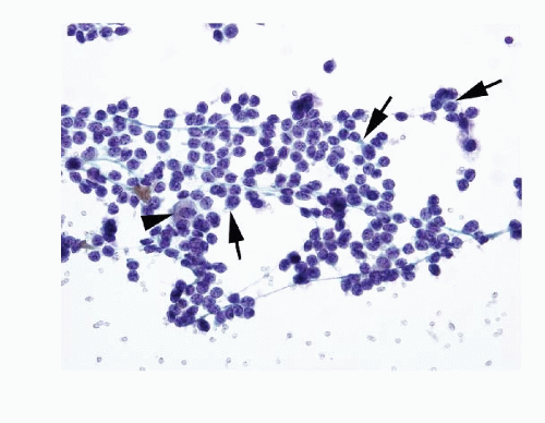
Fig. 8.11: FNA of a Medullary Carcinoma. These medullary carcinoma cells are dispersed, monomorphic, small, round with mostly poorly defined cell borders and scant cytoplasm that is indiscernible to a thin rim of dense cyanophilic (arrows). An occasional cell is large with appreciable cytoplasm. The nuclei are round with coarsely granular chromatin. There is no nuclear molding. With a monomorphic pattern of small round cells, malignant lymphoma must be considered in the differential diagnosis.


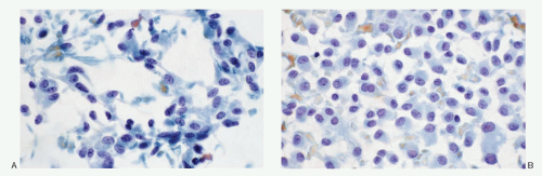
Fig. 8.12: FNA of a Medullary Carcinoma. A: The pleomorphic neoplastic cells are present singly, with many spindle forms possessing cytoplasmic processes. The background contains fluffy acellular material which may represent amyloid. B: Another field from the same case demonstrating plasmacytoid cells with minimal variation in size.


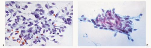
Fig. 8.13: FNA of a Medullary Carcinoma. A: The aspirate is characterized by a pleomorphic cell pattern with predominant spindle cells. Note long cytoplasmic processes, eccentric nuclei, and coarsely granular chromatin. B: Different example of medullary carcinoma with a spindle cell pattern.


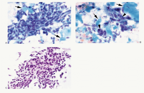
Fig. 8.14: FNA of a Medullary Carcinoma. A: The medullary carcinoma cells are spindle shaped and in fascicles with an admixture of round cells. Note the acellular cyanophilic material in close association with carcinoma cells and probably representing amyloid (arrows). B: A different field showing pleomorphic malignant cells. Note multinucleation and intranuclear inclusions (arrowheads). The background shows abundant dense acellular material that probably represents amyloid (AM). In Papanicolaou-stained smears, amyloid resembles colloid. C: The cell block of the aspirate showing a very pleomorphic, diagnostic cell pattern of medullary carcinoma (H&E).

Only gold members can continue reading. Log In or Register to continue

Stay updated, free articles. Join our Telegram channel

Jul 17, 2016 | Posted by in PATHOLOGY & LABORATORY MEDICINE | Comments Off on Neuroendocrine Tumors of the Thyroid Gland

Full access? Get Clinical Tree

Get Clinical Tree app for offline access