Neuroendocrine Tumors of the Salivary Glands


Neuroendocrine Tumors of the Salivary Glands


Neuroendocrine tumors of the salivary glands include mostly small cell neuroendocrine or poorly differentiated neuroendocrine carcinomas. Other types of neuroen-docrine tumors such as carcinoid tumors and paragan-gliomas of salivary glands are not documented in the literature. Rare examples of large cell neuroendocrine carcinoma have been reported. Salivary glands are more often the sites for metastatic neuroendocrine carcinomas. Neuroendocrine cells have been identified in intercalated ducts and mucoserous acini of the salivary glands.


SMALL CELL CARCINOMA (NEUROENDOCRINE CARCINOMA, HIGH-GRADE)

Small cell carcinomas (neuroendocrine carcinomas, grade III, or poorly differentiated) are rare primary malignant neoplasms of the salivary glands, accounting for <1% of all salivary gland tumors. Small cell carcinomas constitute 2.8% to 1.8% of all malignant tumors of the major and minor salivary gland tumors, respectively. Small cell carcinomas demonstrate cytologic, histologic, ultrastructural, and immunochemical features similar to that of pulmonary small cell carcinomas. Over 80% of these tumors occur in the parotid gland.

Small cell carcinomas typically present as painless rapidly growing mass of short duration. Most patients are in their fifth to sixth decades of life. Small cell carcinoma is slightly more common in males. Cervical lymphadenopathy is common. Primary small cell carcinoma of the salivary glands is an aggressive tumor and can metastasize widely with a fatal outcome.


GROSS AND HISTOLOGIC FEATURES

Grossly, small cell carcinoma occurs as a lobulated, poorly defined mass, white to grayish-white with necrosis and hemorrhage. The tumor may be infiltrating into the adjacent tissues.

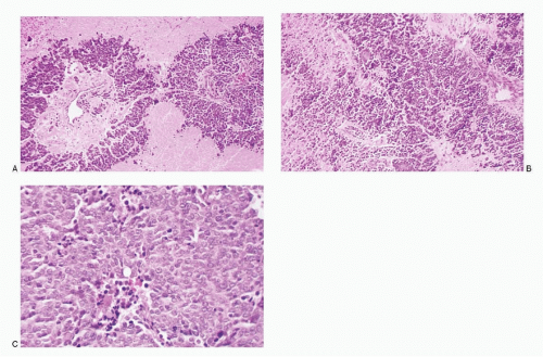
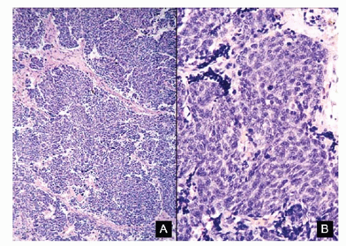
Microscopically, small cell carcinomas show a diffuse growth pattern or nests, large islands, and trabeculae of small cells with areas of necrosis, which can be very extensive (Fig. 7.1A,B). The malignant cells are round, oval to short, and spindle shaped with scant, indiscernible cytoplasm and high N/C ratios (Figs. 7.1C and 7.2). The nuclei have deep-staining, compact chromatin lacking nucleoli. Mitosis, karyorrhexis, and nuclear molding are typically seen. Many carcinomas show focal ductal or squamous differentiation (see Fig. 7.7F).


CYTOPATHOLOGIC FEATURES

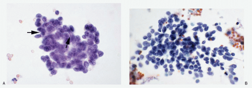
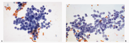
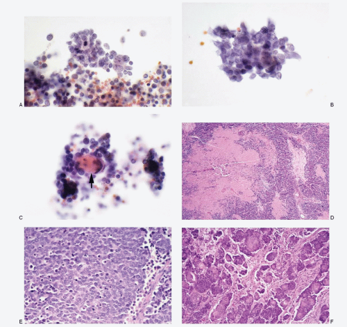
The cytologic specimen is usually a fine needle aspirate of the salivary gland mass. The cytologic features (Table 7.1; Figs. 7.3,7.4,7.5,7.6 and 7.7) are similar to those seen in pulmonary small cell carcinomas. The aspirates of small cell carcinomas present variable cellularity but are usually very cellular with the malignant cells occurring singly, in groups, or in small to large syncytial tissue fragments without any architectural patterns. Rosettes are seen only occasionally. The malignant cells are small, round, and oval to fusiform with molded nuclear borders. Their nuclei have very coarsely granular chromatin of varying intensity, often appearing compact. Nucleoli are absent to inconspicuous. The small cell carcinoma cells contain scant to insignificant cytoplasm with almost invisible cell borders giving an appearance of naked nuclei to the malignant cells. Mitotic figures are easily found. Karyorrhectic and necrotic debris is often present in the background.

The small cell carcinomas may exhibit focal squamous differentiation (Fig. 7.7C,F). Small tissue fragments of squamous cells may be present amidst the malignant cells. The cells of small cell carcinoma with scant cytoplasm tend to air-dry quickly, losing cellular
details, often making cytologic interpretation very difficult. The neoplastic cells are also very fragile and exhibit stretch artifacts that result from the smearing process. In Romanowsky-stained preparations, the cells of small cell carcinoma often demonstrate paranuclear vacuoles.


Fig. 7.1: A: Histologic section of a primary small cell carcinoma of the parotid gland showing large areas of tumor necrosis (medium power, H&E). B: Another field showing islands of small malignant cells with scant cytoplasm and high N/C ratios (medium power, H&E). C: Higher magnification with a solid growth pattern and focal necrosis (high power, H&E).


Fig. 7.2: A: Another example of a primary small cell carcinoma of the parotid gland, showing islands of small cells with peripheral palisading (medium power, H&E). B: Higher magnification of (A) (H&E).


IMMUNOPROFILE

The small cell carcinoma cells of the salivary glands react positively to pan-neuroendocrine markers, pancytokeratin (AE1/AE3, CAM5.2), and CK20. They do not react to TTF-1, S100-protein, and CK7. With cytokeratin, typical punctate pattern of staining is noted.


ULTRASTRUCTURE

Ultrastructurally, the small cell carcinoma cells contain neurosecretory granules.


DIFFERENTIAL DIAGNOSES

The cytomorphology of primary small cell carcinomas (neuroendocrine carcinoma, grade III) of the salivary glands is identical to that of small cell carcinomas occurring at other body sites especially the lung. From morphology alone, these tumors cannot be distinguished from each other. Positive immunoreactivity to


cytokeratin 20 has been used to differentiate primary salivary gland small cell carcinoma and/or metastatic Merkel cell carcinoma from pulmonary small cell carcinoma.


TABLE 7.1 CYTOPATHOLOGIC FEATURES OF SMALL CELL CARCINOMA (NEUROENDOCRINE CARCINOMA) OF THE SALIVARY GLANDS


Cellularity


Scant to very cellular


Presentation


Malignant cells discrete, in aggregates or in syncytial tissue fragments usually without any architectural patterns; rosettes infrequent; crush artifacts may be extensive resulting in poor cytopreparation


Cells


Small, round, 2.5-3 times the size of a resting lymphocyte to slightly larger, oval to short spindle shaped, cell borders poorly defined, and often flush with the nucleus


Nucleus


Round, oval to oblong; multinucleation not observed; N/C ratios very high; nuclear membranes smooth; nuclear chromatin very coarsely granular to compact and deep-staining; nucleoli inconspicuous; nuclear molding +; mitoses +; karyorrhexis +; stretch artifacts +


Cytoplasm


Indiscernible to very scant; paranuclear vacuoles in Romanowsky stained preparations; squamous differentiation present occasionally


Background


Dirty; necrosis; fragmented forms


Immunoprofile


Neuroendocrine markers +; pancytokeratin (AE1/AE3) +; CD56+; CD57+; TTF-1 -; CK20 +; S100 protein –


Differential diagnoses


Metastatic small cell carcinoma


Merkel cell carcinoma


Metastatic poorly differentiated carcinomas


Basaloid squamous carcinoma


Basal cell adenoma/basal cell adenocarcinoma


Cutaneous basal cell carcinoma (from skin overlying parotid gland)


Pleomorphic adenoma with a predominant basaloid cell pattern


Reactive intraparotid lymph node


Malignant non-Hodgkin lymphoma


From Kini SR, Dardick I. Salivary Gland Tumor. Cytopathology. CD ROM, Ottawa, Pathology Images Inc. 2006.


Fig. 7.3: A, B: Fine needle aspiration (FNA) biopsy of a parotid mass showing syncytial tissue fragments and loosely cohesive small cells with round to oval nuclei, compact chromatin, high N/C ratios, and nuclear molding (arrows), consistent with a diagnosis of small cell carcinoma.


Fig. 7.4: A, B: FNA biopsy of a parotid mass showing typical cytomorphology of a small cell carcinoma. Note round, oval to oblong nuclei, poorly defined cell borders, indistinct cytoplasm, and deep-staining coarsely granular to compact chromatin. Nuclear molding is easily appreciated.


Fig. 7.5: A, B: The malignant cells from this case of small cell carcinoma demonstrate intense staining of the nuclear chromatin. Note nuclear molding.


Fig. 7.6: A, B: These malignant cells from another case of primary small cell carcinoma of the parotid are slightly larger but show dark-staining chromatin and nuclear molding.


Fig. 7.7: A, B: The aspiration biopsy specimen from a parotid gland mass shows syncytial tissue fragments of small cells with marked crowding and overlapping. Nuclear molding is evident. Note that the single malignant cells in the background are rounded and strongly resemble the lymphocytes. C: The tissue fragment of small cell carcinoma cells shows squamous differentiation. Note the squamous pearl (arrow). Excision of the parotid mass confirmed a small cell carcinoma. D: A medium power view showing a neoplasm with broad areas of necrosis (medium power, H&E). E: Higher magnification showing carcinoma with peripheral palisading (H&E). F: Areas within the carcinoma also demonstrated marked squamous metaplasia (high power, H&E).

The cytologic diagnosis of small cell carcinoma is easily made because of the characteristic cytomorphology. Since primary small cell carcinomas of the salivary gland are extremely rare (<1% of salivary malignancies) and primary small cell carcinomas of the lung are more common, it is prudent to rule out a metastatic process in the salivary glands before diagnosing the aspirate as a primary lesion. The differential diagnostic considerations include all the lesions that are composed of small cells as listed in Table 7.2.


TABLE 7.2 DIFFERENTIAL DIAGNOSES OF SALIVARY GLAND TUMORS WITH A SMALL CELL PATTERN


Diagnostic Entities


Cytopathology Features


See Figure(s)


Small cell carcinoma; metastatic, from lung


(NEC)


Neoplastic cells discrete, in loosely cohesive groups or in syncytial tissue fragments with no architectural patterns; cells small, round to oval, occasionally oblong to spindle shaped; nuclear membranes smooth to irregular; chromatin deep-staining and coarsely granular; nucleoli inconspicuous to absent; nuclear molding +; stretch artifacts +; mitoses +; karyorrhexis +; cytoplasm scant to indiscernible; cellular necrosis +; Immunoprofile: CK (AE1/AE3) +; CK 20-; neuroendocrine markers +; S100 protein -; LCA -; TTF-1 +


Figure 7.8


Merkel cell carcinoma


Neoplastic cells discrete, in loosely cohesive groups with no architectural pattern; cells small, round to oval; nuclear membranes smooth to irregular; chromatin deepstaining, coarsely granular; nucleoli inconspicuous to absent; nuclear molding +; stretch artifacts +; mitoses +; karyorrhexis +; cytoplasm scant to indiscernible; necrosis +; Immunoprofile: CK20 +; neuroendocrine markers +; TTF-1 -; history of Merkel cell carcinoma helpful


Figure 7.9


Poorly differentiated carcinomas


Malignant cells isolated, in loosely cohesive groups or in syncytial tissue fragments with no architectural patterns; cells small, round, oval to cuboidal; occasionally oblong to spindle shaped; nuclear membranes smooth to irregular; chromatin deep-staining and coarsely granular; parachromatin clearing; nucleoli usually conspicuous; no nuclear molding; mitoses +; karyorrhexis +/-; cytoplasm variable, scant to indiscernible; cellular necrosis +; Immunoprofile: CK +; neuroendocrine markers -; S100 protein -; LCA –


Figure 7.10


Basaloid squamous cell carcinoma


Malignant cells isolated, in loosely cohesive groups or in syncytial tissue fragments with no architectural patterns; cells small, round, oval to cuboidal; occasionally oblong to spindle shaped; nuclear membranes smooth to irregular; chromatin deep-staining and coarsely granular; parachromatin clearing; nucleoli usually conspicuous; no nuclear molding; mitoses -; karyorrhexis +/-; cytoplasm variable, scant to indiscernible; cellular necrosis -; Immunoprofile: CK +; neuroendocrine markers -; S100 protein -; LCA -; p53 +


Figure 7.11


Adenoid cystic Carcinoma


Neoplastic cells mostly in syncytial tissue fragments with intense crowding and overlapping, forming three-dimensional cell balls or in tight aggregates; cribriform pattern +/-; cylindrical cores of acellular, homogeneous hyaline material within the tissue fragments or in extracellular locations, staining intensely magenta pink with Romanowsky stain and pale green with Papanicolaou stain; neoplastic cells small with high N/C ratios; cell borders poorly defined; nuclei round to oval with smooth nuclear membranes; finely granular to compact chromatin; no nuclear molding; nucleoli inconspicuous to absent; no mitoses; no necrosis; background clean; bare nuclei +/-; Immunoprofile: CK +; S100 protein +; antibasement membrane antibody +; calponin +; p63 +


Figure 7.15


Basal cell adenocarcinoma


Aspirate usually cellular; neoplastic cells discrete, in aggregates and in syncytial tissue fragments with or without branching and with crowding and overlapping of nuclei; peripheral palisading of nuclei may be present within the tissue fragments; cells small, uniform to slightly pleomorphic and enlarged; lymphocyte-like; high N/C ratios; scant to indiscernible cytoplasm; cell borders poorly defined; nuclei round to oval with smooth nuclear membranes; finely granular to compact chromatin; no nuclear molding; nucleoli inconspicuous to absent; no mitoses; no necrosis; background clean; bare nuclei +/-; Immunoprofile: CK +; S100 protein +


Figure 7.13


Basal cell adenoma


Aspirate usually cellular; neoplastic cells discrete, in aggregates and in syncytial tissue fragments with crowding and overlapping of nuclei; cells small, uniform, lymphocyte-like; high N/C ratios; scant to indiscernible cytoplasm; cell borders poorly defined; nuclei round to oval with smooth nuclear membranes; finely granular to compact chromatin; no nuclear molding; nucleoli inconspicuous to absent; no mitoses; no necrosis; background clean; Immunoprofile: CK +; S100 protein +


Figure 7.12


Pleomorphic adenoma with a predominant basaloid cell pattern


Small basaloid cells, mostly in syncytial tissue fragments or in tight aggregates, acinar pattern with hyaline globules +/-; cells small with high N/C ratios; poorly defined cell borders; round to oval nuclei with smooth nuclear membranes; coarsely granular to compact chromatin; nucleoli inconspicuous; scant cytoplasm; clean background; fibrillar stroma with aggregates of neoplastic cells in a diagnostic clue; Immunoprofile: CK +; S100 protein +; calponin +; p63 +;


Figure 7.16


Cutaneous Basal Cell Carcinoma (from skin overlying salivary gland)


Aspirate usually cellular; neoplastic cells discrete, in aggregates and in syncytial tissue fragments with or without branching and with crowding and overlapping of nuclei; peripheral palisading of nuclei may be present within the tissue fragments; cells small, uniform to slightly pleomorphic and enlarged; lymphocyte-like; high N/C ratios; scant to indiscernible cytoplasm; cell borders poorly defined; nuclei round to oval with smooth nuclear membranes; finely granular to compact chromatin; no nuclear molding; nucleoli inconspicuous to absent; no mitoses; no necrosis; background clean; bare nuclei +/-


Figure 7.14


Reactive intraparotid lymph node


Polymorphic lymphoid cell population with germinal center cells; tingible body histiocytes


Figure 7.17


Malignant non-Hodgkin lymphoma


Dissociated cell pattern; lymphoma cells discrete and in aggregates; small, monomorphic pattern with well to poorly defined cell borders; high N/C ratios; nuclei round to oval with smooth to irregular nuclear membrane; finely granular chromatin with parachromatin clearing; prominent nucleoli; cytoplasm variable but scant; no nuclear molding; mitoses +; karyorrhexis +; Immunoprofile: CK -; LCA +; B/T-cell markers +; neuroendocrine markers -; S100 protein –


Figure 7.18

Only gold members can continue reading. Log In or Register to continue

Stay updated, free articles. Join our Telegram channel

Jul 17, 2016 | Posted by in PATHOLOGY & LABORATORY MEDICINE | Comments Off on Neuroendocrine Tumors of the Salivary Glands

Full access? Get Clinical Tree

Get Clinical Tree app for offline access