Neuroendocrine Tumors of the Pancreas


Neuroendocrine Tumors of the Pancreas


Pancreatic neuroendocrine neoplasms, previously referred to as pancreatic endocrine tumors (PETs), are uncommon, constituting 1% to 2% of all pancreatic neoplasms. In the United States, their incidence is estimated at <1 per 100,000 people. Pancreatic neuroendocrine neoplasms were previously referred to as “islet cell tumor/islet cell carcinoma,” because of the assumption that they arise from the islets of Langerhans. Pancreatic endocrine neoplasms are now considered to be arising from the multi-potential epithelial cells in pancreatic ductules.


ENDOCRINE PANCREAS


HISTOLOGY AND CYTOLOGY

Majority of the pancreas (85%) consists of exocrine component that includes acinar tissue and ductal system (Fig. 5.1) and the remaining represents the endocrine tissue. Ninety percent of all pancreatic endocrine cells in the adult pancreas reside in the islets of Langerhans (Fig. 5.1 A). Fewer than 10% are distributed singly or in clusters among the ductal cells or paraductular acinar cells. The islets consist of aggregates of lightly staining endocrine cells, well demarcated from the surrounding exocrine tissue. They are embedded in reticulin and collagen fibers and lack a true capsule. Two types of islets are recognized based on their architecture and location: ordinary or regular islets and pancreatic polypeptide (PP)-rich irregular islets. The regular islets are evenly distributed in the anterior part of the head, body, and tail of the pancreas. They are round to ovoid, arranged into ribbons or lobules. The diameter of the ordinary islets ranges from 75 to 225 µm. The PP-rich irregular islets are large, 400 to 500 µm, and have trabecular architecture. The islets are penetrated by a rich network of anastomosing sinusoids. The innervation of the islets are derived from sympathetic and parasympathetic nerve fibers, closely associated with the blood vessels.

Each islet of Langerhans is composed of approximately 1,000 endocrine cells, filled with characteristic neurosecretory granules. Four major cell types are recognized in the islets, namely, alpha secreting glucagons, beta secreting insulin, delta secreting somatostatin, and PP cells secreting PP. Each islet cell produces a single peptide. Ultrastructurally, secretory granules of each type have distinctive appearance: (1) Alpha-cell granules measure 180 to 300 nm. They are round and contain an eccentric electron-dense core within a less dense peripheral region. There is a thin halo beneath the limiting membrane. These constitute 15% to 20% of total endocrine cells. (2) Beta-cell granules measure 225 to 375 nm constituting 60% to 70% of total endocrine cells. They are polymorphous and contain crystalline cores with a wide halo beneath the limiting membrane. (3) Delta-cell granules constitute 2% to 10% of total endocrine cells. The granules measure 170 to 220 nm and are round with a moderately dense core surrounded by a very thin halo. (4) The granules of PP cells constitute 15% to 20% of total endocrine cells and measure 138 to 208 nm. The granules are smaller and have homogeneous hypodense cores.


PANCREATIC NEUROENDOCRINE NEOPLASMS

Pancreatic neuroendocrine neoplasms or tumors (NETs) were classified by the World Health Organization (WHO) in 2010 (see Appendix) on the bases of criteria similar to those for neuroendocrine neoplasms of the gastrointestinal tract (see Chapter 4; Appendixes I and II). This classification also introduces a grading system determined by proliferative rate replacing the grade-and stage-based system as recommended in 2004 WHO classification.


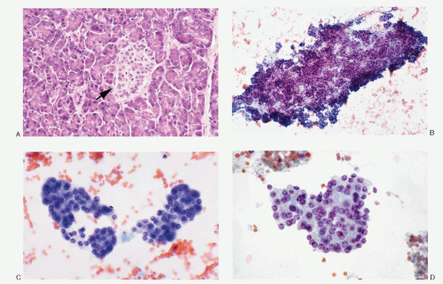
Fig. 5.1: Normal Histology and Cytology of the Pancreas (Exocrine and Endocrine). A: Histologic section of the pancreas showing acinar tissue formed by large triangular cells containing abundant eosinophilic granular cytoplasm, basally located round nuclei. An islet of Langerhans is seen in the center of the field (arrow). The component cells (neuroendocrine) of the islet are smaller, with scant, pale eosinophilic cytoplasm, uniform round nuclei, and high N/C ratios (H&E). B: Fine needle aspiration (FNA) biopsy specimen showing normal pancreatic acinar tissue (low power). C, D: Higher magnification depicting the cytomorphology of the acinar cells. Normal islet cells usually not seen in the aspirated samples.


PANCREATIC NEUROENDOCRINE TUMORS

Majority of the pancreatic neuroendocrine neoplasms fall in the category of pancreatic NET Grade I and Grade II, previously classified (WHO, 2004) as well-differentiated endocrine tumors, well-differentiated endocrine tumor with unknown potential, and well-differentiated carcinoma. Cytologically, these three types cannot be differentiated and are described together.

PETs can be nonfunctioning (30% to 40%) or functioning with elaboration of a number of different hormones. The relative frequency of PETs is estimated to be 30% for insulinoma, 16% to 30% for gastrinoma, 10% for vasoactive intestinal polypeptide-secreting tumor, 5% to 6% for glucagonoma, and 25% to 36% for nonfunctioning tumors.

PETs occur either sporadically or as a part of hereditary syndromes. The sporadic tumors are solitary, whereas pancreatic tumors in hereditary settings such as multiple neuroendocrine neoplasia Type I and von Hippel-Lindau disease are frequently multiple (Table 5.1).


CLINICAL FEATURES

The mean reported age at presentation for pancreatic NET is 58 years, with a range of 12 to 78 years. The patients with hereditary syndromes are usually younger with a mean age of 35 years. There is no gender predilection. The presenting symptoms in nonfunctioning tumors are related to the size of the tumor, which remains silent for a long period of time. The presenting symptoms include abdominal pain, cachexia, weight loss,
and jaundice. In case of functioning tumors, the presenting signs depend on the effects of hormone secreted by the tumor.


TABLE 5.1. PANCREATIC NEUROENDOCRINE TUMORS (NETS) IN HEREDITARY SYNDROMES


Syndrome


Inheritance


Frequency of Pancreatic Tumors


Location of the Gene and (Gene Product)


Sites for Associated Tumors


MEN type 1


Autosomal dominant


More than 60%; usually nonfunctional; multiple microadenomas; functioning tumors— gastrinomas and 20% insulin producing


11q13(610-amino acid protein, MENIN)


Anterior pituitary (pituitary adenoma) Parathyroid glands (hyperplasia and adenoma) GI Tract NET (stomach and duodenum), gastrinomas associated with Zollinger-Ellison syndrome Adrenal cortex


von Hippel-Lindau disease


Autosomal dominant


2%-12%; always nonfunctioning


3p25.5 (213-amino acid protein)


Renal cell carcinoma


Hemangioblastoma


Pheochromocytoma


von Recklinghausen disease


Autosomal dominant


6% somatostatinoma


17q11.2 (2485-amino acid protein, neurofibromin)


Neurofibromas


Most of the pancreatic NETs follow an indolent course; they may metastasize to distant organs several years after the initial diagnosis have been made. The organs most frequently involved are liver, lymph nodes, and lungs. More than 90% of insulinomas are benign and cured by complete surgical removal.


RADIOGRAPHIC FINDINGS

Radiographically, pancreatic neuroendocrine can be detected as localized mass lesions. Magnetic resonance imaging can also detect peripancreatic lymph node and liver involvement.


GROSS AND MICROSCOPIC FEATURES

Regardless of the functioning status of the tumor and the type of hormone elaborated by the tumor, the gross findings and the cytohistomorphology remain similar in all PETs. Grossly, PETs (Fig. 5.2) are well demarcated and well circumscribed, may or may not be encapsulated, and can occur anywhere in the pancreas but commonly involve body and the tail. Their size is variable. The functioning tumors are usually smaller than the nonfunctioning ones. The cut surface may be soft, red to yellow-tan, hemorrhagic, or grey and firm with fibrosis, or may show extensive cystic change. Larger tumors may show lobulations. Cystic change is frequent.

Microscopically, several architectural patterns are recognized (Fig. 5.3): (1) trabecular (gyriform), (2) acinar, and (3) solid or diffuse. The sheets or nests of cells may be separated by strands of fibrous tissue or closely packed with a thin vascular stroma. Frequently cells are arranged around the blood vessels or spaces with eosinophilic material. The growth pattern in these tumors varies


considerably within the same tumor. Although the architectural pattern suggests the endocrine nature, there is no reliable relationship between architectural patterns and the cell type, hormone production, or biologic behavior. The tumor cells are generally uniform in size and shape. They may be round, polygonal, or elongated. The cell borders are usually well-defined. The cytoplasm is eosinophilic or clear. The nuclei are uniform, and the nucleoli range from inconspicuous to prominent. The salt-pepper chromatin is characteristic but not universally present. Pleomorphic large cells may be scattered throughout the tumor. Mitoses are generally rare. The cytoplasm is usually scant but can be appreciable and eosinophilic in oncocytic variants. PETs are richly vascular with a prominent capillary network. Hyalinization of the tumor stroma is common and may be extensive. Amyloid is sometimes present, especially in insulinomas.


Fig. 5.2: Gross Photograph of Pancreatic Neuroendocrine Tumor. Well-defined, large tumor with a bulging cut surface.


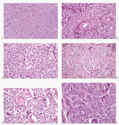
Fig. 5.3: Spectrum of Histologic Features of Pancreatic NETs. A: Histologic section showing a solid growth pattern (low power, H&E). B: Medium-sized cells with modest eosinophilic cytoplasm, central to eccentric uniform nuclei (H&E). C: A closer view of a PET showing round to cuboidal cells with moderate amounts of eosinophilic cytoplasm. The nuclear chromatin is granular (H&E). D: Broad trabeculae separated by abundant stroma (H&E). E: Higher magnification of D showing uniform cells with insignificant cytoplasm (H&E). F: Neuroendocrine tumor with broad trabeculae and insulae consisting of moderately pleomorphic nuclei. The cells are larger with appreciable eosinophilic cytoplasm.


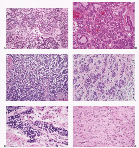
Fig. 5.3: (continued) G: Neuroendocrine carcinoma depicting an insular pattern consisting of small uniform cells (H&E). H: Neuroendocrine tumor presenting an acinar and a pseudopapillary pattern (H&E). I: This neuroendocrine tumor consists of delicate cords with an anastomosing pattern (H&E). J: Fibrous tissue strands separating islands of neoplastic cells (H&E). K: Higher magnification showing smaller neoplastic cells with hyperchromatic nuclei (H&E). L: This pancreatic neuroendocrine tumor shows cords and nests of uniform tumor cells diffusely infiltrating the extensive fibrous stroma (H&E).


HISTOCHEMISTRY

The pancreatic neuroendocrine tumor cells are both argentophilic and argyrophilic and stain positively with Fontana-Masson and Grimelius, respectively. Periodic acid-Schiff (PAS) and mucin stains are negative.


IMMUNOPROFILE

PETs react positively to cytokeratin and pan-neuroen-docrine markers such as chromogranin, synaptophysin, neuron-specific enolase, PGP 9.5, CD56, or CD57. In addition, peptide hormones can be detected by using specific antibodies. Immunohistochemical assessment of specific hormone production is valuable and correlates with the clinical symptoms and the blood profile.


ULTRASTRUCTURE

Ultrastructurally, the pancreatic NETs demonstrate dense core neurosecretory granules, ranging from 80 to 300 nm in diameter and with a tendency to cluster around Golgi complexes. Two subtypes of neurosecretory granules present a characteristic ultrastructural pattern that relates to the product synthesized. The first displays a crystalline core and an irregular submem-branous halo and correspond to insulin granules (see Chapter 1; Fig. 7.1B). The second shows dual density of an internal core structure, with only an indistinct halo and no crystalline configurations. This corresponds to glucagon granules.


CYTOPATHOLOGIC FEATURES

The specimens for cytopathologic diagnosis include mostly fine needle aspiration (FNA) biopsies of the pancreatic mass lesions via endoscopic ultrasound (EUS) guidance. Brushings of the biliary ducts and pancreatic ducts obtained via endoscopic retrograde cholangiopancreatography (ERCP) are also utilized. Metastatic tumors can be identified by fine needle biopsies. The deep-seated organs are sampled mostly under radiologic guidance. Body cavity fluids are also evaluated for the metastatic spread to the serous membranes (Table 5.2).

The cytologic presentation of the pancreatic NETs is characteristic (Table 5.3; Figs. 5.4,5.5,5.6,5.7,5.8,5.9,5.10,5.11,5.12,5.13,5.14,5.15,5.16 and 5.17). The aspirates are generally very cellular. The neoplastic cells are small to medium-sized and are either uniform and monomorphic or may demonstrate pleomorphism. They present themselves as isolated and in loosely cohesive groups forming a dispersed cell pattern (Fig. 5.11D) or in syncytial tissue fragments. The latter may present trabecular pattern, insulae, rosettes or may form no architectural configuration. The neoplastic cells are small to medium-sized, either round, cuboidal to plasmacytoid, and very rarely spindle shaped (Fig. 5.15) with central or eccentric nuclei. Spindle and large giant forms are occasionally present. A given case of pancreatic NETs may show a monomorphic pattern


(Figs. 5.7, 5.9,5.10,5.11 and 5.12, and 5.14) or a pleomorphic pattern (Figs. 5.4,5.5 and 5.6, 5.8, 5.9, and 5.13). The nuclei are round to oval; nuclear membranes are smooth and crisp. The chromatin is characteristic of the neuroendocrine family of tumors, being uniformly and coarsely granular presenting a salt-pepper pattern. Nucleoli are not consistently present. The nuclei tend to be uniform but may be variable in size. The cytoplasm ranges from scant to moderate, pale to dense, and is eosinophilic in oncocytic variant. Capillary network may be prominent in the background and neoplastic cells may be seen in perivascular location. Mitotic activity or necrosis is usually not present but can be seen in widely invasive NETs (Fig. 5.5). The cell block preparations can be extremely useful in the diagnostic evaluation since they allow ancillary testings.


TABLE 5.2. TYPES OF SPECIMENS FOR CYTOPATHOLOGIC DIAGNOSIS OF PANCREATIC ENDOCRINE TUMORS


  • Endoscopic brushings/rinsings of pancreatic duct under


    (1) Endoscopic retrograde cholangiopancreatography (ERCP)


    (2) Endoscopic ultrasound guidance (EUS)


  • EUS-guided FNA biopsy of the pancreatic or peri-pancreatic lesions (lymph nodes)


  • Intraoperative FNA


  • FNA of the metastatic lesions


  • Peritoneal/pleural fluids


TABLE 5.3. CYTOPATHOLOGIC FEATURES OF PANCREATIC NEUROENDOCRINE TUMORS


Cellularity


Usually very cellular


Presentation


Cells discrete, in loosely cohesive groups or in syncytial tissue fragments; dispersed cell pattern frequent; tissue fragments may be trabecular, form well-defined nests (insulae) or acinar pattern; solid cell groups may be traversed by branching blood vessels; cells attached to the blood vessels


Cells


Small to medium-sized with occasional large forms; round to cuboidal to plasmacytoid, sometimes polygonal and rarely triangular or spindle shaped; usually a monomorphic pattern; cell borders poorly defined with high N/C ratios


Nucleus


Central to eccentric; round to oval; smooth nuclear membranes; salt-pepper chromatin; micronucleoli +/-; molding, mitotic activity and karyorrhexis are not the features of well-differentiated neoplasms


Cytoplasm


Variable; insignificant to scant; pale to granular; abundant and eosinophilic in oncocytic variant


Background


Usually clean; necrotic in high-grade lesions or with cystic change; hemorrhagic +/-


Histochemistry


Positive reactivity with silver stains; argentophilic and argyrophilic


Immunoprofile


Positive reactivity with panneuroendocrine markers and cytokeratin; specific peptide markers depending on tumor type


Ultrastructure


Membrane-bound neurosecretory granules


Differential diagnoses


Well-differentiated duct adenocarcinoma with small cell pattern


Acinar cell carcinoma


Solid pseudopapillary tumor


Pancreatoblastoma (rare)


Other NETs


Malignant lymphoma


Serous microcystic adenoma (rare) with cystic NETs


Normal acinar tissue


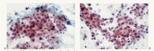
Fig. 5.4: A-C: FNA of a Pancreatic Neuroendocrine Tumor. The cellular aspirate consists of small to medium-sized cells in loosely cohesive groups and syncytial tissue fragments. The cells have poorly defined cell borders, scant cytoplasm, and pleomorphic nuclei containing coarsely granular chromatin. Some contain nucleoli.


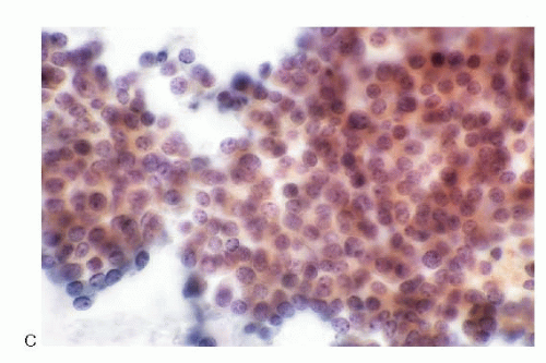
Fig. 5.4: (continued)


Fig. 5.5: FNA of Pancreatic Neuroendocrine Tumor. A: This aspirate consists of discrete cells that are uniform, small with poorly defined cell borders, high N/C ratios. The nuclear chromatin is typical salt-pepper type. B: This field shows an area of necrosis (arrow).


Fig. 5.6: A, B: FNA of a Pancreatic Neuroendocrine Tumor. The aspirate reveals dissociated cell pattern. The neoplastic cells are pleomorphic in size and shape. The nuclear chromatin is salt-pepper type.


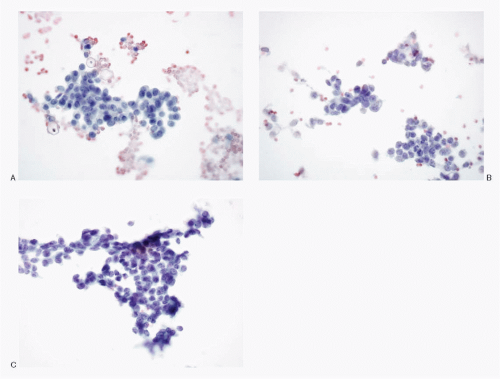
Fig. 5.7: A-C: FNA of a Pancreatic Neuroendocrine Tumor. The cellular aspirate shows syncytial tissue fragments of uniform small cells with bland chromatin pattern.


Fig. 5.8: FNA of a Pancreatic Neuroendocrine Tumor. A: This image shows a dispersed pattern of plasmacytoid cells with occasional giant forms. B: Same case stained by H&E. (Courtesy of Dr. Mithra Baliga, University of Mississippi, Jackson, Mississippi.)

Only gold members can continue reading. Log In or Register to continue

Stay updated, free articles. Join our Telegram channel

Jul 17, 2016 | Posted by in PATHOLOGY & LABORATORY MEDICINE | Comments Off on Neuroendocrine Tumors of the Pancreas

Full access? Get Clinical Tree

Get Clinical Tree app for offline access