NASH
Matthew M. Yeh, MD, PhD
Key Facts
Etiology/Pathogenesis
Associated conditions
Diabetes, obesity, hyperlipidemia, dyslipidemia, drugs, malabsorption, malnutrition
Clinical Issues
Patients often have metabolic syndrome
Transaminases usually elevated
May have positive ANA
Microscopic Pathology
Zone 3 injury pattern
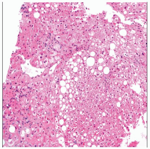
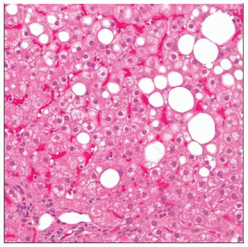
Steatosis (predominantly macrovesicular)
Lobular inflammatory infiltrate
Ballooned hepatocytes
Zone 3 perivenular/pericellular (“chicken wire”) fibrosis
Mallory-Denk bodies
Megamitochondria
Glycogenated nuclei
NASH in pediatric population has different injury pattern
Inflammation, fibrosis accentuated in portal region
Ballooning degeneration and perisinusoidal fibrosis not obvious
Top Differential Diagnoses
Steatosis without specific liver injury
Alcoholic hepatitis
Chronic hepatitis C
Autoimmune hepatitis
Glycogenic hepatopathy
Microvesicular steatosis
Wilson disease
TERMINOLOGY
Abbreviations
Nonalcoholic steatohepatitis (NASH)
Definitions
Steatosis, inflammation, and liver cell injury in absence of excessive alcohol use history
ETIOLOGY/PATHOGENESIS
Mechanism
Abnormal accumulation of lipids in hepatocytes provide source of oxidative stress
Leads to injury/inflammation
Subsequent activation of TGF-β and hepatic stellate cells results in liver fibrosis
Associated Conditions
Diabetes, obesity, hyperlipidemia, dyslipidemia, drugs, malabsorption, malnutrition
CLINICAL ISSUES
Presentation
Hepatomegaly
Metabolic syndrome
Central (visceral) obesity
Type 2 diabetes
Dyslipidemia (hypertriglyceridemia and low HDL)
Systemic hypertension
Laboratory Tests
Elevated transaminases
Treatment
Management of associated metabolic conditions (diabetes, hyperlipidemia, and dyslipidemia)
Exercise
Dietary control and weight reduction
Prognosis
May lead to liver fibrosis, cirrhosis, and hepatocellular carcinoma
MICROSCOPIC PATHOLOGY
Histologic Features
Steatosis (predominantly macrovesicular)
Lobular inflammatory infiltrate
Lymphocytes, pigmented macrophages, Kupffer cells
Typically more severe than portal inflammation
Liver cell injury
Ballooned hepatocytes
Apoptotic bodies
Hepatocyte injury predominantly zone 3
Hepatocytes may contain Mallory-Denk bodies, megamitochondria, glycogenated nuclei
Zone 3 perivenular/pericellular (“chicken wire”) fibrosis
Variants
Pediatric patients
Inflammation and fibrosis accentuated in portal (not centrilobular) region
Ballooning degeneration and perisinusoidal fibrosis not typically obvious
Stay updated, free articles. Join our Telegram channel

Full access? Get Clinical Tree


