Mycetoma
Brian J. Hall, MD
Francisco G. Bravo, MD
Key Facts
Terminology
Localized chronic granulomatous infection involving skin, subcutaneous tissue, and occasionally underlying soft tissue and bone; can be caused by either fungi or bacteria
Clinical Issues
Classic clinical triad of draining sinuses, swollen tissues, and identifiable grains in discharge are classic
Microscopic Pathology
Type 1 reaction: Grains surrounded by layer of neutrophils, then layer of chronic inflammatory cells, and finally layer of fibrous tissue
Type 2 reaction: Very similar to type 1 reaction except innermost neutrophil layer is replaced by giant cells and macrophages engulfing grains
Type 3 reaction: Well-formed epithelioid granulomas including Langhans giant cells occasionally with central remnants of fungi
Eumycetes are filaments wider than 1 µm often forming mass of hyphae in intercellular cement
Top Differential Diagnoses
Botryomycosis (bacterial pseudomycosis)
Sporotrichosis
Chromomycosis
Subcutaneous phaeohyphomycosis
Hyalohyphomycosis
Majocchi granuloma
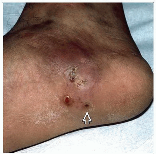 Mycetoma of the right foot from a patient stationed in Guam demonstrates several draining sinus tracts  (present for months) with tissue swelling and slight hyperpigmentation. (Courtesy J. Steger, MD.) (present for months) with tissue swelling and slight hyperpigmentation. (Courtesy J. Steger, MD.) |
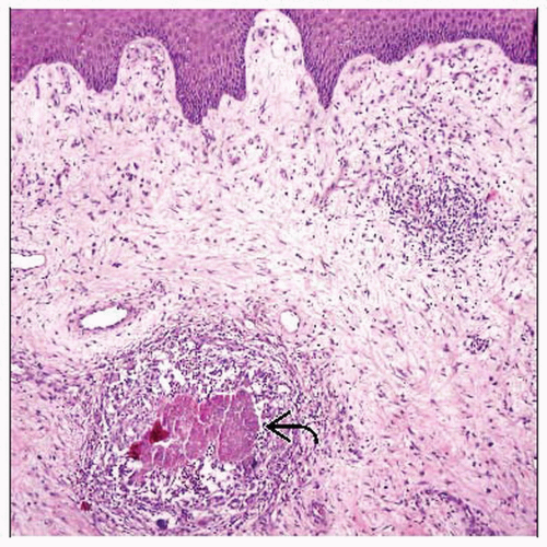 Although usually more deep-seated, characteristic pale grains of eumycetoma  are seen surrounded by inflammation. (Courtesy S. Florell, MD.) are seen surrounded by inflammation. (Courtesy S. Florell, MD.) |
TERMINOLOGY
Synonyms
Madura foot, maduromycosis
Definitions
Localized chronic granulomatous infection involving skin, subcutaneous tissue, and occasionally underlying soft tissue and bone; can be caused by either fungi or bacteria
ETIOLOGY/PATHOGENESIS
Infectious Agents
Most common causal organisms are either true fungi from class Eumycetes (eumycetomas) or filamentous bacteria from class Actinobacteria (actinomycetomas)
In eumycetomas, most common causal agent worldwide is Madurella mycetomatis, although many other species can cause disease
In the USA, Pseudallescheria boydii is most common agent
In actinomycetomas, Nocardia brasiliensis and Actinomadura madurae are 2 most common isolates
CLINICAL ISSUES
Epidemiology
Incidence
Unknown due to slow progression and late presentation in most patients
Age
Most infections occur in patients aged 20-40 years
Gender
M:F ˜ 4:1
Ethnicity
Most cases occur in tropical and subtropical populations
“Mycetoma belt” includes areas between latitudes 15° south and 30° north
Includes India, Mexico, Venezuela, Colombia, Argentina, Sudan Somalia, and Senegal
Occasional case report in USA and Europe
People involved with fieldwork &/or frequent contact with soil (herdsmen, farmers, field laborers) are more commonly affected
Site
Feet most common location (˜ 70%)
Other common sites include hands, legs, knee joints, arms
Actinomycetomas of shoulder are commonly seen in Mexico, especially in lumber workers carrying logs
Presentation
Classic clinical triad of draining sinuses, swollen tissues, and identifiable grains in discharge are classic
Black discharging grains are characteristic of eumycetoma
White/yellow-colored grains (“pale grains”) can be seen in both eumycetoma and actinomycetoma
2 common histories
Initial pain and discomfort upon inoculation
No recollection of any preceding trauma
After inoculation, a painless subcutaneous nodule develops and slowly spreads
Nodule slowly increases in size, developing draining sinuses and sometimes secondary nodules and papules
Some sinuses close and heal, while other new ones are formed
Overlying skin often hyperpigmented, but occasionally hypopigmented
As lesions continue to develop, subcutaneous abscesses develop and lesions extend to involve underlying soft tissue and bone
Stay updated, free articles. Join our Telegram channel

Full access? Get Clinical Tree


