Mucoepidermoid Carcinoma of the Pleura
Key Facts
Terminology
MEC
Clinical Issues
Symptoms
Chest pain
Shortness of breath
Image Findings
Pleural-based mass
Diffuse involvement of pleura has not been recorded for these tumors
Microscopic Pathology
Epidermoid component without keratinization
Mucus-secreting cells admixed with epidermoid component
Mild cellular atypia
Low mitotic activity
Possible sclerotic stroma
Top Differential Diagnoses
Adenocarcinoma
MEC lacks the presence of malignant glandular component
Squamous cell carcinoma
Presence of keratinization unusual in MEC
Mucus-producing cells (mucocytes) admixed with epidermoid component in MEC
Synovial sarcoma
Spindle cell component in synovial sarcoma is malignant
Sclerotic component in MEC is fibroblastic benign
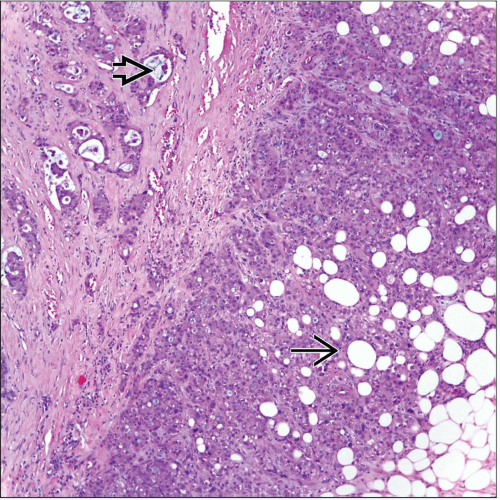 Pleural MEC shows invasion of the tumor into the adipose tissue  . Similar features may also be seen in mesotheliomas. However, note the presence of cystic structures containing mucin . Similar features may also be seen in mesotheliomas. However, note the presence of cystic structures containing mucin  . . |
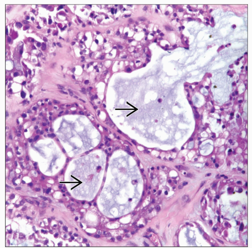 MEC shows gland-like structures of different sizes with mucoid material  . The neoplastic cellular proliferation is rather homogeneous. . The neoplastic cellular proliferation is rather homogeneous. |
TERMINOLOGY
Abbreviations
Pleural mucoepidermoid carcinoma (MEC)
Definitions
Primary salivary gland-type tumor of pleura
ETIOLOGY/PATHOGENESIS
Etiology
Although etiology of this tumor in pleura is unknown, it is possible that it may arise from entrapped glands in pleura
CLINICAL ISSUES
Epidemiology
Incidence
Exceedingly rare tumor in pleura
Age
Has been described only in adults
Gender
No gender predilection
Presentation
Chest pain
Shortness of breath
Treatment
Surgical approaches
Complete resection of tumor
Lobectomy
Decortication
Simple resection of pleural tumor
Adjuvant therapy
Possible chemotherapy
Prognosis
May be good depending on histological grade of tumor
Difficult to assess due to the few cases reported
IMAGE FINDINGS
General Features
Pleural-based mass
Diffuse involvement of pleura has not been recorded for these tumors
MACROSCOPIC FEATURES
General Features
Tumor mass well circumscribed and attached to pleura
Size
May vary from 2 cm to > 5 cm
MICROSCOPIC PATHOLOGY
Histologic Features
Epidermoid component without keratinization
Mucus-secreting cells admixed with epidermoid component
Mild cellular atypia
Low mitotic activity
Possible sclerotic stroma
Stay updated, free articles. Join our Telegram channel

Full access? Get Clinical Tree


