Metastatic Malignancies (Carcinoma, Melanoma)
Rose Anton, MD
Key Facts
Cytopathology
Hyperchromatic nuclei with sharp angulations in squamous cell carcinoma
Dense/glassy cytoplasm in squamous cell carcinoma
Vesicular chromatin and prominent nucleoli in adenocarcinomas in general
Elongated, cigar-shaped hyperchromatic nuclei in large bowel primary
Nuclear molding, increased cellular dyshesion, high N:C ratio, and coarse chromatin in high-grade NECs
Melanoma may have any nuclear pattern but often shows prominent nucleoli or intranuclear cytoplasmic inclusions and binucleation
Melanoma may or may not display cytoplasmic pigment
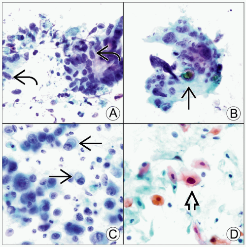 (A) On Pap stain, a smear of a colorectal adenocarcinoma often consists of elongated (cigar-shaped) hyperchromatic nuclei  in a dirty, necrotic background. (B) Malignant cells that are highly pleomorphic, often with associated psammoma bodies in a dirty, necrotic background. (B) Malignant cells that are highly pleomorphic, often with associated psammoma bodies  and cytoplasm that may contain large vacuoles, favor a papillary serous carcinoma, usually from the ovary, as seen in this Pap stain. (C) An adenocarcinoma that consists of relatively small cells and may show intracytoplasmic mucin and cytoplasm that may contain large vacuoles, favor a papillary serous carcinoma, usually from the ovary, as seen in this Pap stain. (C) An adenocarcinoma that consists of relatively small cells and may show intracytoplasmic mucin  suggests stomach or breast origin. (D) This Pap stain shows angulated, hyperchromatic nuclei suggests stomach or breast origin. (D) This Pap stain shows angulated, hyperchromatic nuclei  of squamous cell carcinoma. of squamous cell carcinoma. |
CLINICAL ISSUES


