Mesenchymal Hamartoma
Hanlin L. Wang, MD, PhD
Key Facts
Etiology/Pathogenesis
Developmental anomaly vs. neoplasm
Recurrent balanced translocation involving chromosome band 19q13.4 or 19q13.3
Clinical Issues
Typically occurring in 1st 2 years of life, rarely seen in adults
Excellent prognosis after complete excision
Malignant transformation only in rare case reports
Macroscopic Features
Large, solid, &/or cystic hepatic lesion
Microscopic Pathology
Mixture of varying portions of mesenchymal cells, bile ducts, hepatocytes, blood vessels, and cystic spaces
Top Differential Diagnoses
Hepatoblastoma
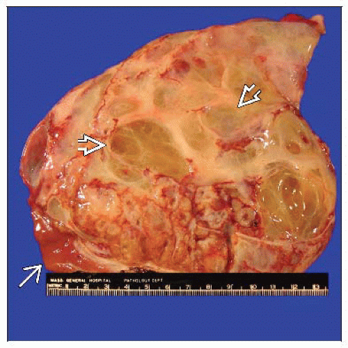 The cut surface of a large mesenchymal hamartoma shows numerous variably sized cystic spaces  admixed with solid areas. Minimal uninvolved liver tissue is present at the resection margin admixed with solid areas. Minimal uninvolved liver tissue is present at the resection margin  . . |
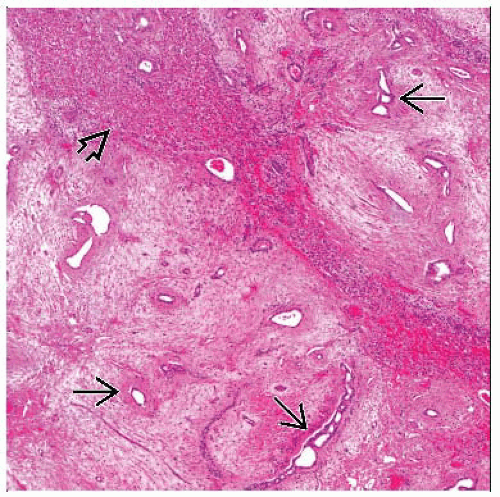 Low-power view shows numerous bile ducts  in a loose, myxoid mesenchymal stroma. Note the presence of a large island of normal-appearing hepatocytes in a loose, myxoid mesenchymal stroma. Note the presence of a large island of normal-appearing hepatocytes  within the tumor. within the tumor. |
TERMINOLOGY
Definitions
Benign mass lesion of liver primarily occurring in young children
ETIOLOGY/PATHOGENESIS


