Mesenchymal Chondrosarcoma
Key Facts
Clinical Issues
Extremely rare tumor
Predilection for young adults (15-35 years)
Slight female predilection
Incidental finding on routine chest x-rays
Aggressive malignancy
Approximately 50% 5-year survival rate
Macroscopic Features
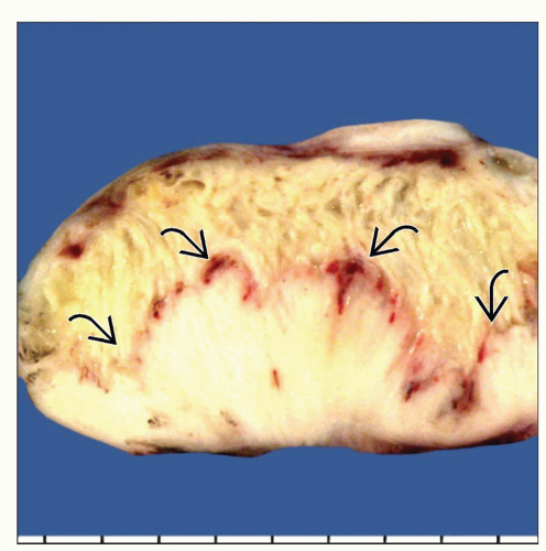
Well-circumscribed, multilobated soft tissue mass
Cut surface shows fleshy, gray-white homogeneous tissue with calcified and cartilaginous foci
May contain small foci of hemorrhage and necrosis
Average size: 5-8 cm
Microscopic Pathology
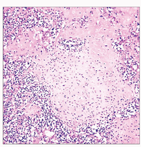
Sheets of primitive round, oval, or spindle cells
Foci of cartilaginous differentiation showing abrupt transitions with surrounding round/spindle cells
Cartilaginous foci may often show areas of calcification and ossification
Primitive spindle cell component may adopt a prominent hemangiopericytic growth pattern
Undifferentiated tumor cell component
Usually negative for S100 protein
Shows CD99 membranous positivity
Primitive small round/spindle cell component shows positivity for SOX9
Some cases show t(11;22)(q24;q12) translocation typical of the Ewing sarcoma/primitive neuroectodermal tumor family
TERMINOLOGY
Abbreviations
Mesenchymal chondrosarcoma (MC)
Definitions
Biphasic malignant neoplasm composed of primitive neoplastic small round blue cell population admixed with islands of mature cartilage
ETIOLOGY/PATHOGENESIS
Pathogenesis
Originally believed to represent a variant of chondrosarcoma due to presence of mature cartilage
Currently regarded as closely related to primitive neuroectodermal tumors (PNET) or Ewing sarcoma
CLINICAL ISSUES
Epidemiology
Incidence
Extremely rare tumor
Age
Predilection for young adults (15-35 years)
May also occur in young children
Gender
Slight female predilection
Presentation
Back pain
Dyspnea
More often asymptomatic
Incidental finding on routine chest x-rays
Treatment
Surgical approaches
Complete surgical excision
Adjuvant therapy
Radiation and chemotherapy
Prognosis
Aggressive malignancy
Approximately 50% 5-year survival rate
Metastasizes in a high percentage of cases
Metastases are most common to lung
IMAGE FINDINGS
General Features
Well-defined soft tissue mass in paraspinal location
May protrude into or involve posterior mediastinum
Irregular radiopaque stippling due to focal calcifications
MACROSCOPIC FEATURES


