Melanotic Schwannoma
Ayca Ersen, MD
Bernd W. Scheithauer, MD
Key Facts
Clinical Issues
Sporadic or syndromic (Carney complex)
Macroscopic Features
No encapsulation; occasionally interrupted, thin collagen layer with soft tissue invasion
Microscopic Pathology
Delicate chromatin/small nucleoli/rare mitoses (benign)
Vesicular nuclei/coarse chromatin/violaceous macronucleoli, frequent mitoses (malignant)
Pigmentation variable or patchy
Plus psammomatous calcifications in psammomatous variant
Ancillary Tests
Reticulin outlines lobules
S100, HMB-45, Melan-A, tyrosinase positive
Collagen IV and laminin outline basal lamina around lobules
Top Differential Diagnoses
Conventional schwannoma
Contain lipofuchsin (PAS[+]) but lack melanin
Pigmented neurofibroma
Typically diffusely infiltrative
Melanocytoma
Lack long-spacing collagen
Primary or metastatic melanoma
Cytologically malignant
Clear cell sarcoma (soft tissue melanoma)
No immunohistochemical or ultrastructural basal lamina
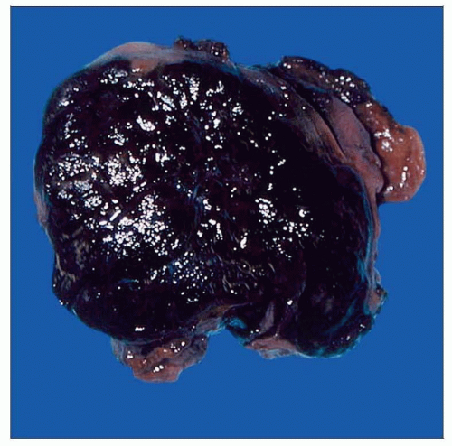 Circumscribed and ovoid like other schwannomas, melanocytic variants are variably pigmented, extremely so in this case. |
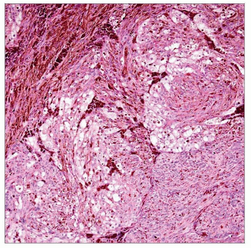 Pigmented spindle-shaped or epithelioid cells often form lobules. |
TERMINOLOGY
Synonyms
Pigmented schwannoma
Melanogenic schwannoma
Melanogenic nerve sheath tumor
Definitions
Usually benign, often circumscribed tumor of melanin-producing Schwann cells
CLINICAL ISSUES
Epidemiology
Incidence
Sporadic
Syndromic (Carney complex)
Frequent mutation of tumor suppressor gene PRKAR1A
Age
Childhood to senescence (mean: 35 years)
A decade younger in patients with Carney complex
Gender
Slight female predominance (1.5:1) in both sporadic and syndromic types
Site
Sporadic
Spinal nerves and ganglia (cervicothoracic)
Rarely multiple
Syndromic (Carney complex)
Alimentary tract
Viscera (heart, liver, lung)
Bone
15% multiple
Presentation
Sporadic
Nerve root associated tumors: Pain or sensory disturbance
Syndromic (Carney complex; autosomal dominant)
Psammomatous melanotic schwannoma: Mass effects
Lentigines (65%) of face, lips, caruncle, and female genitalia
Myxomas of heart (65%), skin (25%), breast (20%)
Blue nevi of extremities and trunk
Endocrine overactivity
Cushing syndrome (25%): Pigmented nodular adrenocortical disease
Acromegaly: Pituitary adenoma, mammosomatotrophic type
Precocious puberty (30%): Large cell Sertoli tumor of testis
Treatment
Surgical approaches
Resection with tumor-free margins for all lesions
Prognosis
Benign
Recurrence in subtotal resection
Malignant
Metastasis
Distinguish metastases from 2nd primary
Prognosis same in conventional and psammomatous variants
15% die of tumor
IMAGE FINDINGS
MR Findings
CT Findings
Calcification/ossification on CT in some psammomatous tumors
MACROSCOPIC FEATURES
General Features
0.5-26 cm (median: 5 cm)
Circumscribed
Round to sausage-shaped
Spinal nerve root tumors: Dumbbell-shaped
Occasionally lobulated or cystic
No encapsulation
Occasionally thin collagen layer with soft tissue invasion
Effects of bone
Erosion (benign tumors)
Invasive destruction (malignant tumors)
Cut surface gray to tar black
Infrequently cystic
Occasional hemorrhage or necrosis
Occasional subcapsular grittiness (psammomatous variant)
Soft to rubbery; occasionally hard
Maybe multifocal, usually when malignant
MICROSCOPIC PATHOLOGY
Histologic Features
Melanotic schwannoma
High cellularity, lobules and fascicles
Spindle to epithelioid cells
Eosinophilic to amphophilic cytoplasm
Clear cells occasional
Occasional multinucleation
Variable pigmentation
Nuclear/cytoplasmic pseudoinclusions
Pigmentation variable or patchy
Thin-walled vessels
Scan often incomplete collagenous “capsule”
Palisading, Verocay bodies, microcysts (very uncommon)
Melanophages frequent and most pigmented of cells
Benign
Delicate chromatin and small nucleoli
Rare mitoses
Malignant
Vesicular nuclei and coarse chromatin
Stay updated, free articles. Join our Telegram channel

Full access? Get Clinical Tree


